Transcription
Elaissi et al. BMC Complementary and Alternative Medicine 2012, ESEARCH ARTICLEOpen AccessChemical composition of 8 eucalyptus species'essential oils and the evaluation of theirantibacterial, antifungal and antiviral activitiesAmeur Elaissi1* , Zyed Rouis2 , Nabil Abid Ben Salem2, Samia Mabrouk5, Youssef ben Salem3, Karima Bel Haj Salah2,Mahjoub Aouni2, Farhat Farhat4, Rachid Chemli1, Fethia Harzallah-Skhiri5 and Mohamed Larbi Khouja6AbstractBackground: In 1957, Tunisia introduced 117 species of Eucalyptus; they have been used as fire wood, for theproduction of mine wood and to fight erosion. Actually, Eucalyptus essential oil is traditionally used to treatrespiratory tract disorders such as pharyngitis, bronchitis, and sinusitis. A few investigations were reported on thebiological activities of Eucalyptus oils worldwide. In Tunisia, our previous works conducted in 2010 and 2011 hadbeen the first reports to study the antibacterial activities against reference strains. At that time it was not possible toevaluate their antimicrobial activities against clinical bacterial strains and other pathogens such as virus and fungi.Methods: The essential oils of eight Eucalyptus species harvested from the Jbel Abderrahman, Korbous (North EastTunisia) and Souinet arboreta (North of Tunisia) were evaluated for their antimicrobial activities by disc diffusion andmicrobroth dilution methods against seven bacterial isolates: Haemophilus influenzae, Klebsiella pneumoniae,Pseudomonas aeruginosa, Staphylococcus aureus, Streptococcus agalactiae, Streptococcus pneumoniae andStreptococcus pyogenes. In addition, the bactericidal, fungicidal and the antiviral activities of the tested oils werecarried out.Results: Twenty five components were identified by GC/FID and GC/MS. These components were used to correlatewith the biological activities of the tested oils. The chemical principal component analysis identified three groups,each of them constituted a chemotype. According to the values of zone diameter and percentage of the inhibition(zdi, % I, respectively), four groups and subgroups of bacterial strains and three groups of fungal strains werecharacterized by their sensitivity levels to Eucalyptus oils. The cytotoxic effect and the antiviral activity variedsignificantly within Eucalyptus species oils.Conclusions: E. odorata showed the strongest activity against S. aureus, H. influenzae, S. agalactiae, S. pyogenes,S. pneumoniae and against all the tested fungal strains. In addition, E. odorata oil showed the most cytotoxic effect.However, the best antiviral activity appeared with E. bicostata. Virus pretreatment with E. bicostata essential oilshowed better antiviral activity (IC50 0.7 mg/ml, SI 22.8) than cell-pretreatment (IC50 4.8 mg/ml, SI 3.33). Theessential oil of E. astringens showed antiviral activity only when incubated with virus prior to cell infection. Thisactivity was dose-dependent and the antiviral activity diminished with the decreasing essential oil concentration.Keywords: Eucalyptus sp., Essential oil, Principal Components Analysis, Hierarchical Cluster Analysis, Antibacterialactivity, Antifungal activity, Antiviral activity* Correspondence: aelaissi@yahoo.fr1Laboratory of Pharmacognosy, Faculty of Pharmacy, University of Monastir,Avenue Avicenne, Monastir 5019, TunisiaFull list of author information is available at the end of the article 2012 Elaissi et al.; licensee BioMed Central Ltd. This is an Open Access article distributed under the terms of the CreativeCommons Attribution License (http://creativecommons.org/licenses/by/2.0), which permits unrestricted use, distribution, andreproduction in any medium, provided the original work is properly cited.
Elaissi et al. BMC Complementary and Alternative Medicine 2012, ackgroundThe Eucalyptus, a native genus from Australia, belongsto Myrtaceae family and comprises about 900 species[1]. More than 300 species of this genus contain volatileoils in their leaves. Fewer than 20, within these species,known for their high content of 1,8-cineole (more than70%), have been commercially used for the productionof essential oils in pharmaceutical and cosmetic industries [2]. Over the past few years, the interest in naturalmedicine has been increasing in industrialized societiesparticularly against microbial agents because of the evergrowing problem of antibiotic resistance [3]. In Tunisianfolk medicine, inhalation of Eucalyptus sp. essential oilhas traditionally been used to treat respiratory tract disorders such as pharyngitis, bronchitis, and sinusitis [4].Consequently, the scientific interest in this field has beenexpanding. Some researchers have demonstrated someefficacy of Eucalyptus globulus essential oil against Haemophilus influenzae and Stenotrophomonas maltophilla[3,5]. Streptococcus pyogenes, Streptococcus pneumoniae,Streptococcus agalctia, Staphylococcus aureus, Pseudomonas aeruginosa, Klebsiella pneumonia and Hemophilisinfluenzae are the most important causes of the respiratory tract infections and the most resistant to antibiotics.Many studies reported the antifungal propriety of plantextracts and essential oils against dermatophytes, filamentous and Candida albicans [6,7]. The essential oils extractedfrom Eucalyptus species, mainly from E. urophylla S.T.Blake, E. grandis, E. camaldulensis, E. citriodora andE. globulus were found to be active on phyto-pathogenicfungi [8-10].Few studies have reported the antiviral activity of Eucalyptus essential oils against Adenovirus, mumps andherpes simplex viruses [11,12]. One of the major challenges is the practical use of these essential oils in vivo.This is difficult and needs to be overcome by trial assays.The importance of the antiviral activity of natural compounds is their perspective uses against these pathogens.Enteroviruses are more prevalent in the environmentthan the viruses discussed above and it is worth studyingthem for any antiviral activity. To the best of our knowledge, there is no published report on the Eucalyptusessential oils’ activities against human fungal andenteroviral infections. As reported previously [13,14],we have studied the antibacterial activity of 35 Eucalyptus essential oils against four reference gastrointestinalstrains (Esherichia coli, ATCC 25922; Pseudomonas aeruginosa, ATCC 227853; Enterococcus faecalis, ATCC292112; Staphylococcus aureus, ATCC 25932) using thedisc diffusion method. On the basis of the best diameterinhibition against these pathogens, eight Eucalyptus essential oils were selected and used to evaluate their activity against bacterial strains isolated from patientssuffering respiratory infections. In addition, these oilsPage 2 of 15were tested for their antifungal and anti-enteroviralactivities.MethodsPlant materialsSamples of clean mature leaves of eight species of thegenus Eucalyptus L’HÉR., five of which were collected inJune 2006 from Souiniat arboreta located in the Northwest of Tunisia (E. bicostata, E. cinerea, E. maidenii, E.odorata and E. sideroxylon); two species were collectedin April 2006 from Korbous arboreta (E. astringens andE. lahmannii); one species was collected from Jbelabderrahaman arboreta (E. leucoxylon). The last twoarboreta were located in Nabeul region (North East ofTunisia with sub-humid bioclimatic stage). Botanicalvoucher specimens have been deposited in thePharmacognosy Laboratory Herbarium in the Facultyof Pharmacy (Monastir, Tunisia), under the followingreferences: 0119, 0120, 0126, 0127, 0129, 0132, 0138and 0154.Extraction of essential oilsThe extraction of essential oils was carried out by hydrodistillation during four hours, using a standard apparatusrecommended in the European Pharmacopoeia. Wemade three attempts for each sample of 100 g of boorishly crushed dried leaves for each species. The oil collected from each plant was then dehydrated with Na2SO4(Sigma-Aldrich, NY, USA) and stored at 4 C until use.Chemical analysisQuantitative and qualitative data for all the essential oilswere performed in triplicate by Gaz Chromatography(GC) and Gaz Chromatography coupled with the MassSpectroscopy (GC/MS), respectively.Gas chromatography analysisGC was carried out using Hewlett-Packard (HP) 6890chromatography apparatus equipped with Flame IonizationDetector (FID) and Carbowax column (30 m x 0.32 mmi.d., film thickness 0.25 μm) under the following analytical conditions: injector and detector temperatures weremaintained at 250 C and 280 C, respectively; oventemperature programmed to rise from 35 C to 250 C at5 C/min, isothermal temperature 35 C for 1 min and250 C for 3 min; the carrier gas was nitrogen with aflow rate of 1.2 mL/min., the injected volume was 1 μLsample of 10% solution of oil in purified hexane. Relativeconcentrations were calculated using the software HPChemstation, which allows the assimilation of the percentages of the peak areas to the percentages of the various constituents. Retention indices were obtained byrunning a series of aliphatic hydrocarbons (C9 - C28) by
Elaissi et al. BMC Complementary and Alternative Medicine 2012, age 3 of 15increasing the number of carbon atoms in the CarbowaxGC column).according to the protocol of Şahin et al. (2004) [16]. The96-well plates were prepared by dispensing into eachwell 95 μL of nutrient broth and 5 μL of the inoculum.One-hundred microliters from each extract were initiallyprepared at a concentration of 0.166 (v/v) and addedinto the first well, followed by two-fold dilution until the9th well. The wells of column 10 were filled with 195 μLof MH broth and reserved for the bacterial growth control, whereas the 11th column wells were reserved forthe control of the broth sterility. The wells of the lastcolumn were used as a negative control, and contained195 μL of nutrient broth and 5 μL of the inoculum. Theplates were screened visually after incubation at 37 Cfor 24 h for broth turbidity. The minimum bactericidalconcentration (MBC) is the lowest concentration of theessential oil that can kill 99.9% of the bacterial population after incubation for 18–24 h at 37 C [17,18]. It wascalculated by inoculating the content of the well indicating the MIC and the wells that precede it in an agarplate.Gas chromatography-mass-spectrometry analysisThe chemical analysis of the essential oils was carriedout using a HP 5890 series II gas chromatography apparatus equipped with a polar column Carbowax(30 m x 0.32 mm i.d., film thickness 0.25 μm) and5972 mass selective detectors. Helium was used as thecarrier gas. The mass spectrometer operating conditions were: ionisation voltage, 70 eV, ion source 230 C.The GC/MS parameters were identical to those for theGC analysis.Compound identificationThe identification of the compounds was based on thecomparison of their retention index (determined relatively to the retention time of aliphatic hydrocarbons(C9 - C28) and of the mass spectra with those of authentic compounds by means of NBS75K.L. and Wiley 275databases and with the literature data) [15].Antifungal testingAntibacterial testingFungal strainsBacterial strainsSeven Eucalyptus essential oils were tested against fivefungal strains that comprise one opportunist pathogenicyeast (Candida albicans), one filamentous (Scopulariopsis brevicaulis) and three dermatophytes (Trichophytonrubrum, Trichophyton soudanense, Microsporum canis).The micro-organisms were obtained from the Laboratory of the Transmissible Diseases and Biological ActiveSubstances LR99ES27 (Faculty of Pharmacy of Monastir,Tunisia).The bacterial strains used in the present study wereseven clinical bacterial isolates: Haemophilus influenzae(11 strains), Klebsiella pneumoniae (13 strains), Pseudomonas aeruginosa (10 strains), Staphylococcus aureus(17 strains), Streptococcus agalactiae (9 strains), Streptococcus pneumoniae (19 strains) and Streptococcus pyogenes (2 strains). These clinical strains were obtainedfrom Microbiology and Immunology Laboratory (EPSFarhat Hachad, Sousse, Tunisia).Agar incorporation methodKirby Bauer paper methodThe antibacterial activity of the different essential oilswas evaluated by the paper-disc agar diffusion methodwith a bacterial inoculum of 0.5 Mcfarland; MuellerHinton (MH) with 5% sheep blood or with MH only forPseudomonas aeruginosa, and Staphylococcus aureus.Absorbent discs (Whatman disc n 3, 6 mm diameter)were impregnated with 10 μL of each oil, and thenplaced on the surface of inoculated plates (90 mm). Positive control discs of antibiotics commonly used for thetreatment of respiratory tract diseases were tested. After24 h of incubation at 37 C, the inhibition zones weremeasured and expressed in mm. All experiments weredone in triplicate.Determination of MIC and MBCThe minimal inhibition concentration (MIC) was studiedonly with oils which were proved to effective using thedisc diffusion method (inhibition zones 17 mm). MICwas determined using micro-well dilution methodAntifungal activity was carried out by the agar incorporation method (dilution in a solid medium) including anegative control, as described previously by Bel HajSalah et al. (2006) [19]. The test was performed in sterilePetri dishes (33 mm) containing Sabouraud GlucoseAgar (SGA). Samples were mixed with ethanol 99% (v/v)to obtain a final concentration of 1000 μL/mL. This solvent was also used as a negative control. After coolingand solidification, the medium was inoculated with asmall amount (5 mm) of a 7 day-old mycelium culture(for dermatophytes), a three days culture suspensionadjusted to 105 conidies/mL (Scopulariopsis brevicaulis)or a 3 day culture suspended in sterile distilled waterand adjusted to 105 spores/ml (for yeasts). The pevarylwas used as anti-fungal reference. The Petri dishes werethen incubated for additional seven days at 24 C fordermatophytes and for the filamentous, 24 h at 37 Cfor Candida. Three replications were carried out foreach concentration and for each micro-organism. Theantifungal activity of the essential oils was evaluated by
Elaissi et al. BMC Complementary and Alternative Medicine 2012, age 4 of 15calculating the percentage of inhibition (% I) from thediameters of colonies in the control plate (dC) and thecolonies in the treated plate (dE); % I (dC - dE)/dC,according to the method of Singh et al. (1993) [20].Virology, Saint-Etienne, France) was propagated in Verocells.Confluent Vero cell cultures were treated with threenon cytotoxic concentrations of the essential oil duringand after virus infection in two sets of experiments asfollows: (1) 5 x 10⁴ TCID50 of the virus was exposed toessential oil for one hr at 37 C. Then 100 μL of the mixture were added to the cells cultured fluently in 96-wellflat-bottom microtiter plate (100 μL); (2) Cells were treated with essential oil (100 μL) for one hr at 37 C. Afterone hr of incubation at 37 C, 5 x 10⁴ TCID50 of the virus(100 μL) were added.All plates were incubated at CO2-incubator for 48 hrs.The viability of the infected and non-infected cells wasevaluated according to the absorbance values of formazan using the MTT inclusion assay, as described in cytotoxicity assay. The percentage of protection wascalculated as follows:Percent protection [(ODT) - (ODC) V]/[(ODC) M (ODC) V] 100Where (ODT), (ODC) V and (ODC) M indicate absorbance of the test sample, the virus-infected control(no compound) and mock-infected control (no virus andno compound), respectively. The 50% inhibition concentration (IC50) was calculated by regression curve analysis, which is defined as the concentration of theessential oil that inhibits the viral infection by 50% [22].Cytotoxicity assayThe evaluation of the cytotoxic effect of samples is basedon the reduction of MTT (3-[4,5-dimethylthiazol-2-yl]2,5-diphenyl tetrazolium bromide), by the mitochondrialdehydrogenase of viable cells, to give a blue formazanproduct which can be measured spectrophotometricallyat 540 nm. The MTT colorimetric assay was performedin 96-well plates. Cells were seeded in 96-well plates at aconcentration of 105 cells per well and incubated for24 h at 37 C in a 5% CO2 humidified atmosphere. Aftertreatment with various concentrations of the test compound, the cells were incubated for an additional 48 h at37 C. The cells were examined daily under a phasecontrast microscope to determine the minimum concentration of compounds that induced alterations in cellmorphology. After that, the medium was removed andcells in each well were incubated with 100 μL of MTTsolution (5 mg/mL) for 3–4 h at 37 C. Fifty microlitersof dimethyl sulfoxide (DMSO) were then added to dissolve insoluble formazan crystal and the plates wereincubated at 37 C for 30 min. Optical density (OD) wasmeasured at 540 nm using a Perkin-Elmer ELISA reader(HTS 7000 plus). Data were obtained from duplicatewells. Cell viability was expressed with respect to the absorbance of the control wells (untreated cells), whichwere considered as 100% of absorbance. The percentageof cytotoxicity is calculated as [(A - B)/A] x 100, whereA and B are the OD540 of untreated and of treated cells,respectively. The percentage of viability was carried outusing the formula: 100 - % cytotoxicity. The 50% cytotoxic concentration (CC50) was defined as the compound’s concentration (μg/mL) required for thereduction of cell viability by 50%, which were calculatedby regression analysis [y f(x); where y % viability andx concentration of extract, μg/mL]. The used definitionof the cytotoxicity, as supported by other reports [21]was; CC50 1.0 μg/mL – high cytotoxicity; CC50 1.010.0 μg/mL – moderate; CC50 10.0 - 20.0 μg/mL –mild cytotoxicity; and CC50 20 μg/mL – non cytotoxic.Antiviral activityCell culture and virus. The Vero cell line was maintained in RPMI 1640 (Gibco, Tunisia) supplemented tofetal bovine serum (10%, v/v), L-Glutamin (2 mM), penicillin (100 U/mL), and streptomycin (100 μg/mL). Cellswere incubated at 37 C in a 5% CO2 humidified atmosphere. Coxsakievirus B3 Nancy strain (kindly providedby Pr. Bruno Pozzetto, Laboratory of Bacteriology-Statistical analysisThe data were analyzed using analysis of variance(ANOVA). The significance of the differences betweenmeans was determined at p 0.05 using Duncan's multiple range tests. Results were expressed as means Standard Deviation (SD). To evaluate whether the identified components may be useful in reflecting chemotaxonomic and biological activities relationships, 25compounds detected in the oil samples at an averageconcentration greater than 0.9% of the total oil wereselected and used for this purpose. Both of these components and all the values of the essential oils zone diameters of bacteria growth inhibition were subjected to aprincipal component analysis (PCA) and to hierarchicalcluster analysis (HCA) using SPSS 12.0 software (SPSSInc. Chicago, IL, USA). To evaluate the antiviral activityin vitro, the selectivity index (SI CC50/IC50) was determined. The selectivity index describes the ratio betweenthe cytotoxic and the antiviral activity of a testedcompound.Results and discussionChemical compositionThe chromatographic analysis (GC retention index (RI)and GC/MS) of the essential oils allowed the identification of 144 components representing 87.40 to 99.37% of
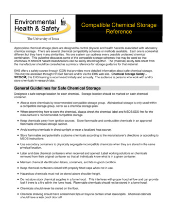
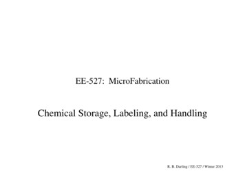
Elaissi et al. BMC Complementary and Alternative Medicine 2012, age 5 of 15the total oil content [23-25]. Twenty five major compounds at an average concentration greater than0.9 0,2% have been retained for the statistical analysis(Table 1). The main components were 1,8-cineole(4.5 1,61 -70.4 2.5%) followed by cryptone (0.0 20.9 1.3%), α-pinene (1.0 0.7 - 17.6 7.5%), p-cymene(0.8 0.1 - 16.7 5.2%), α-terpineol (0,6 0,3 - 10,3 1,1%),trans-pinaocarveol (0.8 0.2 - 7.0 2.5%), phellandral(0.0 - 6.6 0.4%), cuminal (0.0 - 6.6 0,6%), globulol(0.6 0.2 - 6.2 0.9%), limonene (0.4 0.2 - 4.4 0.3%),aromadendrene (0.1 0.0 - 3.6 1.2%), sapthulenol(0.1 0.1 - 3.2 0.9%) and terpinene-4-ol (0.3 0.1 3.0 0.8%).These 25 components were used for the PCA and theHCA analysis. The PCA horizontal axis explained 47.2% ofthe total variance while the vertical axis a further 23.80%(Figure 1). The HCA based on the Euclidean distance between groups indicated three specie groups (A, B and C)(with a dissimilarity of 11.0) (Figure 2), identified by theiressential oil chemotypes. Group A clearly stood out forminga separate group in the PCA (Figure 1) and a deep dichotomy in the HCA (Figure 2). It was correlated positively withthe axes 1 and 2. Groups A and B were negatively correlated, their separation was mainly due to axis 2. Group Aspecies reduced to E. odorata, the essential oil of whichwas characterized by the highest mean percentage ofcryptone (20.9 1.3%), cuminal (6.6 0.6%), phellandral(6.6 0.4%),verbenone(0.9 0.2%),p-cymen-8-ol(2.9 0.6%), sapthulenol (3.2 0.9%), carvacrol (1.7 0.3%),p-cymene (16.7 5.2%), terpinene-4-ol (3.0 0.8%), caryophyllene oxyde (1.7 0.2%), viridiflorol (4.5 1.6%) and byPrincipal components analysis (PCA) and hierarchicalcluster analysis (HCA)The yield content of the 25 selected component was significantly different (p 0.05) among species (Table 1).Table 1 Contents (%) of the 25 major compounds of the essential oils, extracted from the leaves of 8 Eucalyptusspecies, selected for the Principal Components and the Hierarchical Clusters Analyses CompoundsContenta (%)AbbreviationsbE. maidE. cinE. sidE. odoE. bicE. lehE. astrE. leuc6.9 1.11.0 0.73.7 1.217.6 7.522.0 6.07.8 2.3α-Pinene2.Limonenelim3.1 0.23.7 0.54.1 0.10.4 0.20.9 0.54.4 0.31.4 0.32.0 0.53.1.8-Cineole1.8-cin57.8 1.970.4 2.569.2 0.64.5 1.668.0 5.356.6 4.342.0 5.959.2 10.1γ-ter1.7 0.90.1 0.00.1 0.00.2 0.20.1 0.10.9 0.20.1 0.10.2 0.4p-cym7.4 2.91.2 0.10.8 0.116.7 5.21.4 0.52.0 0.20.9 0.22.9 e-4-ol8.Aromadendrene9.tr-Pinocarveolα-pin4.5 0.71.7.3 0.7pin0.5 0.20.3 0.10.2 0.00.2 0.12.2 0.50.2 0.01.8 0.81.2 0.2Ter-4ol1.1 0.30.4 0.10.6 0.03.0 0.80.2 0.10.3 0.10.4 0.10.2 0.2aro1.6 0.80.1 0.00.5 0.20.4 0.02.0 0.80.2 0.03.6 1.22.1 1.4tr-pin2.0 0.81.0 0.21.2 0.10.8 0.24.6 0.61.0 0.27.0 2.54.3 .Phellandralphe-0.2 0.2-0.1 0.0α-tercum2.2 0.2-tr 0.120.9 1.310.3 1.15.4 0.90.8 0.2-tr 0.10.9 0.2-6.6 0.4-6.6 0.60.6 0.30.2 0.2-1.3 0.30.1 0.1--Cuminaltr-p-Mentha-1.7. 8dien-2-ol16.p-Cymen-8-olp-cym0.1 0.00.1 0.00.1 0.02.9 0.60.1 0.0tr 0.10.1 0.00.1 0.117.cis-p-Mentha-1. 7. 8dien-2-olcis-men0.4 0.21.0 0.20.6 0.00.1 0.11.0 0.10.3 0.20.4 0.10.8 0.118.Caryophyllene oxidecar-ox0.1 0.0tr 0.10.1 0.11.7 0.2tr 0.10.1 0.10.1 0.0tr 0.119.Epiglobulolepi0.4 0.20.1 0.10.1 0.0tr 0.11.0 0.20.1 0.01.2 0.31.1 0.620.Globulolglo1.7 1.40.6 0.21.1 0.30.8 0.25.4 1.20.6 0.26.2 0.95.8 2.921.Viridiflorolvir0.7 0.30.2 0.10.4 0.11.0 0.30.8 0.20.2 0.11.1 0.10.8 0.422.Spahulenolspa0.1 0.10.2 0.10.5 0.33.2 0.90.1 0.01.0 0.10.9 0.10.1 0.123.Carvacrolcar0.4 0.10.1 0.00.1 0.01.7 0.30.1 0.00.2 0.00.2 0.10.3 0.30.1 0.00.3 0.10.1 0.00.2 0.1tr 0.10.3 0.10.1 0.00.1 0.224.α-Eudesmolα-eud1.1 0.625.β-Eudesmolβ-eud3.0 1.9ab1.0 0.20.2 0.00.6 0.00.4 0.10.6 0.10.5 0.10.1 0.21.0 0.10.2 0.1-1.6 0.715.0.5 0.1-0.1 0.114.tr-p-men-8.7 2.50.4 0.10.8 0.1Values are means SD of triplicate determination.E.maid E. maidenii, E. cin E. cinerea, E. Sid E. sideroxylon, E. odo E. odorata, E. bic E. bicostata, E. leh E. lehmannii, E. ast E. astringens, E. leuc E. leucoxylon.
Elaissi et al. BMC Complementary and Alternative Medicine 2012, age 6 of 15Figure 1 ACP of the 26 major components of eight eucalyptus essential oils. For the abbreviation of Eucalyptus species ( ) andcomponents ( ) see Table 1.the lowest level of 1,8-cineole (4.5 1.6%). Group B, made upof E. maidenii, E. lehmannii, E. sideroxylon and E. cinerea,has essential oils characterized by the highest amount oflimonene (3.1 0.2 to 4.4 0.2%), α-terpineol (2.2 0.3%for E. maidenii to 10.3 1.1% for E. cinerea). The PCAshowed that the variation between these species is mainlydue to the variation of 1,8-cineole content (57.8 1.9% forE. maidenii to 70.4 2.5 for E. cinerea) and of α-pinene(4.5 0.7% for E. cinerea to 17.6 7.5% for E. lehmannii).Group C, consisting of E. astringens, E. leucoxylon and E.bicostata, has essential oils distinguished by their highestmean percentage of epiglobulol (1.0 0.2 - 1.2 0.3%), globulol (5.4 1.2 - 6.2 0.9%), trans-pinocarveol (4.3 1.0 to7.0 2.5%), aromadendrene (2.0 0.8 - 3.6 1.2%) andpinocarvone (1.2 0.2 - 2.2 0.5%). The E. bicostataand E. leucoxylon oils differed from E. astringens oil bytheir richness in 1,8-cineole (68.0 5.3, 59.2 10.1%, respectively) and their poverty in α-pinene (3.7 1.2,7.8 2.3%, respectively) against 42.0 5.9% and 22.0 6.0%,respectively for the latter species.As in the present study, E. cinerea, E. sideroxylon, E.bicostata, E. maidenii, E. leucoxylon, E. lehmannii and E.astringens have been reported to contain 1,8-cineole as amajor component [26-30]. It was also reported that E.cinerea grown in Morocco contained a higher mean percentage of 1,8-cineole (87.8%) than that from Tunisia(70.4-2.5%) [27], whereas E. sideroxylon acclimated inTunisia was richer in 1,8-cineole (69.2-0.6%) than thatfrom Congo (59.9%) [31]. The Tunisian E. astringens essential oil had the same chemotype as that extractedfrom leaves picked from Moroccan tree plantations withan important difference in their mean percentages. BothFigure 2 Dendrogram obtained by hierarchical cluster analysis based on the Euclidean distance between groups of leaf essential oilsof eight Tunisian Eucalyptus species. Components that characterise the major sub groups, considered as chemotypes, are indicated.
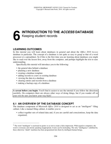
Elaissi et al. BMC Complementary and Alternative Medicine 2012, age 7 of 15of them were different from the Australian E. astringensessential oil [32], which was represented by β-caryophyllene (14.75%), p-cymene (17.72%), and α-pinene (12.53%).The same principal components were also observed in E.leucoxylon essential oil [33,34], whereas the essential oilobtained from Iran was significantly richer in 1,8-cineole(89.8%) than that from Tunisia [33].To evaluate the correlation between the antibacterialactivities and the essential oils of the eight Eucalyptus species, all the mean values of the zone diameters inhibitionwere subject to the PCA and the HCA analysis. The statistical analysis of the antibacterial activities of the oilsshowed a significant difference among Eucalyptus speciesoils and the tested bacterial strains (p 0.05). The PCAhorizontal axis explained 59.39% of the total variance,while the vertical axis a further 13.84% (Figure 3). TheHCA showed two species groups (I and II), identified bytheir bacteria growth inhibition with a dissimilarity 15.0(Figure 4). When the dissimilarity was 5.0, group II wasdivided into three subgroups (IIa, IIb and IIc). The horizontal axis permitted the separation of group I from groupII, however axis II separated all the species of the group IIinto three subgroups. Group I, limited to the Gram negative (Gram( )) bacteria, P. aeruginosa and K. pneumoniae,Antibacterial activityThe essential oils were tested for their putative antibacterialactivity against seven bacterial isolates represented by 81strains (Table 2). As shown in Table 2, E. odorata oil possessed the best activity against S. aureus (27.4 10.7 mm,zdi), followed by S. agalactiae (19.4 5.6 mm, zdi), H. influenzae (19.2 9.6 mm, zdi), S. pyogenes (19.0 0.0 mm, zdi)and S. pneumoniae (17.4 4.1 mm, zdi). E. maidenii oil showeda relatively good activity against S. aureus (22.8 6.8 mm, zdi).Table 2 Diameter of the inhibition of respiratory bacterial growth by essential oils and by antibiotics*)Essential oilsTested microorganisms (number of strains)H. influenzae(11)K. pneumoniae(13)P. aeruginosa(10)S. aureus(17)S. agalactiae(9)E. maidenii14.5 5.7bc )10.7 1.5ab7.1 1.5a22.8 6.8d13.8 2.2bc13.0 2.6bc15.5 4.0cE. odorata19.2 9.6b10.8 1.2a6.0 0.0a27.4 10.7c19.4 5.6b17.4 4.1b19.0 0.0baabbE. lehamnniiE. leucoxylonbc11.5 3.36.8 0.714.2 4.811.1 1.49.8 2.411.5 1.7bc16.4 5.4c12.9 1.8b14.4 3.0bc9.0 0.0a6.6 1.1a6.0 0.0abaa6.0 0.015.6 2.813.8 2.817.0 4.013.0 0.0b7.1 1.6a6.0 0.0a15.5 5.2c12.0 3.2b12.3 12.3bc12.5 1.7bcaabbE. bicostata13.6 5.07.1 1.4E. astringens13.0 3.8bcbS. pyogenes(4)6.5 0.88.1 2.2acS. pneumoniae(19)bcbc6.2 0.612.2 2.811.6 1.411.5 2.812.5 1.7b8.1 1.8a7.0 1.5a13.7 3.8b12.3 1.6b10.7 2.5ab12.0 0.0bNTNTNTNTNTNTNTNTNTE. cinerea13.0 6.38.5 2.2E. sideroxylon12.3 8.4b30.5 6.1cbReference antibioticsAmpicillinBenzylpenicillin(Penicillin inFosfomycinGentamicinb20.7 6.0NTb19.9 4.2aNTNT22.3 7.5a23.8 5.6aNTNTNTNTNTNTNTNTNTNT32.4 2.9b34.7 5.0bNTNTNTNT29.9 5.1ab29.2 4.2b26.3 12.0a29.0 0.0cNT34.3 11.1c27.9 5.9ab35.6 5.5c24.5 0.8aNTabNT22.1 2.9aNTa22.9 5.024.5 7.529.6 6.2aNTNTNTNtNTImipenemNTNT24.6 6.6NTNTLevofloxacinNTNTNTNT24.8 2.8aNTNTNTNTNTNTNTNTNT33.6 4.2c31.7 11.9abc30.0 0.0cLincomycin11.3 1.8aOxacillinNTPiperacillinNTNT25.3 7.3aNTNTRifamycinb23.9 4.7NTNTNT34.4 4.3cTetracyclin29.2 2.6cNT24.4
lyptus essential oils against Adenovirus, mumps and herpes simplex viruses [11,12]. One of the major chal-lenges is the practical use of these essential oils in vivo. This is difficult and needs to be overcome by trial assays. The importance of the antiviral activity of natural com-pounds is their perspective uses against these pathogens.