Transcription
Molecular Mechanisms ofAutophagy in YeastNobel Lecture, December 7, 2016by Yoshinori OhsumiTokyo Institute of Technology, Tokyo, Japan.Science is a system of knowledge that is gradually accumulated over manyyears by society, but it is also an inherently human activity. I believe thatevery scientist is a product of the era in which they live. So I’d like to start froma brief introduction of my life, and then give a historical overview of my scientific work.Early lifeI was born in Fukuoka, southern Japan, in 1945, half a year before the end ofWorld War II. This was a very challenging time in Japan, and everyone haddifficulty getting basic daily necessities, including food. I myself suffered fromsevere malnutrition and was a very sickly child. Around this time my mothergot tuberculosis and spent a long period bed-bound, but she was miraculouslyable to recover thanks to a gift of just-developed antibiotics from a family friendin Hawaii. When I was about 8 years old, my mother was finally able to resumea normal life. My brother, Kazuo, who is 12 and a half years my senior, left hometo enter the Faculty of Literature at the University of Tokyo as I began elementary school. I was brought up through the efforts of my father and two sisters,Reiko and Junko, and many other people throughout what was a difficult timefor our family.277
278The Nobel PrizesMy childhood home was surrounded by nature, with rice paddies, streams,hills and the sea all nearby. I spent a lot of time outdoors catching fish and picking plants. As an elementary school student, I was engrossed in collecting insectsand watching the night sky. I would say that this intimacy with nature had astrong influence on me. My brother, every time he returned home from Tokyoduring university holidays, would bring a book, such as carefully chosen worksof George Gamow and Michael Faraday that broadened my awareness of theworld beyond what we learned at school (Faraday, 1861; Gamow, 1940). Being aweak child, I had no talent for sports and was neither artistic nor gifted in literature. But I managed to achieve good results at school. At the time, I had a vaguelonging to be a scientist that was influenced by the expectations of my parents.University & first experience of researchIn high school, I joined the chemistry club, where I first experienced the wonderof chemical reactions. Partly thanks to this experience, I entered the Universityof Tokyo, where I initially hoped to become a chemist, but I had difficulty finding a field to specialise in. Thankfully, this was the time when the central dogmaof molecular biology was just being established. Molecular biology immediatelyfascinated me, so I decided to join the lab of Kazutomo Imahori, who was one ofthe few scientists running a molecular biology laboratory in Japan at the time.Imahori’s laboratory had adopted a physicochemical approach to assess interactions between proteins and nucleic acids. Under the direction of Akio Maeda,I began to study the role of ribosome subunits in protein synthesis. Although Iwas unable to produce particularly interesting results, my time in the Imahori labwas exciting as we participated in the work of a rapidly developing field and I gotmy first taste of the joy of experimental work. The experience also provided mewith a strong sense of the continuous process of protein synthesis within cells.From the second year of my doctoral studies, I decided to move to KyotoUniversity, where Maeda had joined the just-founded Department of Biophysics as an associate professor. Kyoto University provided a very free environment,and I was stimulated by talented new students and friends I met in the biochemistry lab of the Department of Chemistry. Throughout my graduate studies,the Summer Seminar for Young Biochemists also allowed me to make a largenumber of friends from all over Japan who would have a great impact on mylife. In my research, I became interested in colicin E3, a toxic protein that is ableto pass through the membrane of bacterial cells and instantly inhibits proteinsynthesis. I think I was also influenced by my growing interest in biologicalmembranes during this period.
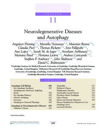
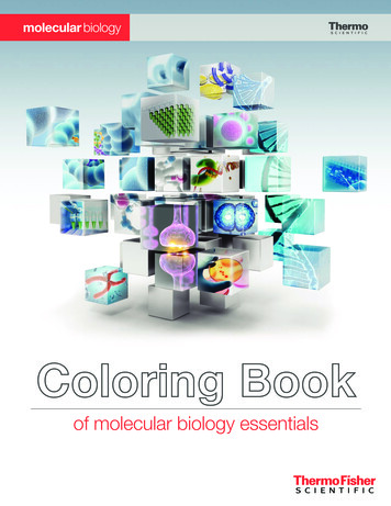
Molecular Mechanisms of Autophagy in Yeast279Post-doc life in New YorkAfter I finished my doctoral studies, I enrolled in the lab of Gerald Edelman atThe Rockefeller University, New York. I was asked was to establish an in vitrofertilisation system for mouse egg cells, which I reluctantly accepted. Experimentally it was not too difficult, but I had no idea how to examine such fascinating yet few early embryos. After some time, Mike Jazwinski joined our groupfrom Arthur Kornberg’s lab, where he had been studying phage DNA duplication. Taking inspiration from the elegant study of the cell cycle through the cdcmutants by Hartwell (Hartwell, 1974), a project to uncover the mechanism ofDNA replication initiation in yeast began in Edelman’s lab, which I joined inmy final year. This was to be my first encounter with yeast as an experimental organism.We had first planned to use isolated nuclei in this project, but it was difficultto obtain these organelles in an undamaged state. During our use of densitygradient centrifugation to purify nuclei, I noticed a white layer at the top ofthe centrifuge tube. Out of mere curiosity I looked at this white layer under themicroscope, and realised that it was a highly-enriched vacuole fraction. The easewith which this organelle could be purified left a strong impression on me, andI think helped to decide the direction of my research.Return to Japan and work on the vacuoleAt the end of 1977 I returned to Japan as an assistant professor in Yasuhiro Anraku’s lab at the University of Tokyo. Anraku’s entire lab was tasked with studying the transporters and the respiratory chain in E. coli. In spite of this, Anrakuallowed me to start a project on yeast, and in retrospect I really appreciate hisdecisiveness in affording me this opportunity, which resulted in the beginningof my nearly 40 years of work in yeast (Figure 1). In those days, the plasmamembrane and its transport mechanisms were the focus of much research. ButI have never been a competitive person, and prefer to work on a subject that fewpeople are interested in. After a period of reflection, I decided instead to work ontransport across the membrane of an intracellular organelle, the vacuole. Whilewe now know that elaborate transport systems exist on the vacuolar membrane,this was by no means a fashionable topic.At that time the vacuole was thought to be no more than a garbage dump inthe cell, but I thought it must play yet unknown roles in cell physiology. First Iestablished procedures for purifying vacuoles and making vacuolar membranevesicles, and was able to show active transport systems of amino acids and calcium ions over the vacuolar membrane, providing evidence that the vacuole is
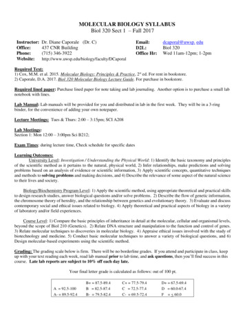
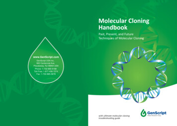
280The Nobel PrizesFIGURE 1. Thin-section electron micrograph of a yeast cell. The vacuole is visible as thelarge, pale region near the centre of each cell.important for homeostasis of metabolites and ions (Ohsumi and Anraku, 1981;Ohsumi and Anraku, 1983). We also described a novel proton-pump on the vacuolar membrane, the V-type ATPase, which generates a proton gradient acrossthe vacuolar membrane as a driving force of these transport systems (Kakinumaet al., 1981; Uchida et al., 1985) (Figure 2). These experiences played an important role in the development of my scientific interests, leading into my continuedstudy of the vacuole and foreshadowing my research on autophagy. Since then,FIGURE 2. Vacuolar membrane transport systems in yeast. The V-type ATPase maintainsthe acidity of the vacuole by pumping H into the vacuole, while various active transporters modulate the flow of calcium, amino acids and ions between the cytoplasm andthis organelle.
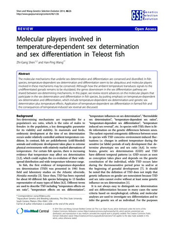
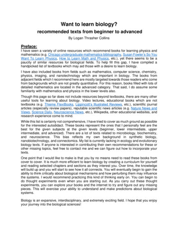
Molecular Mechanisms of Autophagy in Yeast281the structure and function of the V-type ATPase has become a major field of cellbiology research in its own right.STARTING MY OWN LAB AT TOKYO UNIVERSITY, KOMABA CAMPUSIn 1988 I was able to start my own lab—consisting of just myself—at the Collegeof Arts and Sciences at the Komaba campus of the University of Tokyo. My labhere was modestly equipped, without any fancy instruments. I decided to devotemyself to a related but completely novel task: the lytic function of the yeast vacuole. Since the vacuole is an acidic compartment, due to the function of V-typeATPase, and contains various hydrolytic enzymes including proteases, I thoughtit might be homologous to the lysosome of mammalian cells. However, I had nowell-articulated idea how to test this hypothesis.Thinking about the life of proteinsBefore going into experimental details, I would like to briefly outline proteinmetabolism in cells. Proteins, which are a polymer of amino acids, are the keyplayers in all biological processes. The central dogma of molecular biology statesthat protein is synthesised according to the genetic information encoded in DNAthrough RNA. As a consequence of this, many researchers throughout the latterhalf of the 20th century worked diligently to understand gene expression and itsregulation, and the mechanisms of protein synthesis. Another important consideration that subsequently came to scrutiny was the compartmentalisation of thecell, as proteins are able only able to properly function after being delivered to theappropriate part of the cell. This spurred an effort to understand the localisationof each protein within the cell. Thanks to the efforts of researchers, we now knowthat each protein is intricately and precisely trafficked to the correct destinationwithin the cell according to an intrinsic localisation signal (Figure 3).However, one aspect that was missing from this picture when I started myown lab was the fate of proteins, or in other words the question of how intracellular protein degradation occurs. It was known that each protein has a distinctlifetime, ranging from a few minutes to several months. But for many years, thedegradation of protein was thought to be a passive process that is not particularlyimportant or interesting.The significance of protein turnoverI used to begin biology class for first-year undergraduate students with this question: how many red blood cells are made within just one second in your body?
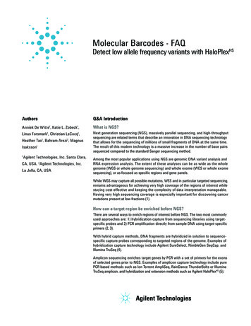
282The Nobel PrizesFIGURE 3. Overview of protein metabolism in cells. In addition to the emphasis on synthesis provided by the central dogma, the delivery, function and eventual degradation ofproteins are important aspects of their life in the cell. The delivery of proteins to variouscellular compartments following translation by the ribosome (purple) is represented atthe bottom of the figure.The calculation is quite simple and gives an answer of about three million cellsper second. How about hemoglobin in the red blood cell? This number can alsobe easily derived, giving about 1x10¹⁵—one million billion—hemoglobin molecules per second. It follows that exactly the same amount of cells and proteinsare degraded. The key message is that living things are extremely dynamicallymaintained.The Japanese climate is characterised by four distinct seasons that influenceour culture, and students at school learn that all things are in a cycle of continuous change. Plant leaves offer an excellent example of this concept. In spring,leaves develop and then actively synthesise starch by sunlight, but in autumnleaves become red or yellow and fall off (Figure 4). What we see as beautifulautumn colours is actually caused by the degradation of the leaf ’s photosynthetic machinery: green chloroplasts are completely degraded, and the resultingamino acids are transported to the trunk, leaving only the red or yellow-coloured
Molecular Mechanisms of Autophagy in Yeast283FIGURE 4. Ginkgo trees throughout the four seasons at the University of Tokyo. Theremoval of green chloroplasts in autumn, leaving golden accessory pigments in the leaves,is one striking example of the importance of degradation in the dynamic cycle of life.accessory photosynthetic pigments in leaves. Similarly, rice leaves turn yellow atharvest time. All the protein in the leaves is degraded and transported to makeproteins in rice grains for the next generation. These examples demonstrate thatdegradation is not an adverse process, but rather is essential for new construction or regeneration. Let’s think about the human body. Our bodies make about2–300 grams of protein every day, but we only intake about 70–80 grams of
284The Nobel PrizesFIGURE 5. Protein dynamics in the human body. The bulk of amino acids required forsynthesis are met not through dietary intake, but through the turnover of existing proteinsin the cell.protein from our diet (Figure 5). The amino acids required for protein synthesismostly come from the degradation of the body’s own proteins. Life is maintainedby a tightly controlled balance between synthesis and degradation. Just two yearsago I experienced this personally when I learned that my wife Mariko’s motherhad lived without any food and even water for 13 days. This was another shocking reminder that the recycling of cellular constituents is a potent force in therobust nature of life.Historical perspectives on protein turnoverThe establishment of the central role of proteins in biology was a very importantdevelopment in modern biological science. In contrast to the process of synthesisemphasised by the central dogma, when I began my research the degradationof proteins was seen as a passive process that was not particularly important bymany of my contemporaries. In this regard Rudolph Schoenheimer was a pioneerin the field, as he had proposed the concept of “protein turnover” in the 1930sfollowing his development of isotopes to follow metabolic processes (Schoenheimer et al., 1939; Schoenheimer, 1942) (Figure 6A). Unfortunately, however,his contention was controversial and not widely accepted. After this, Christiande Duve, recipient of the Nobel Prize in 1974, used cell fractionation to discoverthe lysosome, an organelle containing a range of degradative enzymes (De Duve
Molecular Mechanisms of Autophagy in Yeast(a)285(b)FIGURE 6. (a) Rudolph Schoenheimer and (b) Christian de Duve.et al., 1955) (Figure 6B). Researchers at The Rockefeller University subsequentlyobserved the delivery of extracellular material to the lysosome using electronmicroscopy (Novikoff et al., 1956). The delivery of intracellular material including organelles such as mitochondria to the lysosome was also observed. De Duvereferred to these processes as ‘heterophagy’ (now known as endocytosis) and‘autophagy’, respectively (de Duve, 1963). Autophagy, from the Greek for ‘selfeating,’ was described by The Rockefeller University researchers as having severalhallmarks. Firstly, a membrane sac appears and enwraps a portion of the cytoplasm, forming a double-membrane structure, the autophagosome. Next, theouter membrane of the autophagosome fuses with the lysosome, and finally theinner membrane and its contents are degraded within the lysosome.Autophagy research was continued by two key groups: one, led by GlennMortimore (Figure 7A), conducted detailed physiological and biochemical analyses of the phenomenon using liver perfusion in rats (Mortimore and Ward,1976; Mortimore et al., 1983), while a second group, led by Per Seglen, examinedthe effect of nutrient starvation on autophagy and the regulation of autophagy,mainly using cultured cells (Seglen and Gordon, 1982; Seglen et al., 1980) (Figure7B). However, as electron microscopy was the only means of assessing autophagyat the time and the lysosome is a dynamic organelle, most works focused on theanalysis of membrane dynamics in autophagy, leaving the genes and proteinsimplicated in the autophagy mechanism unidentified for many years. A simplermodel system was required to make further progress in the field.
286(a)The Nobel Prizes(b)FIGURE 7. (a) Glenn Mortimore and (b) Per Seglen.In the meantime, another intracellular degradation system, the ubiquitin/proteasome system, was discovered. As it became clear that this system playsimportant roles in the regulation of a variety of cellular processes, protein degradation suddenly attracted a huge amount of attention in biological research. Thiswas ultimately recognized by the Nobel Prize in Chemistry in 2004, which wasawarded to Aaron Ciechanover, Avram Hershko and Irwin Rose, but the fieldof lysosomal degradation and autophagy remained quiet. Now it is thought thatthese two systems function together as the major means of cytoplasmic proteindegradation.Discovering autophagy in yeast by light microscopyAssuming that the vacuole functions as a lytic compartment, the questions Ihad were when, what and how cytoplasmic constituents go across the vacuolarmembrane and become accessible to vacuolar enzymes. First, I searched for acondition where massive protein degradation might occur in the yeast life cycle.Spore formation is a meiotic process that generates four spores and is induced bythe depletion of nitrogen, the essential element of amino acids (Figure 8). I reasoned that this dramatic cellular remodelling should require the degradation ofpre-existing proteins to make the proteins necessary for this cell differentiation.I have to admit that I’ve always loved to observe yeast cells by light microscopy. The vacuole is relatively large, is the only clearly visible organelle in the celland contains a salt-like solution that has a very low concentration of protein, so Iknew that it would be easy to see structures if they were inside. I probably spentmore time sitting at the light microscope than anyone else during my studies of
Molecular Mechanisms of Autophagy in Yeast287FIGURE 8. Morphological changes associated with sporulation in yeast. The onset of nitrogen starvation causes diploid cells to undergo meiosis and cell division, forming fourspores in a process that requires vast remodelling of the protein component of the cell.N nucleus, V vacuole, CW cell wall, M mitochondrion.the vacuole, looking for morphological changes that might provide hints aboutthe function of this dynamic organelle.I carefully observed the early stages of the sporulation process, but couldn’tsee any obvious changes in the vacuole. I thought that this may be due to thedegradation of structures in the vacuole. I conceived that if I were to use a celllacking vacuolar proteases, structures delivered to the vacuole for degradationwould remain in this organelle and might be visible. Fortunately, the well-knownyeast geneticist Elizabeth Jones had donated many proteinase mutants to theYeast Genetic Stock Center at UC Berkeley, so I obtained these strains and shiftedto nitrogen depleted medium.Looking down the microscope, I saw many spherical structures vigorouslymoving around inside the vacuole. These structures appeared within 30 minutes and almost completely filled the vacuole after 3–4 hours (Figure 9). Forme, this observation marked the exact starting point of more than 27 years ofautophagy research. The reason that I was able to discover this phenomenonwith only a common low magnification light microscope owes to the vigorousmovement of these structures by Brownian motion. I was also fortunate that thestructures, which we now call autophagic bodies, were large enough to be visible under a light microscope. In the otherwise tranquil yeast cell, this dramatic
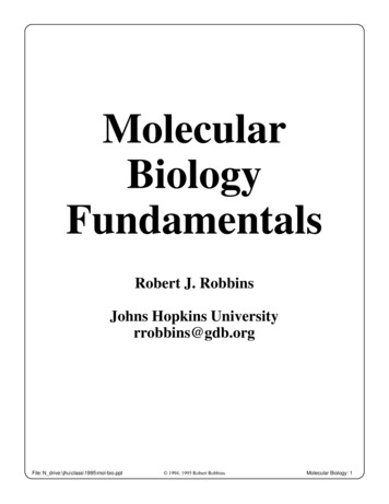
288The Nobel PrizesFIGURE 9. Accumulation of autophagic bodies within the vacuole under nitrogen starvation, as observed by bright-field (top) and phase contrast (bottom) light microscopy.phenotypic change really struck me, and I remember staring down the microscope for some hours, unable to tear my gaze away. I knew that I had encounteredan unknown and fascinating phenomenon; this was the starting point of myautophagy research.Electron microscopy and the morphology of autophagyI next called on the excellent electron microscopists Misuzu Baba and MasakoOsumi to capture the morphology of this phenomenon by electron microscopy.Their efforts produced beautiful images that provided irrefutable evidence ofthe topological features of the entire autophagy process (Baba et al., 1994), asshown in Figure 10.A thin section image of a nitrogen starved cell is shown in Figure 10A. Spherical structures can be seen within vacuole, and the high-magnification imagepresented in Figure 10B shows that these structures are bound by a single membrane. We can see that their contents are exactly the same as the cytoplasm, containing the same density of ribosomes and various cytoplasmic structures, suchas the rough ER observed in Figure 10C, suggesting nonselective sequestrationof cytoplasmic components. A membrane sac next to the vacuole, the isolationmembrane, is also visible in Figure 10C enwrapping a portion of the cytoplasm.After completion of membrane expansion and sealing, a double membranestructure, the autophagosome, is formed (Figure 10D). Freeze-fracture imaging (Figure 10E) shows the fusion event of an autophagosome with the vacuolar membrane, where the outer membrane of the autophagosome is continuous with the vacuolar membrane (Baba et al., 1995), and the inner membranestructures that we named autophagic bodies are also present. Autophagic bodies
Molecular Mechanisms of Autophagy in Yeast289FIGURE 10. Electron micrographs showing topological phenomena associated with nitrogen starvation-induced autophagy. (A) Autophagic bodies within the vacuole, (B) highmagnification image of autophagic bodies, (C) cup-shaped isolation membrane adjacentto the vacuole, (D) an autophagosome adjacent to the vacuole, (E) freeze-fracture imageof an autophagosome fusing with the vacuole, (F) mitochondria within autophagic bodiesdelivered to the vacuole.occasionally contain mitochondria, as can be seen in Figure 10F, indicating thatautophagy can degrade not only cytosolic protein but also supramolecular structures, such as ribosomes and even entire organelles, which is a characteristicfeature of autophagy. This is in contrast to the specific recognition of smaller,individual targets by the ubiquitin/protease system. Autophagosomes are transient, ephemeral structures, and autophagic bodies are degraded immediatelyin the vacuole in wild-type cells, features that prevented their identification formany years.A scheme of the autophagic process in yeast is presented in Figure 11. Whencells are faced with starvation conditions, a small membrane sac appears next tothe vacuole and expands to enwrap a portion of cytoplasm. Then, a double membrane structure, the autophagosome, targets the vacuole and fuses with the vacuolar membrane, releasing the inner membrane structure into the vacuole. In wildtype cells these structures are immediately disrupted by vacuolar enzymes, andtheir contents are degraded for reuse. The proteinase-deficient mutant allowed usto follow the progression of autophagy as the accumulation of autophagic bodies.
290The Nobel PrizesFIGURE 11. Schematic diagram of nitrogen starvation-induced autophagy in yeast.Importantly, while the vacuole is a vastly larger organelle than the mammalian lysosome, the entire process that we observed was topologically the sameas macroautophagy in mammalian cells, confirming that yeast could be usedas a model organism in the study of autophagy (Baba et al., 1994). KazuhikoTakeshige was also able to show early on that this phenomenon is not restrictedto sporulation, but is a general cellular response to a range of nutrient starvationconditions (Takeshige et al., 1992). These factors gave the first indication thatautophagy is a general process that is induced in diverse organisms in responseto a range of stimuli, further convincing me that we had discovered an importantcellular process.Identifying autophagy-related genes in yeastYeast is a very good organism for the dissection of complicated biological phenomena through genetic analyses. In this regard I was inspired by the beautifulworks of Lee Hartwell, whose group uncovered details of the cell cycle throughtheir analysis of the cdc mutants (Johnston et al., 1977), and also by the intricateanalysis of the secretory pathway using sec mutants by Randy Schekman’s group(Novick et al., 1980). These contributions were recognized with the Nobel Prizein Physiology or Medicine in 2001 and 2013, respectively.As we knew nothing of the genes and proteins that are involved in the autophagy mechanism, I thought that the isolation of mutants defective in autophagy
Molecular Mechanisms of Autophagy in Yeast291was the first step in opening up the field of autophagy. However, we had noidea what an autophagy-defective phenotype would look like. At this point, wedecided to use autophagic body accumulation in the vacuole as a morphological indicator of autophagy progression. My first Master’s course student, MikiTsukada, was central at this stage of our work and did an excellent job isolatingautophagy defective mutants. We took mutagenised cells, individually subjectedthem to starvation and checked for the accumulation of autophagic bodies,aiming to isolate cells in which autophagic bodies were not observed. Using thisapproach, we identified the first autophagy mutant strain, which we called apg1,now referred to as atg1 (Tsukada and Ohsumi, 1993). We then confirmed, bythe failure to induce protein degradation in this strain, that the autophagic bodyis indeed an intermediate structure of the degradation process. We also foundthat, as expected, the homozygous diploid mutant was not able to sporulate.However, under standard growth conditions in the nutrient rich medium YEPD,no difference was observed between the atg1 strain and wild-type cells, and thereappeared to be no obvious defect in vacuolar function or secretion.It seemed impossible that such an intricate pathway of delivery to the vacuole could be controlled by a single gene, and I was convinced that we should beable to identify many more. We found that atg1 cells that were subjected to prolonged nitrogen starvation lose viability faster than their wild-type counterparts.Assuming this loss of viability phenotype was caused by the defect in autophagy,we next conducted a primary screen looking at the viability of mutant strains. Byisolating strains in which viability was reduced and then performing a secondaryscreen examining these strains for vacuolar morphology defects, we were able toidentify roughly 100 autophagy-defective strains at once (Tsukada and Ohsumi,1993) (Figure 12). Further genetic analysis showed that 14 complementationgroups were among these mutants. As we now know that in nitrogen starvedcells 18 genes are essential for autophagy, I can say that this refined approachwas extremely effective. We initially predicted that these mutant strains wouldbe characterised by defects at various stages of the autophagy mechanism, but asit turned out all of these genes were essential for the formation of the autophagosome, the most important process in autophagy.This was a morphological screen, based on the inability to accumulateautophagic bodies in the vacuole. We therefore eliminated mutants characterised by a severe growth defect, cells lacking essential genes and also strains withabnormal vacuolar morphology. In addition, as we used the decline in viabilityas a primary screen, which only appears with a complete block of autophagyfunction, we only identified autophagy null mutants, to the exclusion of partiallydefective mutants.
292The Nobel PrizesFIGURE 12. Overview of the experimental approach used to isolate autophagy-defectivemutants. This screen allowed us to identify most of the ATG genes in a short period of time.Next, we set out to clone and identify the genes implicated in the mutantphenotypes observed in the screen. Tsukada was the first to succeed, cloning theATG1 gene. Through the sequencing of ATG1, we learned that this gene encodesa protein kinase (Matsuura et al., 1997). However, as we identified the remaining genes, starting with Kametaka’s work on ATG5 (Kametaka et al., 1996) andATG6, we found that all the other ATG genes encoded unidentified proteins thatwere otherwise not essential for growth in nutrient replete conditions and hadno phenotypic implications for cells other than a defect in autophagy. Therefore, although we had identified nearly all the genes responsible for autophagy,we were unable to deduce anything about their function from their amino acidsequences and went through a difficult period where we were unable to officiallyannounce our findings.Yeast had been the subject of comprehensive genetic analysis throughoutthe world for many years at this point, and it was very surprising that autophagygenes had somehow escaped the attention of other researchers. I suspect that thisis because researchers were interested in essential genes, and the conventionaldefinition of an essential gene was the cell’s inability to grow in nutrient richmedium when the gene in question was disrupted.
Molecular Mechanisms of Autophagy in Yeast293One important product of our time at the College of Arts and Sciences ofTokyo University that deserves mention was Takeshi Noda’s establishment of anassay of autophagic activity (Noda and Ohsumi, 1998; Noda and Klionsky, 2008).Noda constructed a strain in which the precursor of the vacuolar alkaline phosphatase Pho8 lacks its N-terminal vacuolar targeting sequence, and expressedthis precursor in the cytosol. Upon the induction of autophagy, a portion of themodified version of Pho8 would be taken up by the non-selective activity ofautophagy and delivered to the vacuole. Here, the precursor is processed to yieldthe mature, enzymatically active form of Pho8. Therefore, we were able to quantitatively measure the extent of autophagy by assessing Pho8 activity. This assay,in which degradation can be quantitativel
players in all biological processes. e central dogma of molecular biology states that protein is synthesised according to the genetic information encoded in DNA through RNA. As a consequence of this, many researchers throughout the latter half of the 20th century worked diligently to understand gene expression and its