Transcription
HindawiEvidence-Based Complementary and Alternative MedicineVolume 2018, Article ID 8265150, 11 pageshttps://doi.org/10.1155/2018/8265150Research ArticleSmall Peptides Compound Isolated from Agkistrodon withAntiarthritic Effect in Collagen-Induced Arthritis RatsLijun Mei,1 Chen Lin ,1 Shanshan Lei,2 Li Xu1,1 and Yong-Sheng Fan1Zhejiang Chinese Medical University, Hangzhou, Zhejiang 310053, ChinaZhejiang University of Technology, Hangzhou, Zhejiang 310014, China2Correspondence should be addressed to Li Xu; xulihhb@163.com and Yong-Sheng Fan; applezjtcm@126.comReceived 30 November 2017; Revised 28 February 2018; Accepted 22 March 2018; Published 29 April 2018Academic Editor: Antonella FioravantiCopyright 2018 Lijun Mei et al. This is an open access article distributed under the Creative Commons Attribution License, whichpermits unrestricted use, distribution, and reproduction in any medium, provided the original work is properly cited.Agkistrodon in Chinese medicine has long been used as an effective treatment against rheumatoid arthritis (RA). The presentresearch further investigated the effects of peptides extracted from the crude Agkistrodon on the RA rat model. Extracted peptideswere separated by parameter-optimized ion-exchange chromatography (IEC), peptide fractions were further analysed by MALDITOF/TOF MS, and nano-LC-MS/MS acquired mass spectra were further characterized using Mascot software, which ranks the bestmatches in the NCBI database. RT-PCR results in RAW264.7 cells indicated that Agkistrodon peptide components had inhibitoryeffects against inflammatory cytokines. The therapeutic efficacy of Agkistrodon peptides was evaluated on the Wistar rats withcollagen-induced arthritis. Symptom relief and reduced cartilage destruction and bone erosion were observed, which can beexplained by the direct suppression of inflammatory cytokines in the joints. Agkistrodon peptides downregulate the expressionof TNF-𝛼, IL-1𝛽, and IL-6, which may alleviate cartilage destruction and bone erosion, thus relieving symptoms of RA.1. IntroductionRheumatoid arthritis (RA) is a chronic inflammatory disorder characterized by progressive destruction of patients’joints caused by cellular infiltration and proliferation ofsynovium [1]. Proinflammatory cytokines including TNF𝛼, IL-1𝛽, and IL-6 are overproduced in the rheumatoidjoints and are critical mediators in the pathogenesis of RA,promoting leukocytes, and osteoclasts activation, drivingdestruction of bone and cartilage [2]. Inhibiting of proinflammatory cytokines has been shown to improve diseasesymptomatology and outcome in rodent arthritis models andhuman clinical trials [3, 4].The prognosis of RA so far is discouraging, given thatcommon treatments including disease-modifying antirheumatic drugs (DMARDs) and nonsteroid anti-inflammatorydrugs (NSAIDs) partially relieve patients’ symptoms withlimited and short-term effectiveness [5, 6], and prolongedusage might cause serious adverse effects such as leukopenia,elevated transaminases, and infection [7–9]. On the otherhand, it is increasingly acknowledged that natural productswith antiarthritic effects might be a good choice for potentand safe treatment of RA [10]. Insects as natural productresources have since ancient times been used for antiarthritictherapy [11]. Successful isolation of bioactive molecule fromsecretions, excretions, and bodies of insects further encourages the in-depth exploration of their functional mechanisms[12].A wide range of small peptide toxins have been separatedfrom venoms and larvae. Bioactive peptides inhibit synovialangiogenesis by binding to the vasculature endothelial cells[13] or suppressing immune response by inhibiting inductionof T-cell mediated inflammation [14]. Recent studies searching from snake venoms have successfully identified antiinflammatory and antiarthritic activities components andfurther demonstrated that their beneficial effects might beattributed to the inhibition of NF-𝜅B pathway and suppression of T-cell/FLSs proliferation [15–17]. Preclinical modelsof peptide immunotherapy have demonstrated efficacy inautoimmunity, shedding light on the potential as pharmaceutical agents [18, 19].In the present study, peptides isolated from the crudemedicine of snake Agkistrodon acutus were analysed for theirpotency/efficacy on/against arthritis. The dried powder of
2Agkistrodon acutus has been successfully applied in Chinese medicine as an antiarthritic drug, and previous workexplored and determined the optimal treatment dosage [20].Peptides’ cytotoxicity and cytokine-inhibiting effects weretested and affirmed by both in vitro and in vivo experiments. Possible mechanism in association with the cytokineinhibiting effect was discussed.2. Materials and Methods2.1. Animals and Cells. Female Wistar rats (𝑛 30, 6-7weeks of age) were obtained from the Laboratory AnimalResearch Center of Zhejiang Chinese Medical University(China; rodent license number SYXK (Zhe) 2003-0184).After acclimatization for 7 days, four rats were housed ineach Individual Ventilation Cage (IVC) and were maintainedin accordance with national standards (Laboratory AnimalRequirements of Environment and Housing Facilities, GB14925-2001). Six rats were randomized into a normal controlgroup and all others were used for the CIA model. RAW264.7cells were purchased from the Shanghai Institutes for Biological Sciences, Chinese Academy of Sciences.2.2. Reagents. The Agkistrodon for the Agkistrodon powderand the Agkistrodon peptide preparation was purchasedfrom the Chinese Herbal Medicine Co., Ltd., of ZhejiangChinese Medical University (Hangzhou, China). Glycosideof Tripterygium wilfordii (number 1507101B) was purchasedfrom Zhejiang Deende Pharmaceutical Co., Ltd., China.Foetal bovine serum (FBS, number 10099141) was purchased from Gibco Life Technologies., Inc., USA, and highglucose Dulbecco’s modified Eagle’s medium (DMEM) waspurchased from Hangzhou Sijiqing Biological EngineeringMaterials Co., Ltd., China. The Cell Titer 96 AQueous OneSolution Cell Proliferation Assay (MTS, number G3581) andlipopolysaccharide (LPS, number L2880) were purchasedfrom Sigma, USA. TRIzol reagent (number 9109) and PrimeScript RT Master Mix (number RR036A) were purchasedfrom TaKaRa, Japan. SYBR-Green Supermix (number 1708882AP) was purchased from Bio-Rad, USA. The specificprimers for the target genes and GAPDH were synthesizedby and purchased from Shanghai Shenggong Co., China.Incomplete Freund’s adjuvant (IFA, number SLBK3881V)was purchased from Sigma, USA. Bovine type II collagen(CII, number 20021) and the enzyme-linked immunoassay(ELISA) kit (number 2042) were purchased from Chondrex,USA. The TNF-𝛼 polyclonal antibody (number YT4689) andIL-𝛽 polyclonal antibody (number YT5201) were purchasedfrom ImmunoWay, USA. The IL-6 polyclonal antibody (number BS6419) was purchased from Bioworld, USA.2.3. Peptide Separation. Extraction of crude peptides wasprocessed following instructions of the boiling method withdilute ethanol recorded in the Chinese Pharmacopeia (2010edition). Briefly, 10 grams of crude Agkistrodon medicine waspowdered and sifted through a 149 𝜇m mesh filter and thenwas further dried for 30 min to remove any additional water.The powder was then soaked in 50% dilute ethanol, with itsweight/volume percentage concentration to be 10%. The totalEvidence-Based Complementary and Alternative Medicinecrude peptides were heat reflux extracted for 1 h and then aftercooling to room temperature, the solution was centrifuged at10000 g for 20 min. Supernatants were collected and testedfor protein/peptide concentration and then lyophilized at 56 C and stored for further tests.Lyophilized crude peptides, 1.8 grams, were measuredand dissolved in pure water. Ion-exchange chromatographywas used for separation. An XK 26/20 column was packedwith Resins purchased from GE, USA, and separation wasperformed with a 0–0.5 M NaCl gradient and monitored ata 215 nm wavelength. Eluted peptide peaks were collectedand desalted by dialysis with a 500 Da cutoff before beinglyophilized and further analysed by an AB SCIEX 5800MALDI-TOF/TOF mass spectrometer (AB Sciex, Canada) inlinear positive mode. Mass spectra were analysed using DataExplorer 4.5 software (Perspective Bio Systems). Spectra wereacquired over the mass range of 600–4,000 Da using 50 lasershots in the positive ion mode at a laser power of 25%.2.4. MALDI-TOF Analysis. The molecular mass of lyophilized peptides was analysed by MALDI-TOF/TOF on anAB SCIEX 5800 Proteomics Analyser (AB Sciex, Canada).Samples were mixed thoroughly at a ratio of 1 : 1 (v/v) with asaturated matrix solution (𝛼-cyano-4-hydroxycinnamic acid,5 mg/mL prepared in 50% acetonitrile/0.1% trifluoroaceticacid), and 0.5 𝜇l of the mixture was spotted onto a target plate.The mixture was fully dried before analysis. The instrumentwas calibrated externally using a mixture of peptide standardsobtained from Sigma-Aldrich (MS-Cal1). Positive ion (MH )spectra were acquired in the linear MALDI-TOF mode. Them/z signals were recorded between 1,000 and 10,000 Da forpeptide detection.2.5. LC-MS/MS Analysis. To explore and identify speciesspecific peptides, LC/MS/MS analysis was performed. Thecollected peptide sample was analysed by a Thermo QExactive mass spectrometer (Thermo, San Jose, CA) witha nanospray ion source. The lyophilized peptides were dissolved in FA–H2 O (0.1 : 99.9, v/v) and preconcentrated on a2 cm trap column (100 𝜇m i.d.) that was packed with C18 AQbeads (5 𝜇m, 120 Å, Daison, Osaka, Japan). A second 10 cmcapillary analysis column (75 𝜇m i.d.) was packed with C18AQ beads (3 𝜇m, 120 Å, Daison, Osaka, Japan) and was usedto separate the peptides. Eluting buffers were 99.9% H2 O with0.1% FA (buffer A) and 84% ACN with 0.1% FA (buffer B).The separation system was equilibrated initially with buffer A,and a linear elution gradient (at flow rate of 320 nil/min) wasperformed, from 4 to 50% buffer B (formic acid-acetonitrile,0.1 : 99.9) in 20 min and from 50% to 100% buffer B (FAacetonitrile, 0.1 : 99.9) in 5 min, and then held at 100% bufferB for another 5 min. The MS/MS spectra were obtained incollision-induced dissociation (CID) mode, and a full massscan was acquired from m/z 400 to 2000 with a resolution of70,000. The 10 most intense ions with a charge above 2 andintensity threshold higher than 104 were selected for MS/MS.Dynamic exclusion was set at 30 s.2.6. Data Analysis. Raw mass spectrometry data were compared with that of the Squamata in the NCBI database using
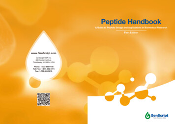
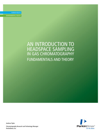
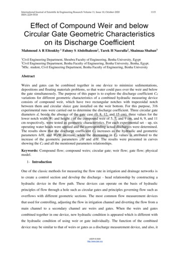
Evidence-Based Complementary and Alternative Medicine3Table 1: Primers for the target genes used in RT-PCR.Target genePrimer5 -CCACATCTCCCTCCAGAAAAGA -3 (F)5 -GCTGGGTAGAGAATGGATGAAC -3 (R)5 -GCTTCAGGCAGGCAGTAT -3 (F)5 -ACAAACCGCTTTTCCATCT -3 (R)5 -GAAATCGTGGAAATGAG -3 (F)5 -TAGGTTTGCCGAGTAGA -3 (R) 5 -TGCACCACCAACTGCTTAG -3 (F)5 -GGATGCAGGGATGATGTTC -3 8472455177TreatmentBooster7Length of target fragment (bp)1417232935 (Days)Arthritis score and paw volume testFigure 1: Scheme of animal experiment protocol. From day 14, rats with CIA were treated with Glycoside of Tripterygium wilfordii, Agkistrodonpowder, and Agkistrodon peptides.the Mascot search algorithm (version 2.2), which ranks thebest fit peptide matches based on a cumulative protein scoreof the unique peptides identified.The criteria used for the Mascot search are as follows:trypsin was used as the protease with a maximum of 1missed cleavage allowed; the protein oxidation of methionine was fixed as a dynamic modification whereas thecarbamidomethyl of cysteine was retained as a static modification; and the protein list was further filtered by applyinga false discovery rate cutoff of 1% at the protein level. Thedata were filtered for ion scores of at least 60. For each file,an ion score 20 indicated identity or extensive homologywith 𝑃 0.05.2.7. Cell Culture and Cytotoxicity Assay. The RAW264.7 cellswere cultured in high-glucose DMEM with 10% FBS in a 37 Cincubator with 5% CO2 . The viability of RAW264.7 cells wasevaluated with the MTS assay. The cells were seeded in 96well plates at a density of 10 104 cells/well for 24 h and thentreated with various concentrations of separated peptides (0.1,1, 10, 100, or 1000 𝜇g/mL) for 24 h. Optical density (OD) wasmeasured at 450 nm using a Multiskan Spectrum MicroplateSpectrophotometer (Bio-Rad, California, USA). Cell viabilitywas expressed as a percentage of the control.2.8. RNA Extraction and Real-Time Polymerase Chain Reaction (RT-PCR) Analysis. The RAW264.7 cells were culturedin 6-well plates at a density of 10 104 cells/mL for 24 h.Following pretreatment with LPS (10 𝜇g/mL) for 4 h, the cellswere treated with different concentrations of Agkistrodonpeptides (1, 10, or 100 𝜇g/mL) for 24 h.Total RNA was extracted using TRIzol reagent. Realtime PCR was performed using SYBR-Green Supermix ona Bio-Rad iQ5 Real-time PCR system (Bio-Rad, California,USA). The mouse primer sequences were used as describedin Table 1. Data were calculated using the formula 2 ΔΔCt andall values were normalized to the level of GAPDH.2.9. Induction of Collagen-Induced Arthritis (CIA). CII wasdissolved in 100 mM acetic acid at the concentration of4 mg/mL by stirring overnight at 4 C. Dissolved CII wasemulsified with an equal volume of incomplete Freund’sadjuvant (IFA) on ice using a T-branch pipe and two syringesto make the final concentration of 2 mg/mL CII/IFA emulsion. For the primary immunization, CII/IFA emulsion wasinjected subcutaneously at the base of the rat tail, as the firstsite, and on the back, as the second site, totalling 200 𝜇l ofemulsion containing 400 𝜇g of CII. A booster injection wasgiven on day 7 after the initial immunization with 100 𝜇l of theemulsion containing 200 𝜇g of CII. Visually apparent arthritiswith swollen joints appeared approximately 11–13 days afterthe first injection.2.10. Animal Grouping and Treatment. After the joints swelling was successfully established in at least one paw, therats were randomized into groups, with the mean of theirhind paw volumes and arthritis scores in each groupapproximately equal. Rats with CIA were randomly dividedinto 4 groups: Glycoside of Tripterygium wilfordii (GTW,0.72 mg/kg body weight, administered by gastric gavagedaily), Agkistrodon powder (APO, 580 mg/kg body weight,administered by gastric gavage daily), Agkistrodon peptides(APE, 4 mg/kg body weight, administered by intraperitonealinjection daily), and the CIA model group (Figure 1). Inaddition, the normal control group received nothing. Inthis study, there were six mice per group and a total of 5groups.2.11. Evaluation of Paw Swelling Degree. The paw swellingdegree was determined by measuring the change in the hindpaw volume (mL) with a plethysmometer (type: YLS-7C, Yanyi Technology Development Co., Ltd., Jinan, China). The pawswelling rate (%) was expressed as increased multiples of thehind paw volume by subtracting the paw volume before theprimary immunization.
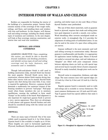
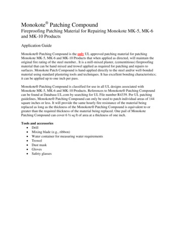
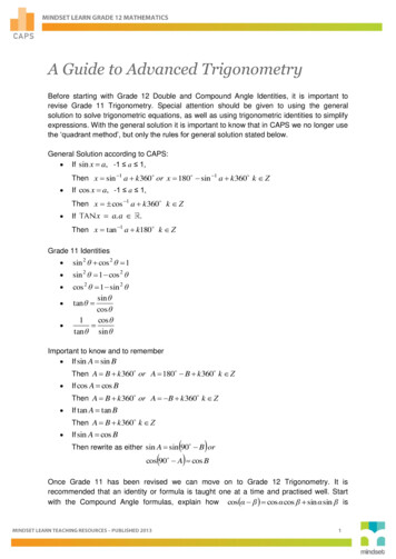
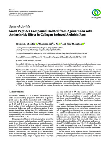
4Evidence-Based Complementary and Alternative MedicineTable 2: Separated peptide fractions of Agkistrodon identified by PK.GR.GFSGLDGAK.GR.DGAAGPK.GK.GEPGDAGAK.GMW (MH . Estimation of Arthritis Indexes. The development ofarthritis in the hind paws of rats was evaluated before treatment and on days 14, 17, 23, 29, and 35 by using a qualitativescoring system: 0, normal; 1, mild, but definite redness andswelling of the ankle or wrist or the apparent redness andswelling are limited to individual digits; 2, moderate rednessand swelling of the ankle; 3, severe redness and swelling of theentire paw including the digits; and 4, maximally inflamedlimb involving multiple joints.2.13. Determination of Type II Collagen IgG Antibodies. IgGantibodies against type II collagen in serum was assessed withELISA kits. According to the manufacturer’s protocol, serumsamples from arthritic rats were centrifuged at 10,000 rpmfor 3 minutes and diluted with sample dilution buffer from1 : 20,000 to 1 : 80,000. The OD values were read at 490 nmusing a Multiskan Spectrum Microplate Spectrophotometer(Bio-Rad, California, USA) and expressed as units per millilitre.2.14. Radiographic Examination. At the end of the study (day35), the hind limbs were collected for radiographic evaluation. X-ray radiography of the ankle joint was performedusing an in vivo Imaging System FX (Carestream Health,Rochester, USA) with a 35 kW exposure for 2 min. Thethree-dimensional reconstruction and images were obtainedby using high-resolution in vivo X-ray microtomography(Bruker, Germany).2.15. Histologic Assessments. At the end of the study (day 35),the hind limbs were preserved in 10% neutral buffered formalin fixative and then placed in decalcifying solution (a mixturecomprising formic acid, hydrochloric acid, and distilledwater) for 50 days (decalcifying solution was exchanged every5 days). After that, all ankles were longitudinally trimmed,embedded in paraffin, sectioned at 5 𝜇m, and stained withhaematoxylin and eosin (H&E), safranin O, and toluidineblue.2.16. Immunohistochemical Analysis. For immunohistochemistry, sections were prepared from the same preparations used for H&E staining. The sections were incubatedwith specific antibodies for rat TNF-𝛼, IL-1𝛽, and IL-6.Images were observed with light microscopy (Nikon, Tokyo,Japan) and representative images were revealed.2.17. Statistical Analysis. Data were expressed as the mean SEM or mean SD. For comparisons among groups,a one-way analysis of variance (ANOVA) followed by aDiff (MH ) 0.00007 0.000220.00022 75.7666.9660.84𝑃 value6.08𝐸 084.16𝐸 074.13𝐸 061.37𝐸 055.36𝐸 05Games-Howell test was performed to test significant differences. All data were analysed using SPSS 17.0. 𝑃 0.05 wasconsidered to be statistically significant.3. Results3.1. Agkistrodon Peptides Compound Isolation and Identification. Following instructions of the Chinese Pharmacopeia(2010 edition), crude peptides were extracted and werefurther separated by taking advantage of ion-exchange chromatography. Initial separation using a 0–100% linear gradientof NaCl was unsuccessful (data not shown), which mightbe attributed to the existence of components not responsiveto the NaCl gradient. The separation protocol was thereforemodified by increasing the NaCl to 0.5 M after the elutionof detected peaks of NaCl-unresponsive. An elution peakresponding to the increased NaCl was observed, and repeattests performed demonstrated similar results (Figure 3(a)).The NaCl responsive eluate was collected and furtherdialyzed for mass determination by MALDI-TOF/MS. Twopeaks (m/z 1022.33 and 1097.87) over 50% intensity wereobserved within the mass range from 1,000 Da to 10,000 Da,indicating small molecular weight peptides 10 amino acidslong (Figure 3(b)).Liquid chromatography tandem mass spectrometry (LCMS/MS) was used to further identify the peptides. Outputof the high-resolution MS/MS spectra, generated by thefragmentation of the precursor, with corresponding intensities was searched against the NCBI protein database ofthe taxonomy of Squamata (158856 entries, downloadedDecember 2016) using the Mascot search engine (version2.2) (Table 2), revealing that the signals at m/z 927.4531[M H ] and m/z 769.38389, m/z 851.42576, m/z 615.30966, and m/z 801.37372 [M H ] corresponded tothe single-protonated fragments of collagen alpha-1(I) chainfrom Ophiophagus hannah.3.2. Effect of Agkistrodon Peptides Compound on Cell Viability and the mRNA Expression of TNF-𝛼, IL-1𝛽, and IL6 in RAW264.7 Cells. To investigate the cytotoxicity ofAgkistrodon peptides, the viability of RAW264.7 cells wasevaluated by using the MTS assay (Figure 4(a)). Treatment with Agkistrodon peptides (0.1–100 𝜇g/mL) did nothave a marked cytotoxic effect on the RAW264.7 cells.Furthermore, the anti-inflammatory effect was evaluated bystimulating the RAW264.7 cells with LPS. The stimulationof LPS promoted the expression of inflammatory cytokinesat the mRNA level in RAW264.7 cells. However, treatmentwith Agkistrodon peptides suppressed the mRNA expression
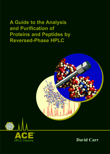
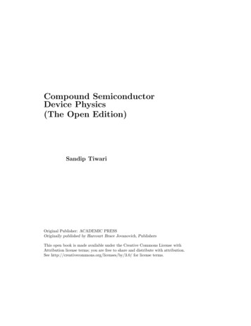
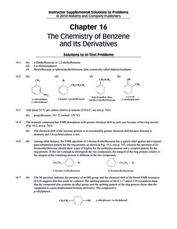
Evidence-Based Complementary and Alternative Medicine5TNF- IL-6IL-1 BoneCartilageAPETNF- APEIL-6IL-1 MacrophagesM Figure 2: A schematic diagram of the effects and the principle molecular mechanisms of APE on Rheumatoid arthritis. APE reduces therecruitment of macrophages and thus downregulates the production of TNF-𝛼, IL-6, and IL-1𝛽, which are mainly released by macrophages.1004700 Reflector Spec #1 MC [BP 1097.9,485]% 21.01136.61352.21567.8Mass (m/z)1783.41999.0567Sodium chloride gradientfrom 0–0.5 -1234(a)(b)Figure 3: Agkistrodon peptides isolation and identification. (a) Seven repeats of fast separation of crude peptide mixture using ion-exchangechromatography. The dotted line refers to the sodium chloride gradient from 0 to 0.5 M. (b) Agkistrodon peptides identification by MALDIspectra.of TNF-𝛼, IL-1𝛽, and IL-6 in a dose-dependent manner(Figures 4(b)–4(d)).3.3. Effect of Agkistrodon Peptides Compound on Paw Swellingand Ankle Changes in Groups with CIA. The effect of Agkistrodon peptides on CIA was evaluation with the hind pawswelling rate and arthritis index. The degree of swelling inthe normal control group showed no significant increase.Compared with the normal control group, the degree ofswelling in all other groups showed significant increases(data not shown). At the end of the experiment, comparedwith the model group the degree of swelling in the rats oftreated groups showed significant reductions (𝑃 0.05or 𝑃 0.01). The degree of swelling in rats from theAPE group showed a continuously significant reduction atdays 9 and 21 of treatment (𝑃 0.01). The strong antiinflammatory potency of the Chinese medicine Tripterygium wilfordii Hook (TWHF) has been verified [21–23],and the active component Tripterygium glycoside (GTW)[24–27] was used as the positive control in the presentstudy.Compared with those in either the GTW or APO group,those in the APE group had significantly reduced swelling.Additionally, there was no significant difference in pawvolumes between the groups treated with GTW and APO(Figure 5(a)).
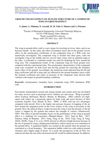
6Evidence-Based Complementary and Alternative Medicine2.5100 806040200Agkistrodon Peptides( g/ml)00.11101001000Relative mRNA expression(TNF- /GAPDH)Relative cell survival(% of control)120 2.0 1.51.00.50.0LPS (10 g/ml)Agkistrodon Peptides( g/ml) (a)2.5 1.5 1.0 0.50.0LPS (10 g/ml)Agkistrodon Peptides( g/ml) 1 10 100 10 100(b) 1 10 100(c)Relative mRNA expression(IL-6/GAPDH)Relative mRNA expression(IL-1 /GAPDH)2.0 2.01.51.00.50.0LPS (10 g/ml)Agkistrodon Peptides( g/ml) 1(d)Figure 4: Effect of Agkistrodon peptides on the inflammatory cytokines mRNA expression in RAW264.7 cells. (a) The cytotoxic effect ofAgkistrodon peptides on the viability of RAW264.7 cells. Cell viability was expressed as percentage of the control. Data were expressed asmean SD. 𝑃 0.05 versus control. (b–d) The effect of Agkistrodon peptides on the mRNA expression levels of TNF-𝛼, IL-1𝛽, and IL-6 inLPS-stimulated RAW264.7 cells. Data were expressed as mean SD. 𝑃 0.05; 𝑃 0.01 versus control. 𝑃 0.05; 𝑃 0.01 versus theLPS-treated cells.Visually apparent arthritis, with swollen joints in CIArats, suggested that the CIA model was successfully established. At the end of the experiment, compared with themodel group, all treated groups showed significant reductions(𝑃 0.05 or 𝑃 0.01) in clinical arthritis indexes. Comparedwith the model group, a significant reduction was observedin the APE group. In addition, the arthritis index of the APEgroup was significantly different compared to the APO group(Figure 4(b)).Plain radiographic analysis revealed normal bone structure with smooth and intact sharp and bone substance inthe normal rats. However, signs of arthritis with soft-tissueswelling, uniform joint space narrowing, and bone damagewere observed in the joints of rats in the model group.Compared with the model group, the soft-tissue swelling intreated groups was minimal, and the bone damage was notsevere. In addition, markedly alleviated plain radiographicchanges were observed in the joints of rats in the APE group(Figure 6(a)). Similar results showing the severe joint destruction were obtained with the three-dimensional images. Theimages showed smooth bone surface and intact joint architecture in normal rats and notable bone destruction in the CIAmodel rats while the bone destruction was much less robustin the APE group, suggesting that Agkistrodon peptides canprotect bone integrity induced by CIA (Figure 6(b)).3.4. Anti-Inflammatory Effect of Agkistrodon Peptides on TypeII Collagen Antibodies Expression in Serum and TNF-𝛼, IL1𝛽, and IL-6 Expression in Joints. The levels of anti-typeII collagen IgG in treated groups was significantly reducedcompared to the model group. The levels of anti-type IIcollagen IgG in the APE group were significantly lower thanthose in the GTW group. There was no significant differencebetween the APO and APE group (Figure 7).Immunohistochemistry analysis revealed a higherexpression of the inflammatory cytokines TNF-𝛼, IL-1𝛽, andIL-6 in the model group compared to the normal group.However, the expression level of these three cytokineswere markedly reduced in the APE group compared to themodel group or to the other treated groups, suggesting thatAgkistrodon peptides could suppress the expression of theinflammatory cytokines TNF-𝛼, IL-1𝛽, and IL-6 under thearthritic conditions caused by CIA (Figures 8(a)–8(c)).3.5. Improvement Effect of Agkistrodon Peptides on Pathological of Joints. The sections stained with H&E from the normalrats revealed that synovial cells on the surface of the anklejoints were well organized in the absence of inflammation.However, the images from rats in the model group revealedsigns of severe arthritis with synovial hyperplasia, inflammatory cell infiltration, vascular pannus formation, and
Evidence-Based Complementary and Alternative Medicine7905.0804.54.060 50Arthritis indexHind paw swelling degree (%)70 40 ##30 ##‰3.5 3.0 ## #‰2.520 ##2.01001.514172329Days post immunization (d)35APOAPENormalCIAGTW14172329Days post immunization (d)CIAGTW35APOAPE(b)(a)Figure 5: Paw swelling changes in different groups of CIA. (a) Paw swelling changes in different groups of CIA. Data were expressed as mean SEM (𝑛 12 hind paws). 𝑃 0.05; 𝑃 0.01 versus the model group. ‰ 𝑃 0.05 versus the GTW group. # 𝑃 0.05; ## 𝑃 0.01 versusthe APE group. (b) Arthritis index changes in different groups of CIA. Data were expressed as mean SEM (𝑛 12 hind paws). 𝑃 0.05; ‰###𝑃 0.01 versus the model group. 𝑃 0.05 versus the GTW group. 𝑃 0.05; 𝑃 0.01 versus the APO gure 6: Plain radiographs and three-dimensional images of rats’ joints. (a) The plain radiographs showed that soft-tissue swelling (yellowarrows), uniform joint space narrowing (triangles), and damaged bone substance (red arrows) were markedly alleviated in the joints of ratsin the APE group. (b) The three-dimensional images showed that bone destruction (red arrows) was much less robust in the APE groupcompared with the model and other treated groups.
8Evidence-Based Complementary and Alternative MedicineAnti-collagen IgG level in serum( 104 units/ml)300250 ‰200150CIAGTWAPOAPEAPOAPECIAGTWFigure 7: Expression of collagen type II IgG in serum in different groups of CIA. Data were expressed as mean SD. 𝑃 0.05; 𝑃 0.01versus the model group. ‰ 𝑃 0.05 versus the GTW group.NormalCIANormalCIAGTWAPOAPEAPOAPEAPOAPETNF- (a)GTWIL-1 (b)NormalCIAGTWIL-6(c)Figure 8: Immunohistochemistry analysis in different groups of CIA. (a) TNF-𝛼, (b) IL-1𝛽, and (c) IL-6 expression in joints (originalmagnification, 200). The expressions of TNF-𝛼, IL-1𝛽, and IL-6 were decreased in joints of the treated groups compared with the modelgroup. The arrows indicated locations of TNF-𝛼, IL-1𝛽, or IL-6.cartilage destruction. The extent of synovial hyperplasia andinflammatory cell infiltration in the joint of rats in the treatedgroups was comparatively reduced. In contrast, intact articular cartilage and slight inflammatory cell infiltration wereobserved in the joints of rats in the APE group (Figure 9(a)). Aloss of safranin O and toluidine blue indicated severe cartilagedestruction and bone erosion in rats from the model group.However, the damage was improved in the treated groupsand the improvement was more prominent in the joints ofrats from the APE group (Figures 9(b) and 9(c)). Collectively,
Evidence-Based Complementary and Alternative MedicineCIAGTWAPOAPEAPOAPEAPOAPEH&E StainNormal9(a)CIAGTWToluidine Blue StainNormal(b)CIAGTWSafranin-O StainNormal(c)Figure 9: Representative images of joint tissues stained with H&E, safranin O, and toluidine blue (original magnification, 200). (a) Imagesstained with H&E revealed that synovial hyperplasia, inflammatory cell infiltration, the vascular pannus formation, and cartilage destructionwere inhibited in the treated groups, especially the APE group when compared with the model group. The arrows indicated synovium. (b, c)Images stained with safranin O and toluidine blue showed that cartilage destruction and bone erosion were ameliorated in the treated groups,especially the APE group when compared with the model group. The triangles indicated articular cartilage.these results demonstrated that Agkistrodon peptides caneffectively attenuate inflammation, cartilage destruction, andbone erosion caused by CIA.4. DiscussionProinflammatory cytokines such as TNF-𝛼, IL-1𝛽, and IL6 found in high concentrations in the synovial fluid andtissues of RA patients are crucial m
2.5. E ect of Agkistrodon Peptides Compound on Cell Via-bility and the mRNA Expression of TNF-,IL-1,andIL-7 in RAW576.8 Cells. To investigate the cytotoxicity of Agkistrodon peptides, the viability of RAW. cells was evaluated by using the MTS assay (Figure (a)). Treat-ment with Agkistrodon peptides (.- g/mL) did not