Transcription
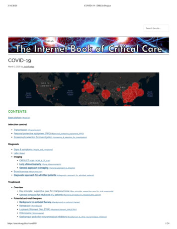
3/16/2020COVID-19 - EMCrit ProjectSearch the site .TOCABOUT THE IBCCAIRWAYTWEET USIBCC PODCASTARCHIVESCOVID-19March 2, 2020 by Josh FarkasCONTENTSBasic biology (#biology)Infection controlTransmission (#transmission)Personal protective equipment (PPE) (#personal protective equipment (PPE))Screening & selection for investigation (#screening & selection for investigation)DiagnosisSigns & symptoms (#signs and symptoms)Labs (#labs)ImagingCXR & CT scan (#CXR & CT scan)Lung ultrasonography (#lung ultrasonography)General approach to imaging (#general approach to imaging)Bronchoscopy (#bronchoscopy)Diagnostic approach for admitted patients (#diagnostic approach for admitted patients)TreatmentOverviewKey principle: supportive care for viral pneumonia (#key principle: supportive care for viral pneumonia)General template for intubated ICU patients (#general template for intubated ICU patient)Potential anti-viral therapiesBackground on antiviral therapy (#background on antiviral therapy)Remdesivir (#remdesivir)Lopinavir/Ritonavir (KALETRA) (#lopinavir/ritonavir (KALETRA))Chloroquine (#chloroquine)Oseltamavir and other neuraminidase inhibitors (#oseltamavir & other neuraminidase inhibitors)https://emcrit.org/ibcc/covid19/1/24
3/16/2020COVID-19 - EMCrit ProjectImmunomodulatory therapySteroid (#steroid)Tocilizumab (#tocilizumab)Anti-bacterial therapy (#anti-bacterial therapy)Cardiovascular (#cardiovascular)PulmonaryNoninvasive respiratory supportHigh ow nasal cannula (HFNC) (#high ow nasal cannula)BiPAP (#noninvasive ventilation (BiPAP))Intubation procedure (#intubation procedure)Invasive mechanical ventilation (#invasive mechanical ventilation)Disaster ventilation strategies (#disaster ventilation strategies)Renal failure (#renal failure)ECMO (#ECMO)Prognosis (#prognosis)Disposition (#disposition)Questions & discussion (#questions & discussion)Pitfalls (#pitfalls)PDF of this chapter ID-19-3-6.pdf) (or create customized PDF (https://emcrit.org/ibcc/about-guide/#pdf) )biology(back to contents) (#top)basicsCOVID-19 is a non-segmented, positive sense RNA virus.COVID-19 is part of the family of coronaviruses. This contains:(i) Four coronaviruses which are widely distributed and usually cause the common cold (but can cause viral pneumonia in patients with comorbidities).(ii) SARS and MERS – these caused epidemics with high mortality which are somewhat similar to COVID-19. COVID-19 is most closely related to SARS.It binds via the angiotensin-converting enzyme 2 (ACE2) receptor located on type II alveolar cells and intestinal epithelia (Hamming 2004(https://www.ncbi.nlm.nih.gov/pubmed/15141377) ).This is the same receptor as used by SARS (hence the technical name for the COVID-19, “SARS-CoV-2”).When considering possible therapies, SARS (a.k.a. “SARS-CoV-1”) is the most closely related virus to COVID-19.COVID-19 is mutating, which may complicate matters even further ( gure below). Virulence and transmission will shift over times, in ways which we cannotpredict. New evidence suggests that there are roughly two different groups of COVID-19. This explains why initial reports from Wuhan described a highermortality than some more recent case series (Tang et al. 2020 10.1093/nsr/nwaa036/5775463#.XmA64GbsBuI.twitter) ; Xuet al 2020 (https://www.bmj.com/content/368/bmj.m606) ). Image showing evolution of COVID-19 here 020/03/covid88evo.jpg?resize 1536%2C1218&ssl 1) .Ongoing phylogenetic mapping of new strains can be found here (https://nextstrain.org/ncov) .nomenclature used in this chapterTechnically, the virus is supposed to be called “SARS-CoV-2” and the clinical illness is called “COVID-19.” This gets confusing, so for this chapter the termCOVID-19 will be used to refer to both entities.The term “SARS” will be used to refer to the original SARS virus from 2003 (which has currently been renamed SARS-CoV-1).pathophysiology(1) ARDSThe primary pathology is ARDS, characterized by diffuse alveolar damage (e.g. including hyaline membranes). Pneumocytes with viral cytopathic effectare seen, implying direct virus damage (rather than a purely hyper-in ammatory injury; Xu et al 2/17) (https://www.thelancet.com/action/showPdf?pii S22132600%2820%2930076-X) .(2) Cytokine stormEmerging evidence suggests that some patients may respond to COVID-19 with an exuberant “cytokine storm” reaction (with features of bacterial sepsisor hemophagocytic lymphohistiocytosis ndrome-shlhos/) ).Clinical markers of this may include elevations of C-reactive protein and ferritin, which appear to track with disease severity and mortality (Ruan 00134-020-05991-x) ).https://emcrit.org/ibcc/covid19/2/24
3/16/2020COVID-19 - EMCrit Projectstages of illness ?There seem to be different stages of illness that patients may move through.(#1) Replicative stage – Viral replication occurs over a period of several days. An innate immune response occurs, but this response fails to contain thevirus. Relatively mild symptoms may occur due to direct viral cytopathic effect and innate immune responses.(#2) Adaptive immunity stage – An adaptive immune response eventually kicks into gear. This leads to falling titers of virus. However, it may alsoincrease levels of in ammatory cytokines and lead to tissue damage – causing clinical deterioration. There is a suggestion that this could lead to virusinduced hemophagocytic lymphohistiocytosis (HLH)(Mehta et al. 36(20)30628-0) ). More discussion about thisentity and possible therapy here (https://emcrit.org/ibcc/in uenza/#virus-associated hemophatocytic syndrome (VAHS)) .This progression may explain the clinical phenomenon wherein patients are relatively OK for several days, but then suddenly deteriorate when they enter theadaptive immunity stage (e.g. Young et al. 3/3/2020 /2762688) ).This has potentially important clinical implications:Initial clinical symptoms aren't necessarily predictive of future deterioration. Sophisticated strategies may be required to guide risk-strati cation anddisposition (see below section on prognosis (#prognosis) ).Anti-viral therapies might need to be deployed early to work optimally (during the replicative stage).Immunosuppressive therapy (e.g. low-dose steroid) might be best initiated during the adaptive immune stage (with a goal of blunting thisimmunopathologic response slightly, in the sickest patients). But this is purely speculative.transmission(back to contents) (#top)large droplet transmissionCOVID-19 transmission can occur via large droplet transmission (with a risk limited to 6 feet from the patient)(Carlos del Rio icle/2762510) ).This is typical for respiratory viruses such as in uenza.Transmission via large droplet transmission can be prevented by using a standard surgical-style mask.airborne transmission ?It's controversial whether COVID19 can be transmitted via an airborne route (small particles which remain aloft in the air for longer periods of time). Airbornetransmission would imply the need for N95 masks (“FFP2” in Europe), rather than surgical masks. This controversy is explored further in Shiu et al /tada2019.pdf) .Airborne precautions started being used with MERS and SARS out of an abundance of caution (rather than any clear evidence that coronaviruses aretransmitted via an airborne route). This practice has often been carried down to COVID19.Guidelines disagree about whether to use airborne precautions:The Canadian Guidelines ofessionals.html) and World HealthOrganization guidelines ssuspected-20200125) bothrecommend using only droplet precautions for routine care of COVID19 patients. However, both of these guidelines recommendairborne precautions for procedures which generate aerosols (e.g. intubation, noninvasive ventilation, CPR, bag-mask ventilation, and bronchoscopy).The United States CDC recommends .html?CDC AA refVal v%2Fhcp%2Finfection-control.html) usingairborne precautions all the time when managingCOVID19 patients.Using airborne precautions for all patients who are de nitely or potentially infected with COVID19 will likely result in rapid depletion of N95 masks. This willleave healthcare providers unprotected when they actually need these masks for aerosol-generating procedures.In the context of a pandemic, the Canadian and WHO guidelines may be more sensible in countries with nite resources (i.e. most locales). However, infectioncontrol is ultimately local, so be sure to follow your hospital's guidance regarding this.contact transmission (“fomite-to-face”)This mode of transmission has a tendency to get overlooked, but it may be incredibly important. This is how it works:(i) Someone with coronavirus coughs, emitting large droplets containing the virus. Droplets settle on surfaces in the room, creating a thin lm ofcoronavirus. The virus may be shed in nasal secretions as well, which could be transmitted to the environment.(ii) The virus persists on fomites in the environment. Human coronaviruses can survive on surfaces for up to about a week (Kampf et al 2020(https://www.ncbi.nlm.nih.gov/pubmed/32035997) ).It's unknown how long COVID-19 can survive in the environment, but it might be even longer (some animalcoronaviruses can survive for weeks!).(iii) Someone else touches the contaminated the surface hours or days later, transferring the virus to their hands.(iv) If the hands touch a mucous membrane (eyes, nose, or mouth), this may transmit the infection.Any effort to limit spread of the virus must block contact transmission. The above chain of events can be disrupted in a variety of ways:https://emcrit.org/ibcc/covid19/3/24
3/16/2020COVID-19 - EMCrit Project(a) Regular cleaning of environmental surfaces (e.g. using 70% ethanol or 0.5% sodium hypochlorite solutions; for details see Kampf et al /10.1016@j.jhin .2020.01.022-2.pdf)and CDC guidelines .html?CDC AA refVal v%2Fhcp%2Finfection-control.html) ).(b) Hand hygiene (high concentration ethanol neutralizes the virus and is easy to perform, so this might be preferable if hands aren't visibly soiled)(Kampf 2017 (http://www.fha.org/ les/JohnW/EM/Ethanol-hand-sanitizer-and-HAV.pdf) ).(c) Avoidance of touching your face. This is nearly impossible, as we unconsciously touch our faces constantly. The main bene t of wearing a surgicalmask could be that the mask acts as a physical barrier to prevent touching the mouth or nose.Any medical equipment could become contaminated with COVID-19 and potentially transfer virus to providers (e.g. stethoscope earpieces and shoes). Arecent study found widespread deposition of COVID-19 in one patient's room, but fortunately this seems to be removable by cleaning with sodiumdichloroisocyanurate (Ong et al 2020 /2762692) ).when can transmission occur?(#1) Asymptomatic transmission (in people with no or minimal symptoms) appears to be possible (Carlos del Rio icle/2762510) ).(#2) Transmission appears to occur over roughly 8 days following the initiation of illness.Patients may continue to have positive pharyngeal PCR for weeks after convalescence (Lan 2/27 /2762452) ).However, virus culture methods are unable to recover viable virus after 8 days of clinical illness (Wolfel 03.05.20030502v1.full.pdf) ).This implies that prolonged PCR positivity probably doesn't correlate with clinical virustransmission. However, all subjects in Wolfel et al. 5.20030502v1.full.pdf) had mild illness, so it remainspossible that prolonged transmission could occur in more severe cases.CDC guidance nical-guidance-management-patients.html) is vague on how long patients with known COVID-19should be isolated. It may be advisable to obtain two paired RT-PCR tests (one of the nasopharynx and one of the pharynx), with each pair collected 24hours apart, prior to discontinuing precautions.R R is the average number of people that an infected person transmits the virus to.If R is 1, the epidemic will burn out.If R 1, then epidemic will continue at a steady pace.If R 1, the epidemic will increase exponentially.Current estimates put R at 2.5-2.9 (Peng PWH et al, 2/28 0098-2/pdf) ). This is a bit higher than seasonal in uenza.R is a re ection of both the virus and also human behavior. Interventions such as social distancing and improved hygiene will decrease R .Control of spread of COVID-19 in China proves that R is a modi able number that can be reduced by effective public health interventions.The R on board the Diamond Princess cruise ship was 15 – illustrating that cramped quarters with inadequate hygiene will increase R (Rocklov doi/10.1093/jtm/taaa030/5766334) ).personal protective equipment (PPE)(back to contents) (#top)gear(1) Contact precautions (waterproof gown and gloves)(2) Some sort of mask (discussed above in the transmission (#transmission) section)N95 mask or a powered, air-purifying respiratory (“PAPR”)Surgical mask for patients not undergoing aerosol-generating procedures (based on WHO & Canadian guidelines)(3) Goggles or eye shieldNote: The exact gear used is probably less important than using it correctly.applying and removing PPE (donning & do ng)Understanding how to put on (don) and remove (doff) personal protective equipment is extremely important (especially if contact transmission is a dominantmode of transmission).Removing soiled PPE is the most critical and di cult aspect.Applying and removing PPE should ideally be practiced before patients arrive (e.g. using simulation).The video below describes how to use PPE (you may skip the rst 5 minutes).https://emcrit.org/ibcc/covid19/4/24
3/16/2020COVID-19 - EMCrit ProjectNETEC: Personal Protective Equipment for COVID-19some pearls about personal protective equipmentPay attention to the junction between gloves and gowns. The gown should be tucked into the gloves (leaving no gap in-between). Using gloves with extendedcuffs facilitates this (similar to sterile surgical gloves). Gloves with long cuffs may facilitate removal of the gown and gloves as a single unit (see 12:30 in theabove video if this doesn't make sense).When removing PPE, always start by rst applying alcohol-based hand sanitizer to your gloves.After fully removing PPE, sanitize hands and wrists with alcohol-based hand sanitizer again.Create a step-wise protocol for PPE removal. Two examples are shown below, but this may very depending on your exact gear. Follow the steps slowly. Checklist 020/03/pperemovegood.jpg?w 1484&ssl 1) for removing PPE, using a tear-away gown here (this typeof gown is preferred) Checklist 020/03/pperemove2.jpg?resize 1520%2C1536&ssl 1) for removing PPE, using a gown withxed neckties.Consider do ng with someone watching you (to ensure good technique). If this isn't possible, do ng in a mirror may be helpful.screening & selection for investigation(back to contents) (#top)key considerations include(1) Recent travel to affected areas.Areas with community-based transmission are increasing rapidly.The incubation time is up to 14 days, so travel within that window is relevant.The importance of travel will decrease over time, as coronavirus starts being transmitted within the community.(2) Contact with anyone with known COVID-19 (de ned as a prolonged period of time spent 6 feet apart).(3) As community acquisition emerges, broader testing will be needed. This will be based on a more detailed clinical evaluation, weighing:i) How well patients meet the clinical features of Coronavirus (e.g. laboratory and imaging features explored further below).ii) Presence or absence of alternative diagnoses (e.g. if patient tests positive for in uenza, this would make it less likely that they simultaneouslycontracted in uenza and coronavirus).approach to isolation and testingBelow is a general strategy aimed at rapid isolation of potentially infected patients, although it's already out of date for the following reasons:(1) Travel risk has been updated by the CDC to include South Korea, Iran, Italy, and Japan (and at this point might also be appropriate to include someother areas in Europe).(2) Many areas with community spread are starting to screen patients without de ned epidemiological exposure.This is only intended as a general rubric. Be sure to follow your institutional protocols. Close communication with infection control, infectious diseasespecialists, and the local health department is essential.Note that some patients may present with gastrointestinal symptoms. Unfortunately, most diagnostic algorithms will fail to detect and isolate thesepatients.signs and symptoms(back to contents) (#top)signs & symptomshttps://emcrit.org/ibcc/covid19/5/24
3/16/2020COVID-19 - EMCrit ProjectCOVID-19 may cause constitutional symptoms, upper respiratory symptoms, lower respiratory symptoms, and, less commonly, gastrointestinal symptoms.Most patients will present with constitutional symptoms and lower respiratory symptoms (e.g. fever and cough). Table 020/03/sxjpg.jpg?resize 1536%2C934&ssl 1) of symptoms described by various studies.Fever:The frequency of fever is variable between studies (ranging from 43% to 98% as shown in the table above). This may relate to exact methodology usedin various studies, different levels of illness severity between various cohorts, or different strains of the virus present in various locations. Additionally,some studies de ned fever as a temperature 37.3 C (Zhou et al. 3/9/20 (https://www.thelancet.com/action/showPdf?pii S0140-6736%2820%2930566-3) ).Regardless of the exact numbers – absence of a fever does not exclude COVID-19.Gastrointestinal presentations: up to 10% of patients can present initially with gastrointestinal symptoms (e.g. diarrhea, nausea), which precede thedevelopment of fever and dyspnea (Wang et al. 2/7/20 /2761044) ).“Silent hypoxemia” – some patients may develop hypoxemia and respiratory failure without dyspnea (especially elderly)(Xie et al. /s00134-020-05979-7.pdf) ).Physical examination is generally nonspeci c. About 2% of patients may have pharyngitis or tonsil enlargement (Guan et al 02032?articleTools true) ).typical disease courseIncubation is a median of 4 days (interquartile range of 2-7 days), with a range up to 14 days (Carlos del Rio icle/2762510) ).Typical evolution of severe disease (based on analysis of multiple studies by Arnold Forest (https://www.youtube.com/watch?v zFrghcp5pbY&feature youtu.be) )Dyspnea 6 days post exposure.Admission after 8 days post exposure.ICU admission/intubation after 10 days post exposure. However, this timing may be variable (some patients are stable for several days afteradmission, but subsequently deteriorate rapidly).labs(back to contents) (#top) Table 020/03/labsjpg.jpg?resize 1536%2C1115&ssl 1) of general laboratoryndings described in several studies.complete blood countWBC count tends to be normal.Lymphopenia is common, seen in 80% of patients (Guan et al 2/28 2?articleTools true) , Yang et al 2/21(https://www.thelancet.com/action/showPdf?pii S2213-2600%2820%2930079-5) ).Mild thrombocytopenia is common (but platelets are rarely 100). Lower platelet count is a poor prognostic sign (Ruan et al 34-020-05991-x) ).coagulation studiesCoagulation labs are generally fairly normal upon admission, although elevated D-dimer is commonly seen (table above).Disseminated intravascular coagulation may evolve over time, correlating with poor prognosis ( gure below)(Tang et al. 2020(https://www.ncbi.nlm.nih.gov/pubmed/32073213) ). Image of DIC labs in survivors versus non-survivors over time here 020/03/dicevo.jpg?resize 636%2C600&ssl 1) .in ammatory markersProcalcitoninCOVID-19 does not appear to increase the procalcitonin. For example, the largest series found that procalcitonin levels were 0.5 in 95% of patients(Guan et al 2/28 2?articleTools true) ).Elevated procalcitonin may suggest an alternative diagnosis (e.g. pure bacterial pneumonia). For patients who have been admitted with COVID-19,procalcitonin elevation may suggest a superimposed bacterial infection.C-reactive protein (CRP)COVID-19 increases CRP. This seems to track with disease severity and prognosis. In a patient with severe respiratory failure and a normal CRP,consider non-COVID etiologies (such as heart failure).Young et al. 3/3 /2762688) found low CRP levels in patients not requiring oxygen (mean 11 mg/L, interquartilerange 1-20 mg/L) compared to patients who became hypoxemic (mean 66 mg/L, interquartile range 48-98 mg/L).Ruan et al 3/3 020-05991-x) found CRP levels to track with mortality risk (surviving patients had a median CRP of 40 mg/L with an interquartile range of 10-60 mg/L, whereas patients who died had a median of 125 mg/L with an interquartile range of 60-160https://emcrit.org/ibcc/covid19/6/24
3/16/2020COVID-19 - EMCrit Projectmg/L)( gure below in the section on prognosis (#prognosis) ).evaluation for competing diagnosesPCR for in uenza and other respiratory viruses (e.g. RSV) may be helpful. Detection of other respiratory viruses doesn't prove that the patient isn't co-infectedwith COVID-19 ( 5% of patients may be co-infected with both COVID-19 and another virus)(Wang et 2.12.20022327v2) ).However, an alternative explanation for the patient's symptoms will reduce the index of suspicionfor COVID-19 substantially.Conventional viral panels available in some hospitals will test for “coronavirus.”This test does not work for COVID-19!This PCR test for “coronavirus” is designed to evaluate for four coronaviruses which usually cause mild illness.Ironically, a positive conventional test for “coronavirus” actually makes it less likely that the patient has COVID-19.Blood cultures should be performed as per usual indications.speci c testing for COVID-19(back to contents) (#top)Currently in the United States, all testing is done by state reference labs. Specimen collection and testing should be coordinated with the department of health.specimens(1) Nasopharyngeal swab should be sent.(2) If intubated, tracheal aspirate should be performed.(3) Bronchoalveolar lavage or induced sputum are other options for a patient who isn't intubated. However, obtaining these specimens may pose substantialrisk of transmission.It's dubious whether these tests are bene cial if done for the sole purpose of evaluating for coronavirus (see the section below on bronchoscopy(#bronchoscopy) ).limitations in determining the performance of RT-PCRThere are several major limitations, which make it hard to precisely quantify how RT-PCR performs.(1) RT-PCR performed on nasal swabs depends on obtaining a su ciently deep specimen. Poor technique will cause the PCR assay to under-perform.(2) COVID-19 isn't a binary disease, but rather there is a spectrum of illness. Sicker patients with higher viral burden may be more likely to have a positiveassay. Likewise, sampling early in the disease course may reveal a lower sensitivity than sampling later on.(3) Most current studies lack a “gold standard” for COVID-19 diagnosis. For example, in patients with positive CT scan and negative RT-PCR, it's murkywhether these patients truly have COVID-19 (is this a false-positive CT scan, or a false-negative RT-PCR?).(Convalescent serologies might eventually solve this problem, but this data isn't available currently.)speci citySpeci city seems to be high (although contamination can cause false-positive results).sensitivity may not be terri cSensitivity compared to CT scansIn a case series diagnosed on the basis of clinical criteria and CT scans, the sensitivity of RT-PCR was only 70% (Kanne 2/28).Sensitivity varies depending on assumptions made about patients with con icting data (e.g. between 66-80%)(Ai et 020200642) ). Image of analysis of Ai et al to determine sensitivity & speci city of PCR here 020/03/perfctpcr.jpg?resize 732%2C600&ssl 1) .Among patients with suspected COVID-19 and a negative initial PCR, repeat PCR was positive in 15/64 patients (23%). This suggests a PCR sensitivity of 80%. Conversion from negative to positive PCR seemed to take a period of days, with CT scan often showing evidence of disease well before PCR positivity(Ai et al. 200642) ).Bottom line?PCR seems to have a sensitivity somewhere on the order of 75%.A single negative RT-PCR doesn't exclude COVID-19 (especially if obtained from a nasopharyngeal source or if taken relatively early in the diseasecourse).If the RT-PCR is negative but suspicion for COVID-19 remains, then ongoing isolation and re-sampling several days later should be considered.https://emcrit.org/ibcc/covid19/7/24
3/16/2020COVID-19 - EMCrit ProjectCXR & CT scan(back to contents) (#top)general description of imaging ndings on chest x-ray and CT scanThe typical nding is patchy ground glass opacities, which tend to be predominantly peripheral and basal (Shi et al 2/24 (https://www.thelancet.com/action/showPdf?pii S1473-3099%2820%2930086-4) ).The number of involved lung segments increases with more severe disease. Over time, patchy ground glass opacities maycoalesce into more dense consolidation.In ltrates may be subtle on chest X-ray (example above from Silverstein et al (https://www.thelancet.com/action/showPdf?pii S0140-6736%2820%2930370-6) ). Image of example chest X-ray here 020/02/covidcxr.jpg?resize 750%2C380&ssl 1) . Image of example CT scans here 020/02/covct.jpg?resize 768%2C809&ssl 1) .Findings which aren't commonly seen, and might argue for an alternative or superimposed diagnosis:Pleural effusion is uncommon (seen in only 5%).COVID-19 doesn't appear to cause masses, cavitation, or lymphadenopathy.sensitivity and time delayLimitations in the dataData from different studies con ict to a certain extent. This probably re ects varying levels of exposure intensity and illness severity (cohorts withhigher exposure intensity and disease severity will be more likely to have radiologic changes).Sensitivity of CT scanning?Sensitivity among patients with positive RT-PCR is high. Exact numbers vary, likely re ecting variability in how scans are interpreted (there currentlydoesn't seem to be any precise de nition of what constitutes a “positive” CT scan).Sensitivity of 86% (840/975) in Guan et al. 2?articleTools true)Sensitivity of 97% (580/601) in Ai et al 200642) .Among patients with constitutional symptoms only (but not respiratory symptoms), CT scan may be less sensitive (e.g., perhaps 50%)(Kanne 200527) ).CT scan abnormalities might emerge before symptoms?Shi et al. (https://www.thelancet.com/action/showPdf?pii S1473-3099%2820%2930086-4) performed CT scanning in 15 healthcare workers who were exposed toCOVID-19 before they became symptomatic.Ground glass opaci cation on CT scan was seen in 14/15 patients! 9/15 patients had peripheral lung involvement (some bilateral, some unilateral).Emergence of CT abnormality before symptoms could be consistent with the existence of an asymptomatic carrier state (discussed above).Chest X-raySensitivity of chest X-ray is lower than CT scan for subtle opacities. In Guan et al. 2?articleTools true) , thesensitivity of chest x-ray was 59%, compared to 86% for CT scan.Martin Schranz@martinpschranzQuality CT/XR images on proven COVID-19 provided by Prof.Dr. Filippo Cademartiri, Chairman of Radiology , Marche - Italy.603 4:45 PM - Mar 5, 2020345 people are talking about thislung ultrasonography(back to contents) (#top)https://emcrit.org/ibcc/covid19/8/24
3/16/2020COVID-19 - EMCrit ProjecttechniqueIn order to achieve sensitivity, a thorough lung examination is needed (taking
adaptive immunity stage (e.g. Young et al. 3/3/2020 (h t t p s : // j a ma net w o r k . c o m/ j o u r na l s / j a ma / f u l l a r t i c l e/ 2762688) ). This has potentially impor tant clinical implications: Initial clinical symptoms aren't necessarily predictive of future deterioration.