Transcription
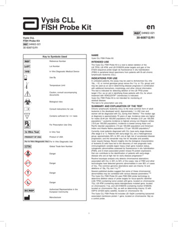
Vysis CLLFISH Probe Kiten04N02-02130-608712/R1Vysis CLLFISH Probe Kit04N02-02130-608712/R1NAMEKey to Symbols UsedVysis CLL FISH Probe KitReference NumberINTENDED USEThe Vysis CLL FISH Probe Kit is a test to detect deletion of theLSI TP53, LSI ATM, and LSI D13S319 probe targets and gain of theD12Z3 sequence probe target via fluorescence in situ hybridization(FISH) in peripheral blood specimens from patients with B-cell chroniclymphocytic leukemia (CLL).Lot NumberIn Vitro Diagnostic Medical DeviceUse ByINDICATIONS FOR USEIn untreated patients, the assay may be used to dichotomize CLL (the13q-, 12, or normal genotype group versus the 11q- or 17p- group) andmay be used as an aid in determining disease prognosis in combinationwith additional biomarkers, morphology, and other clinical information.The test is indicated for detecting deletion of the LSI TP53 probetarget (17p-) as an aid in identifying those patients with CLL for whomtreatment with VENCLEXTA (venetoclax) is indicated.Vysis CLL FISH Probe Kit is not intended for monitoring ofresidual disease.The test is for prescription use only.Temperature LimitCaution, consult accompanyingdocumentsBiological risksSUMMARY AND EXPLANATION OF THE TESTChronic lymphocytic leukemia (CLL) is the most common form of adultleukemia in the developed world. Approximately 1 in 202 men andwomen will be diagnosed with CLL during their lifetime.1 The median ageat diagnosis is approximately 70 years of age. Incidence rates are higherfor males (6.44 per 100,000 population) than females (3.51 per 100,000population).1 Leukemia incidence is highest among non-Hispanic whites(13.6 per 100,000 population); incidence is lowest among Asian andPacific Islander populations (7.4 per 100,000 population) and AmericanIndian and Alaska Native populations (7.3 per 100,000 population).2Currently, most patients diagnosed with CLL have early stage-disease(Rai stage 0 or 1). Patients with early-stage CLL are a heterogeneousgroup; approximately 30% to 50% are at high risk of accelerated diseaseprogression, and the remainder may live for decades and possiblynever require therapy. Recent insights into the biological characteristicsof leukemic B cells have led to the discovery of new prognostic tools(immunoglobulin variable-region heavy chain gene mutation status,cytogenetic abnormalities assessed by fluorescence in situ hybridization[FISH], and Z-chain-associated protein kinase-70 protein expression)that can contribute to the identification of patients with early-stagedisease who are at high risk for early disease progression.3Routine karyotype analysis only detects chromosomal aberrationsassociated with CLL in 40% to 50% of the cases. Use of FISH and othertechnologies have detected genomic abnormalities in over 80% of casesof CLL. The common genomic aberrations seen are trisomy 12 anddeletions of 13q, 17p, and 11q.4-6Several published studies suggest that some of these chromosomalabnormalities may be correlated with various disease parameters.7-10The Vysis CLL FISH Probe Kit uses FISH DNA probe technology todetermine deletion status of probe targets for locus-specific identifier(LSI) TP53 (containing tumor protein p53 gene, located on chromosome17p), LSI ATM (containing ataxia telangiectasia mutated gene, locatedon chromosome 11q), and LSI D13S319 (containing marker D13S319,located on chromosome 13q), as well as determining trisomy 12 withCEP12 (D12Z3 alpha satellite, located on chromosome 12).The Vysis CLL FISH Probe Kit includes LSI 13q34 (containing lysosomalassociated membrane protein 1 gene, located on chromosome 13q) asa control probe.Consult instructions for useContains sufficient for n testsFor Prescription Use OnlyIn Vitro TestProduct of USAFor In Vitro Diagnostic UseGlobal Trade Item NumberDangerDangerDangerDangerAuthorized Representative in theEuropean CommunityManufacturer1
The National Comprehensive Cancer Network (NCCN) PracticeGuidelines for Non Hodgkins Lymphoma (v.2.2016) which are theconsensus recommendations of leading US oncology experts, statesFISH (including abnormalities tested for by this kit) is informative forboth prognosis and therapy determination. The guidelines recommenduse of FISH at the time of diagnosis as well as re-evaluation by FISH atthe time of relapse to direct treatment options (including abnormalitiestested for by this kit).11The tumor suppressor protein, p53, has been shown to play a criticalrole in oncogenesis and response to chemotherapy in a variety ofhuman cancers. In humans the TP53 gene is found on the short arm ofchromosome 17 (17p13)12 and is reported to be suppressed or mutatedin a large number of human cancers.13 Deletions of the 17p regionresulting in abnormalities of the tumor suppressor protein p53 havebeen identified as one of the poorest prognostic factors for CLL as it ispredictive of short time to disease progression, short response duration,lack of response to therapy and short overall survival (OS).7,14The 17p deletion is more frequently observed in treated patients than inpreviously untreated patients, increasing in frequency during the courseof the disease with up to 50% of patients with relapsed or refractorydisease having the deletion.14 Approximately 8 to 12% of patients withCLL in the first line treatment carry del 17p.14 It is widely accepted thattreatment outcomes in patients with del 17p are poor.15,16 Clinical trialsof venetoclax in patients with refractory and relapsed CLL have shownthat patients with 17p deletion are likely to experience clinical benefit.Refer to the VENCLEXTA (venetoclax) label for more information.BIOLOGICAL PRINCIPLES OF THE PROCEDUREDNA Probe DescriptionGeneral Reagents DescriptionVysis LSI TP53 SpectrumOrange/ATM SpectrumGreen ProbesThe SpectrumOrange-labeled LSI TP53 probe, approximately 172 Kb inlength (chr17:7494395-7666098; February 2009 Assembly University ofCalifornia, Santa Cruz [UCSC] Human Genome Browser17), is located at17p13.1 and contains the complete TP53 gene.The SpectrumGreen-labeled LSI ATM probe, approximately 731 Kb inlength (chr11:107801007-108531514; February 2009 Assembly UCSCHuman Genome Browser), is located at 11q22.3 and contains theATM gene.Vysis LSI D13S319 SpectrumOrange/13q34 SpectrumAqua/CEP 12SpectrumGreen ProbesThe SpectrumOrange-labeled LSI D13S319 probe, approximately 135 Kbin length (chr13:50602368-50737301; February 2009 Assembly UCSCHuman Genome Browser), is located at 13q14.3.The SpectrumAqua-labeled LSI 13q34 probe, approximately 612 Kb inlength (chr13:113691322-114303435; February 2009 Assembly UCSCHuman Genome Browser), is located at 13q34.The SpectrumGreen-labeled CEP 12 probe is located at the centromereof chromosome 7p13.1 RegionTelomereDAPI II CounterstainDAPI II Counterstain consists of DAPI (4′,6-diamidino-2phenylindole 2HCl) (a DNA-specific fluorophore) and1,4-phenylenediamine (an antifade compound used to reduce thetendency of the fluorophores to diminish in intensity) in a glycerol andphosphate buffered saline mixture.NP-40NP-40 is a non-ionic surfactant that is used in the aqueous posthybridization wash solution.20X Standard Sodium Citrate (SSC) Salt20X SSC is a salt composed of sodium chloride and sodium citrate. It isused to make 20X SSC solution and subsequent dilutions for denaturingand wash solutions.Technique DescriptionFISH is a technique that allows visualization of specific nucleic acidsequences within a cellular preparation. Specifically, FISH involvesprecise annealing of a single-stranded, fluorophore-labeled DNA probeto a complementary target sequence. Hybridization of the probe withthe cellular DNA site is visible by direct detection using fluorescencemicroscopy.Peripheral blood cells from CLL patients are attached to microscopeslides using standard cytogenetic procedures. The resulting specimenDNA is denatured to single-stranded form and subsequently allowed tohybridize with the probes of the Vysis CLL FISH Probe Kit. Followinghybridization, the unbound probe is removed by a series of washes,and the nuclei are counter-stained with DAPI, a DNA-specific stain thatfluoresces blue. Hybridization of the Vysis LSI TP53 SpectrumOrange,LSI ATM SpectrumGreen, LSI D13S319 SpectrumOrange, LSI 13q34SpectrumAqua, and CEP 12 SpectrumGreen probes is viewed usinga fluorescence microscope equipped with appropriate excitation andemission filters, allowing visualization of the orange, green, and aquafluorescent signals.In a cell with normal copy numbers of the Vysis LSI TP53SpectrumOrange and Vysis LSI ATM SpectrumGreen probe targets, twoorange and two green signals will be expected. In a cell with normalcopy numbers of Vysis LSI D13S319 SpectrumOrange and CEP 12SpectrumGreen probe targets, two orange signals and two green signalswill be expected. Enumeration of the Vysis LSI TP53 SpectrumOrange,LSI ATM SpectrumGreen, LSI D13S319 SpectrumOrange, and CEP12SpectrumGreen signals provides a mechanism for determining absolutecopy number of the probe targets and the presence of the aberrationsof interest. The LSI 13q34 SpectrumAqua probe functions as an assayprocess control and is described further in the section on QualityControls later in this document. 172 kbLSI TP53 SpectrumOrange Probe11q22.3 Region5’TelomereEXPH5ATMCUL5RAB39Centromere3’ 731 kbLSI ATM SpectrumGreen Probe2
REAGENTSThese precautions include, but are not limited to the following: Wear gloves when handling specimens or reagents. Do not pipette by mouth. Do not eat, drink, smoke, apply cosmetics, or handle contactlenses in areas where these materials are handled. Clean and disinfect spills of specimens by including the use of atuberculocidal disinfectant such as 1.0% sodium hypochlorite orother suitable disinfectant.18 Decontaminate and dispose of all potentially infectious materialsin accordance with local, state and federal regulations.21Material ProvidedThis kit contains five reagents sufficient to process 20 assays. An assayis defined as one 22 mm x 22 mm Vysis LSI TP53 SpectrumOrange/ATM SpectrumGreen probe hybridization area and one 22 mm x 22mm Vysis LSI D13S319 SpectrumOrange/13q34 SpectrumAqua/CEP 12SpectrumGreen probe hybridization area.Vysis LSI TP53 SpectrumOrange/ATM SpectrumGreen ProbesPart No.30-241051Quantity1 vial, 200 µL/vial (300 and 200 ng/10 µL)Storage–20 C ( 5 C) and protected from lightComposition SpectrumOrange and SpectrumGreen fluorophorelabeled DNA probes in hybridization bufferHazard-determining components of labeling: Formamide Polyethylene glycol octylphenyl etherThe following warnings apply:DangerH302Harmful if swallowed.H315Causes skin irritation.H318Causes serious eye damage.H411Toxic to aquatic life with long lasting effects.P280Wear protective gloves / protectiveclothing / eye protection.P264Wash hands thoroughly after handling.P273Avoid release to the environment.P305 P351 IF IN EYES: Rinse cautiously with water for P338several minutes. Remove contact lenses, ifpresent and easy to do. Continue rinsing.P310Immediately call a POISON CENTER/doctor.P301 P312 IF SWALLOWED: Call a POISON CENTER/doctor if you feel unwell.P302 P352 IF ON SKIN: Wash with plenty of water.Vysis LSI D13S319 SpectrumOrange/13q34 SpectrumAqua/CEP 12 SpectrumGreen ProbesPart No.30-241050Quantity1 vial, 200 µL/vial (200, 400 and 25 ng/10 µL)Storage–20 C ( 5 C) and protected from lightComposition SpectrumOrange, SpectrumAqua, and SpectrumGreenfluorophore-labeled DNA probes in hybridization bufferDAPI II CounterstainPart No.30-804811Quantity1 vial, 600 µL/vial (125 ng/mL)Storage–20 C ( 5 C) and protected from lightComposition DAPI (4′,6-diamidino-2-phenylindole 2HCl) and1,4-phenylenediamine in a glycerol and phosphatebuffered saline mixtureNP-40Part No.QuantityStorageComposition30-8048102 vials, 2000 µL/vial–25 C to 30 CNP-40 (non-ionic detergent)20X SSC SaltPart No.QuantityStorageComposition30-8048121 bottle, 66 g–25 C to 30 CSodium chloride and sodium citrateP332 P313P330P362P501REAGENT STORAGE AND HANDLING INSTRUCTIONS-15 C-25 CDangerH360P201P202The Vysis CLL FISH Probe Kit must be stored at-20 C ( 5 C) and protected from light when notin use.The NP-40 and 20X SSC Salt may be storedseparately at room temperature.P280Shipping ConditionsP308 P313The Vysis CLL FISH Probe Kit is shipped on dry ice.If you receive reagents that are in a condition contrary to labelrecommendation, or that are damaged, contact Abbott MolecularTechnical Services.P405P501WARNINGS AND PRECAUTIONSIn Vitro Diagnostic Medical Device.For in vitro diagnostic use.CAUTION: United States Federal Law restricts this device to saleand distribution to or on the order of a physician or to a clinicallaboratory; use is restricted to, by, or on the order of a physician.If skin irritation occurs: Get medical advice/attention.Rinse mouth.Take off contaminated clothing.Dispose of contents / container inaccordance with local regulations.May damage fertility or the unborn child.Obtain special instructions before use.Do not handle until all safety precautionshave been read and understood.Wear protective gloves / protectiveclothing / eye protection.IF exposed or concerned: Get medicaladvice/attention.Store locked up.Dispose of contents / container inaccordance with local regulations.Safety Data Sheet Statement: Important information regarding the safehandling, transport, and disposal of this product is contained in theSafety Data Sheet.NOTE: Safety Data Sheets (SDS) for all reagents provided in thekits are available upon request from the Abbott MolecularTechnical Service Department.Safety PrecautionsCAUTION: This preparation contains human sourced and/orpotentially infectious components. No known test method canoffer complete assurance that products derived from humansources or inactivated microorganisms will not transmit infection. Thesereagents and human specimens should be handled as if infectious usingsafe laboratory procedures, such as those outlined in Biosafety inMicrobiological and Biomedical Laboratories,18 OSHA Standards onBloodborne Pathogens,19 CLSI Document M29-A3,20 and otherappropriate biosafety practices.21 Therefore all human sourced materialsshould be considered infectious.3
Assay Precautions Coplin jars/Vertical staining jar Fluorescence microscope equipped with recommended filters (seeMicroscope Equipment and Accessories section) Calibrated thermometer Microscope slide box with lid Magnetic stirrer pH meter pH indicator strips capable of reading pH 7.0 to 8.0 Vortex mixer 0.45 µm filtration units 37 C incubator Humidified chamber Slide warmer (45 to 50 C) Cytogenetic drying chamber (optional) Microcentrifuge tubes Phase contrast microscope Oven capable of heating to 90 C or equivalent The Vysis CLL FISH Probe Kit is only for use with specimens thathave been handled and stored as described in this package insert. Do not use the Vysis CLL FISH Probe Kit beyond its Use By date. Package insert instructions must be followed. Failure to adhere topackage insert instructions may yield erroneous results. Fluorophores are readily photo bleached by exposure to light. Tolimit this degradation, store slides and probe kits in the dark, andhandle all slides and probe kits containing fluorophores in reducedlight. This includes all steps involved in handling the hybridizedslide. Carry out all steps which do not require light for manipulation(incubation periods, washes, etc.) in the dark or reduced light. Probe mixtures are formulated at intended use concentrations andshould not be diluted. Always verify the temperature of the wash solutions prior to eachuse by measuring the temperature of the solution in the Coplin jarwith a calibrated thermometer.Laboratory Precautions All biological specimens should be treated as if capable oftransmitting infectious agents. Because it is often impossible toknow which might be infectious, all human specimens should betreated with universal precautions. Hemolyzed specimens may prevent proper culture for standardcytogenetic analysis. Exposure of the specimens to acids, strongbases, or extreme heat should be avoided. Such conditions areknown to damage DNA and may result in FISH assay failure. Failure to follow all procedures for slide denaturation, hybridization,and detection may cause unacceptable or erroneous results. The DAPI II Counterstain contains DAPI and 1,4-phenylenediamine. DAPI is a possible mutagen based on positive genotoxic effects.Avoid inhalation, ingestion, or contact with skin. Refer to SDS forspecific warnings. 1,4-phenylenediamine is a known dermal sensitizer and a possiblerespiratory sensitizer. Avoid inhalation, ingestion, or contact with skin.Refer to SDS for specific warnings. The Vysis CLL FISH Probe Kit contains formamide, a teratogen.Avoid contact with skin and mucous membranes. Calibrated thermometers are required for measuring temperatures ofsolutions and water baths. All hazardous materials should be disposed of according to theinstitution’s guidelines for hazardous disposal.Microscope Equipment and AccessoriesMicroscope An epi-illumination fluorescence microscope is required forviewing the hybridization results. The microscope should be checkedto confirm it is operating properly to ensure optimum viewing of FISHassay specimens. A microscope used with general DNA stains such asDAPI, propidium iodide, and quinacrine may not function adequately forFISH assays. Routine microscope cleaning and periodic maintenance bythe manufacturer’s technical representative, especially alignment of thelamp, are advised.Excitation Light Source A 100 watt mercury lamp or other lamp withsimilar intensity and spectral output is the recommended excitationsource. The manufacturer’s technical representative should be consultedto assure that the fluorescence illumination system is appropriate forviewing FISH assay specimens. Record the number of hours that the bulbhas been used and replace the bulb before it exceeds the rated time.Ensure that the lamp is properly aligned.Objectives Use oil immersion fluorescence objectives with numericapertures 0.75 when using a microscope with a 100 watt mercurylamp or other lamp with similar intensity and spectral output. A 40Xobjective, in conjunction with 10X eyepieces, is suitable for scanning thespecimen to select regions for enumeration. For enumeration of FISHsignals, satisfactory results can be obtained with a 60/63X or 100X oilimmersion achromat-type objective.Immersion Oil The immersion oil used with oil immersion objectivesshould be one formulated for low auto fluorescence and specifically foruse in fluorescence microscopy.Filters Hybridization of the Vysis LSI TP53 SpectrumOrange, LSIATM SpectrumGreen, LSI D13S319 SpectrumOrange, LSI 13q34SpectrumAqua, and CEP 12 SpectrumGreen probes to their targetregions of the DNA is marked by orange, aqua, and green fluorescence.The DNA which has not hybridized to the probes will fluoresce blue as aresult of the DAPI II Counterstain.The recommended filters for use with the Vysis CLL FISH Probe Kit arethe Vysis Dual Band Green/Orange Filter V2, the Vysis Single BandDAPI Filter, the Vysis Single Band Orange Filter, the Vysis Single BandAqua Filter, and the Vysis Single Band Green Filter or equivalents.ASSAY PROCEDUREMaterials Provided Vysis CLL FISH Probe Kit (List No. 04N02-021)Materials Required But Not Provided Immersion oil appropriate for fluorescence microscopyEthanol (100%). Store at room temperatureMethanolGlacial acetic acidPurified waterRubber cement12 N Hydrochloric acid (HCl)1 N or 2 N Sodium hydroxide (NaOH)Formamide, Ultra Pure0.075 M Potassium chloride (KCl)ASSAY PROTOCOLRefer to the WARNINGS AND PRECAUTIONS section of this packageinsert before preparing samples.Laboratory Equipment Working Reagent PreparationGlass microscope slides, precleaned22 mm x 22 mm glass coverslipsMicroliter pipettor (1 to 10 µL) and sterile tipsTimersMicrocentrifugeClinical centrifuge capable of reaching 250 x g and accommodatinga 15 mL centrifuge tube15 mL conical tubesGraduated cylinders (100 mL to 1000 mL)Water bath (70 to 80 C)Water bath (37 C)ForcepsDisposable syringeDisposable pipettes (5 mL to 20 mL)Ethanol Solutions (70%, 85%, and 100%)Prepare v/v dilutions of 70% and 85% ethanol using 100% ethanoland purified water. Store the prepared solutions at room temperaturein tightly capped containers. Discard stock solution after 6 months orsooner if solution appears cloudy or contaminated. Dilutions in Coplinjars may be used for up to one week unless evaporation occurs or thesolution becomes diluted or cloudy due to excessive use.20X SSC SolutionDissolve 66 g of 20X SSC salt using 200 mL of purified water in a suitablevessel. Measure pH at room temperature using a pH meter and adjust pHto 5.3 0.2 using 12 N HCl, if necessary. Transfer solution to a graduatedcylinder and add purified water until a final volume of 250 mL is reached.Mix and filter the prepared solution through a 0.45 μm filtration unit. Storeprepared stock solution at room temperature. Discard the stock solutionafter 6 months or sooner if solution appears cloudy or contaminated.4
Slide Preparation2X SSC SolutionMix thoroughly 100 mL of 20X SSC with 850 mL purified water in asuitable vessel. Measure pH at room temperature using pH meter toverify pH is 7.0 0.2. If necessary adjust the pH using 1N or 2 N sodiumhydroxide. Add purified water to bring final volume of the solution to1 L. Mix and filter through a 0.45 μm filtration unit. Store prepared stocksolution at room temperature. Discard stock solution after 6 months orsooner if solution appears cloudy or contaminated. Discard solution thatwas used in the assay at the end of each day.Denaturation Solution (70% formamide/ 2X SSC Solution)Mix thoroughly 49 mL formamide, 7 mL 20X SSC Solution, and 14 mLpurified water. Measure pH at room temperature using pH indicator stripsto verify pH is 7.0 to 8.0. Store prepared stock solution at 2 to 8 C. VerifypH is 7.0 to 8.0 using a pH indicator strip prior to each use. Discardsolution after one week.0.4X SSC / 0.3% NP-40 Wash SolutionMix thoroughly 20 mL 20X SSC Solution and 950 mL purified water ina suitable vessel. Add 3 mL of NP-40 and mix thoroughly until NP-40 iscompletely dissolved. Measure pH at room temperature using a pH meterand adjust pH to 7.4 0.1 with 1 N or 2 N sodium hydroxide. Add purifiedwater to bring final volume of the solution to 1 L. Mix and filter through a0.45 μm filtration unit. Store prepared stock solution at room temperature.Discard stock solution after 6 months or sooner if solution appears cloudyor contaminated. Discard solution that was used in the assay at the end ofeach day.2X SSC / 0.1% NP-40 Wash SolutionUsing a suitable vessel, mix thoroughly 100 mL 20X SSC Solution with850 mL purified water. Add 1 mL NP-40 and mix thoroughly until NP-40is completely dissolved. Measure pH at room temperature using a pHmeter and adjust pH to 7.0 0.2 with 1 N or 2 N sodium hydroxide. Addpurified water to bring final volume to 1 L. Mix and filter through a 0.45 μmfiltration unit. Store prepared stock solution at room temperature. Discardstock solution after 6 months or sooner if solution appears cloudy orcontaminated. Discard solution that was used in the assay at the end ofeach day.12. For optimal results, a cytogenetic drying chamber may be used. Setthe unit to a temperature of 20 to 30 C with a relative humidity of 30to 50%. If a cytogenetic chamber is not available, other means ofachieving the optimal temperature and humidity for target preparationmay be used.13. Label each slide appropriately.14. Ensure the sample is mixed adequately before preparing the slide.Using a transfer pipette, expel 5 to 25 µL of cell suspension ontothe left half of a pre-cleaned microscope slide and 5 to 25 µL of cellsuspension onto the right half of the slide, making two hybridizationareas. Allow the slide to dry.15. Using a phase contrast microscope, examine the number ofinterphase nuclei per field, under low power (10X objective).A minimum of 100 cells per low power field is suggested foroptimum assay results. Adjust the cell suspension to achieve therecommended number of interphase nuclei.NOTE: An optimal specimen will contain little to no debris and/orcytoplasm. Nuclei should appear dark, gray, flat, and non-refractile.Adjust temperature, humidity and volume to achieve optimal nucleimorphology.16. Bake slides at 90 C for 10 minutes (optional).Specimen Target PreparationNOTE: Initiate Manual Probe Denaturation procedure prior tocompleting Step 19 of this section to ensure the materials haveadequate time to thaw.17. Transfer Denaturation Solution and 2X SSC Solution to individualCoplin jars or remove previously prepared Denaturation Solution from2 to 8 C storage.18. Transfer the Coplin jar(s) with Denaturation Solution into a waterbath. Heat until the internal temperature of the Denaturation Solutionis 73 1 C.19. Transfer 2X SSC Solution Coplin jar(s) to a water bath. Warm tothe internal temperature 37 1 C. Age specimen slides in 2X SSCSolution for 30 minutes at 37 1 C. Alternatively, slides can be agedfor 2 minutes in 2X SSC Solution heated to 73 C 1 C.NOTE: Confirm solutions temperature using a calibratedthermometer. Immerse no more than 4 slides simultaneously in eachCoplin jar.20. Remove slides from the 2X SSC Solution and immediately transferto Coplin jar(s) containing 70% ethanol for a minimum of 1 minute.Following 70% ethanol, transfer to 85% ethanol for a minimum of 1minute, and then to 100% ethanol for a minimum of 1 minute.21. Following dehydration in ethanol, immerse the specimen slides in theDenaturation Solution at 73 1 C for 5 minutes. Start timing whenthe last slide has been added to the Coplin jar.NOTE: To maintain the proper temperature in Denaturation Solution,denature 4 slides simultaneously. If there are less than 4 slides, addblank slides that are at room temperature to bring the total to 4.22. Remove specimen slides from the Denaturation Solution andimmediately transfer to the Coplin jar containing 70% ethanol for aminimum of 1 minute. Following 70% ethanol, transfer to 85% ethanolfor a minimum of 1 minute, and then to 100% ethanol for a minimumof 1 minute.NOTE: Keep the specimen slides in 100% ethanol until ready to dryall slides and apply the probe mixture.Specimen Collection and TransportPeripheral blood collections should be performed according to thelaboratory’s institution guidelines. The AGT Cytogenetics LaboratoryManual contains recommendations for specimen collection andprocessing.22 It states that it is acceptable to collect the peripheralblood specimen in a sodium heparin blood collection tube. One (1) mLof peripheral blood is required for performing the assay. Specimens canbe processed immediately or shipped on cold packs and stored at 2 to8 C up to 96 hours prior to the start of sample preparation. Specimensshould never be iced or frozen. Specimens that are clotted or notshipped as indicated should not be used.Specimen ProcessingNOTE: Fixative (3:1 or 2:1 methanol:glacial acetic acid) should beprepared fresh daily.1. Add 1 mL of peripheral blood to a 15 mL conical centrifuge tube.2. Slowly add 10 mL of 0.075 M KCl. Mix gently by inverting the tubeseveral times. Optional: incubate tube(s) for 10 to 20 minutes atroom temperature or 37 C.3. Slowly add 2 mL of fixative to the cell suspension. Mix gently byinverting the tube several times.4. Centrifuge 8 to 12 minutes at 1000 to 1200 RPM (225 to 250 x g).5. Remove and discard all but approximately 0.5 to 1.0 mL of thesupernatant carefully without disturbing the cell pellet. Resuspendthe cell pellet by gently agitating the tube.6. Slowly add 10 mL fixative to the cell suspension. Mix by inverting thetube several times.7. Centrifuge 8 to 12 minutes at 1000 to1200 RPM (225 to 250 x g).8. Repeat the fixative wash (Steps 5 to 7) two more times or until thefixative is clear.9. Resuspend the cell pellet in fixative. Adjust the volume of cellsuspension to a density appropriate for slide preparation. If desired,prepare a test slide to verify the cell density.10. Allow specimen to stand in fixative at room temperature (15 to 30 C)for a minimum of 10 minutes before proceeding to Slide Preparation.11. Proceed to Slide Preparation or, if necessary, fixed pellets can bestored at -20 C ( 5 C) for up to 24 months. If needed, particularlyafter a prolonged storage, wash pellets with fixative before slidepreparation.Probe DenaturationNOTE: Probe mixtures are formulated at intended use concentrationsand should not be diluted. Doing so may lead to erroneousassay results.23. Remove the preformulated probe mixtures from storage and allowthe reagents to reach room temperature.24. Centrifuge probes for 2 to 3 seconds. Vortex and centrifuge themixtures again briefly.25. Place tubes at 73 1 C for 5 minutes.26. Centrifuge tubes for 2 to 3 seconds.27. Place tubes on 45 to 50 C slide warmer until ready to apply probe totarget specimen.NOTE: If specimen slides are ready when probes are denatured,apply probes immediately to the hybridization areas on the slide(s).5
HybridizationRoutine slide evaluation and enumeration is conducted using a DualBand filter. For an individual nucleus, if specific probe signal(s) appearweak with a Dual Band filter, it is recommended to use a single bandfilter to assist in enumeration.28. Remove specimen slides from the 100% ethanol.29. Dry each slide by touching the bottom edge to a paper towel (orequivalent) and wiping the underside (the side that does not containspecimen).30. Transfer slides to a 45 to 50 C slide warmer for up to 2 minutes toevaporate remaining ethanol.31. Using a microliter pipettor, apply 10 µL of the Vysis LSI TP53SpectrumOrange/ATM SpectrumGreen probe to one target area andapply coverslip without introducing bubbles. Repeat procedure forVysis LSI D13S319 SpectrumOrange/13q34 SpectrumAqua/CEP 12SpectrumGreen probe mixture.32. Seal coverslips
emission filters, allowing visualization of the orange, green, and aqua fluorescent signals. In a cell with normal copy numbers of the Vysis LSI TP53 SpectrumOrange and Vysis LSI ATM SpectrumGreen probe targets, two orange and two green signals will be expected. In a cell with normal copy numbers of Vysis LSI D13S319 SpectrumOrange and CEP 12


