Transcription
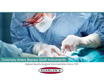
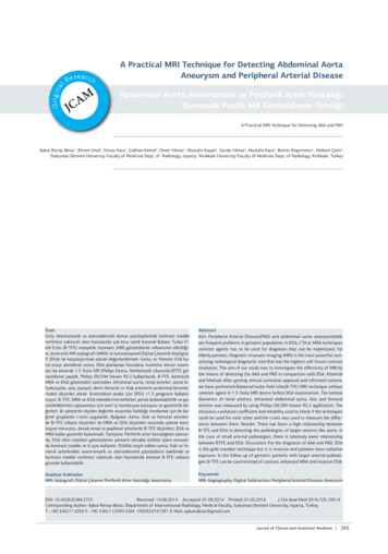
CORONARY ARTERY BYPASS GRAFTING (CABG)(Part 1)Mark Shikhman, MD, Ph.D., CSAAndrea Scott, CSTI have constructed this lecture based on publications by leading cardiothoracic Americansurgeons: Timothy J. Gardner, MD; Brian F. Buxton, MD; Patrick G. Magee, MD.***History. Coronary Artery Bypass Grafting (CABG) began in the late 1960s along twoparallel paths that included bypassing coronary artery obstructions using either theInternal Mammary artery (IMA) as the bypass conduit or reversed saphenous veingraft (SVG) from the leg. Each approach had early proponents, but the use of saphenousvein graft became the dominant approach by the majority of cardiac surgeons in 1970s.This preference was based on ease use of larger and technically less demandingsaphenous vein graft, as well as the grater versatility of the vein graft. Saphenous veinscould be used to graft any coronary artery site. By the mid- 1980s, with CABG beingdone increasingly often throughout the world and with 10- to 15-year follow-upexperience available, two extremely important observations were made. Many of theearliest patients to receive bypass grafts were returning 5-10 or 12 years after operationwith progression of atherosclerosis in their native coronary arteries and, even morealarming, had severe obstructive atherosclerosis in the vein grafts that were used in theoriginal procedure. A second unexpected observation was that in patients who had IMAbypass grafts performed previously, graft atherosclerosis and graft occlusion were rarelyencountered. These surprising finding led to changes in the approach in CABG procedurein the mid- to late 1980s, which has resulted in the current standard approach to coronaryartery bypass surgery. The majority of patients who undergo CABG surgery todayreceive a left IMA (LIMA) graft to the left anterior descending coronary artery(LAD). Other additional bypasses are constructed using reversed saphenous vein graftswith proximal aortic anastomoses.***Preoperative assessment. All patients with considerations of CABG will have acoronary angiogram performed, which will give information about coronary arterialbranches are completely or partially occluded and not visualizes. Another importantcomponent of preoperative assessment is the availability of suitable conduits. But beforewe will analyze importance of such assessment let’s list and discuss variety of arterialand vinous conduits.Arterial Conduits. The most usable arterial conduit in coronary grafting procedure isLeft Internal Thoracic (Mammary) Artery (LIMA). It was originated by work ofDr.Vineberg in 1946. The ITA arise from subclavian artery just above and behind thesternal end of the clavicle (Pic.1)1
Pic.1The artery descends vertically 1cm lateral to the sternal border, behind the first six costalcartilages. Pic.2 shows the ITA (IMA) position regarding pectoralis muscle, sternum, andpleura with endothoracic fascia.Pic.22
The next three pictures (pic3b, 3c, 3d) demonstrate stages of artery harvesting.Surgeons prefer to harvest artery with a pedicle which means together withsurrounding tissues that provide protection for the artery.3
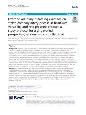
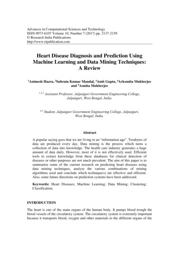
The second artery that can be used as arterial conduit for coronary graft is RadialArtery (RA). The RA arises from the bifurcation of the brachial artery in thecubital fossa and terminates by forming the deep palmar arch in the hand. Themain concern using RA is blood supply to the wrist and hand but in recentanatomical study was found that superficial palmar arch of the ulnar arteryprovides the blood supply to all fingers I 67% of hands. Additionally to it in87.5% of hands there is intense communication between deep palmar branches ofthe radial and ulnar arteries (pic.4).Pic.44
Pictures below demonstrate location of incision and expose of the Radial artery.Pic.5Occasionally, when surgeons do not have other choice they use Ulnar Artery asarterial conduit.The Gastroepiploic artery is sometimes used as an arterial graft when the IMAcannot reach the posterior surface of the heart or when other conduits are notavailable. As you remember, there are two Gastroepiploic arteries: left and right.Both arteries are active participants in stomach vascularisation, but because ofwide system of anastomosis between them and existence of other major gastricarteries, using the bigger – right Gastroepiploic artery as conduit does not5
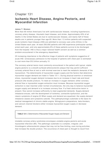
compromise stomach blood supply. Anatomy of Gastroepiploic arteries ispresented on the picture 6.Pic.6It is not necessary to perform additional incision for harvesting this type ofconduit; the median sternotomy incision is extended inferiorly for a further 5cmfor performing laparotomy and Right Gastroepiploic artery harvesting.The last artery that can be used as arterial conduit in the coronary bypass surgeryis Inferior Epigastric Artery. In many patients, when it is used alone, the length6
of this artery is not sufficient for an independent graft. It is used as a compositegraft with the LIMA as extension graft. You are familiar with this artery regardinganatomy of inguinal triangle. The inferior epigastric artery arises from theexternal iliac artery and gives branches to the spermatic cord, pubis, abdominalmuscles, and skin (pic.7). Unfortunately this artery is positioned far away fromeven extended sternotomy incision and its harvesting requires additional eitherlow midline, or paramedian, or oblique inguinal approach.Pic.7Saphenous Vein Graft. Despite of the progressive atherosclerosis that has beenfounded in the vein graft, it is one of the major conduits for coronary arteriesbypass surgery. The Greater Saphenous Vein (GSV) of the lower extremity is thebest choice of this type of graft based on: There are two independent types of low extremity vein system andremoval of superficial one (GSV) does not jeopardize the vinous flowfrom the leg Position, diameter and length of the GSV are in constant pattern whichsimplifies its harvestGSV is harvested in two different ways: Directly through multiple incisions and tunneling over the vein along themedial thigh and leg (Pic.8).7
Pic.8 Endoscopic vein harvest (Pic.9). In many patients it is possible to maketwo small incisions, one above the knee, and the second upper thigh forthis type of harvest. Patient, leg and surgical team positions are presentedon pictures below.Pic.98
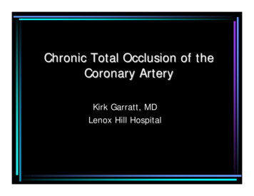
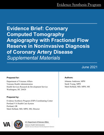
CORONARY ARTERY BYPASS GRAFTING (CABG)(Part 1)Mark Shikhman, MD, Ph.D., CSAAndrea Scott, CSTIncisionThe next picture (Pic.10) shows few different types of surgical accesses thatcommonly used in cardiothoracic surgeryPic.10A – SternotomyB – Anterior ThoracotomyC – Sternotomy with Subxiphoid LaparotomyD – Distal SternotomyE – Posterior ThoracotomyF – Transverse Curved LaparotomyIn majority of the cases Sternotomy is the approach of the choice since entiresternum must be divided for the traditional multi-vessel CABG operation.
Cardiopulmonary bypassCardiopulmonary bypass (Pic.11) has to be established by arterial cannulationonto aortic arch and venous cannulation through the right atrial appendage intothe inferior vena cava.Pic.11In the conventional CABG procedures construction of the Distal VenousAnastomoses should be performed first to avoid possible traction on themammary artery graft. Coronary artery is stabilized, the epicardium overlying theartery is incised, and arteriotomy is performed for a distance of 6-8 mm (Pic.12).
Pic.12The reversed saphenous vein is slightly beveled and anastomosed to the coronaryartery with a single 7/0 polypropylene suture (Pic.13).Pic.13Next, the Internal Mammary Artery (IMA) is anastomosed to the LeftAnterior Descending Coronary Artery (LAD). Once an adequate pedicle lengthis determined, the LAD is incised and the IMA incised on an angle at anappropriate distal site. A 7/0 or 8/0 polypropylene suture is used to construct this
type of anastomosis of approximately 3-4 mm in length with the continuous stitch(Pic.14).Pic.14Final view of the anastomosis is presented below (Pic.15)Pict.15Now surgeon can go back and complete proximal vein graft anastomosis withaorta. But in instances in which available vein for grafting is limited or whenthere is concern about multiple anastomoses on aorta, surgeon can construct two
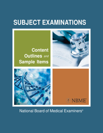
or more distal anastomoses with a single vein graft. It will be called SequentialDistal Vein Graft Anastomoses (Pic.16).Pic.16The final stage of the grafting procedure is construction of the Proximal VeinGraft Anastomoses. The Aorta Wall Punch is used to create openings in aorta foranastomoses (Pic.17) and distal segment of the vein graft is anastomosed withaorta (Pic.18).Pic.17
Pic.18The final view of the completed CABG procedure is presented below (Pic.19).Pic.19***
1 CORONARY ARTERY BYPASS GRAFTING (CABG) (Part 1) Mark Shikhman, MD, Ph.D., CSA Andrea Scott, CST I have constructed this lecture based on publications by leading cardiothoracic American