Transcription
4/8/2021Systemic Lupus Erythematosus: Diagnosisand ManagementJudith Lin, MDAssistant Professor of MedicineDivision of RheumatologyDepartment of Internal MedicineThe Ohio State University Wexner Medical CenterDisclosures None1
4/8/2021Objectives1.Identify clinical features and common manifestations of SLE2.Identify immunologic findings of SLE3.Recognize common SLE treatments and associated side effects4.Recognize complications that may be seen with SLE and theimportance of health maintenance managementWhat is SLE? Systemic autoimmune disease characterized byheterogenous multisystem involvement and production ofautoantibodies Driven by loss of immune tolerance and abnormal innate andadaptive immune function Immune complex mediated reactions and tissue destruction Variable clinical presentation and clinical course2
4/8/2021Risk factors for SLE Women of childbearing age More in African American, Hispanic, other ethnicminorities Genetics Polygenic Early complement deficiencies Family history Environment Infections, smoking, UV exposure, drugs, stress Genetics environment Immune dysregulationDiagnosis vs ClassificationDiagnosisSeveral Classification criteria ACR/EULAR, SLICC based on clinical presentation For categorizing patients forcombined with serologicresearch purposesfindings ANA is not enough No diagnostic criteria Diagnosis made by experiencedphysician/rheumatologist Not intended as diagnosticcriteria Can be used as guide toorganize thoughtsExclude alternative diagnosis3
4/8/20211997 ACR Classification criteria4 or more criteria, excluding other causesCriteriaMalar rashDiscoid rashPhotosensitivityOral ulcersArthritisPleuritis or pericarditisRenal disorderNeurologic disorderHematologic disorderImmunologic disorderPositive ANADefinitionFixed erythema over malar eminences sparing nasolabial foldErythematous raised patches with adherent keratotic scale and follicularplugging, often with atrophic scarsRash from unusual reaction to sunlightOral or nasopharyngeal ulcers, usually painless, observed by physicianNonerosive, 2 or more peripheral joints with tenderness, swelling or effusionConvincing history or objective evidencePersistent proteinuria, 0.5g/24hr or 3 on dipstick, cellular castsSeizures or psychosis in the absence of offending drugs or metabolicderangementsHematolytic anemia, leukopenia, lymphopenia, or thrombocytopeniaAnti‐dsDNA, Sm, or antiphospholipid antibodiesAbnormal titer at any point in time, in absence of drugs known to beassociated with drug induced lupusArth Rheum 19972012 SLICC ClassificationcriteriaImmunologic criteriaClinical CriteriaAcute cutaneous lupus: malar rash, SCLE, othersChronic cutaneous lupus Discoid, panniculitis, lupus tumidus,chillblainsOral ulcers: palateNonscarring alopeciaSynovitis involving 2 or more jointsSerositis: Pleuritis, pericarditisRenal disorder UPCR or 24hr urine protein 500mg/24hr RBC castsNeurologic Seizures, psychosis Mononeuritis multiplex Myelitis Peripheral or cranial neuropathy Acute confusional stateHemolytic anemiaLeukopenia ( 4000/mm3) or lymphopenia( 1000/mm3)Thrombocytopenia ( 100,000/mm3)ANA above lab reference rangeAnti‐dsDNA above lab reference range, exceptELISA: twice above lab reference rangeAnti‐SmithLow complement (C3, C4, CH50)Direct Coombs test in the absence of hemolyticanemia 4 criteria: 1 clinical and 1immunologic exclude othercausesLupus nephritiscan be made bybiopsy and ANAaloneAntiphospholipid antibody: any of the following: Lupus anticoagulant False positive RPR Medium or high titer anticardiolipin (IgG, IgM,or IgA) Anti‐B2 glycoprotein I (IgG, IgM, or IgA)Arth Rheum 20124
4/8/20212019 EULAR/ACR SLE Classification CriteriaClinical domains and criteriaEntry criteria: ANA 1:80Additive criteria: at least1 clinical and 10 points Only the highest weightedcriteria is scored withineach ombocytopeniaAutoimmune hemolysis344NeuropsychiatricDelirium Criteria does not need to bePsychosissimultaneousSeizureExclude alternative causesMucocutaneousNon‐scarring alopeciaOral ulcersSubacute cutaneous ORdiscoid lupusAcute cutaneous lupusMusculoskeletalJoint involvementClinical domains and criteriaWeight235WeightSerosalPleural or pericardialeffusionAcute pericarditis56RenalProteinuria 0.5g/24hrRenal biopsy class II or V LNRenal biopsy Class III or IVLN4810Immunology domains and criteriaWeightAntiphospholipid antibodiesAnti‐cardiolipin antibodies ORAnti‐B2GPI antibodies ORLupus anticoagulant22246ComplementLow C3 OR low C4Low C3 AND low C4346SLE‐specific antibodiesAnti‐dsDNA or Smith antibody6Aringer M. Arth Rheum 2019Antinuclear antibody (ANA) Antibodies against proteins or nucleic acidsin nucleus Found in 95% of SLE but only 57% specific Detection assays Indirect immunofluorescence (IIF) Gold standardTiterStaining pattern may guide clinical thinkingTime consuming, labor intensive, may have falsepositive ELISA Antibodies to different nuclear antigens Faster, detect specific antibodies High sensitivity but less specificBy Simon Caulton ‐ Own work, CC BY‐SA d 20521932ANA more likely to haveclinical significance withtiters 1:80ANA is sensitive but not specific forSLE, higher titer more likely to beassociated with autoimmune disease5
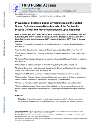
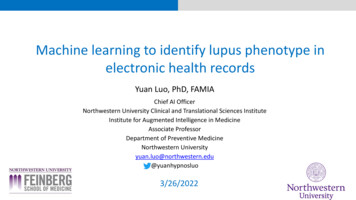
4/8/2021ANA titer and prevalenceANA Prevalence increases with age ANA common in generalpopulation 25‐30% have 1:40 titer 10‐15% have 1:80 titer 5% have 1:160 titer or higherSolomon DH et al. Arth&Rheum 2002ANA is common in healthy subjectsSatoh M, Chan EK, Ho LA, et al. Prevalence and sociodemographic correlates ofantinuclear antibodies in the United States. Arthritis Rheum. 2012;64(7):2319‐2327.Copyright obtained from publisher.ANA is common and nonspecific Can be triggered by Infections Smoking Silica, other chemicals andpollutants Medications: Hydralazine Procainamide Isoniazid Minocycline TNF alpha inhibitorsANA is seen in various conditions Can be seen in other conditions: Other autoimmune disease Other systemic autoimmunerheumatic disease Hashimoto’s thyroiditis Multiple sclerosis Psoriasis Autoimmune hepatitis Idiopathic thrombocytopenic purpura Atopic diseasesInfectionsMalignanciesLiver diseaseFamily history of autoimmunediseaseSoloman DH et al. Arth Care & Res 20026
4/8/2021Recognize common autoantibodies in SLEANA subsets/Extractable nuclear antigen antibodies (ENAs)AntibodyFrequencyANA patternClinical associationsSmith30%SpeckledHigh specificity, low sensitivity. More in African Americans.More organ damage.dsDNA70%Homogenous Can fluctuate with disease activity. Gold standard Crithidia assayis very specific, but common ELISA assay not very specificSSA30%SpeckledSicca, photosensitivity. Seen with Sjogren’s, SCLE, NNL, CHBSSB20%SpeckledSCLE, Sjogren’s, NNL, CHBU1RNP25‐40%SpeckledMCTD, Raynaud, ILD, pulmonary hypertensionHistone50%Homogenous SLE and DIL (75%)NNL: neonatal lupus, CHB: congenital heart blockAntiphospholipid antibodies(aPL) Lupus anticoagulant B2glycoprotein I IgG, IgM Cardiolipin IgG, IgMComplementsC3C4CH50C1qAntibody specificity and sensitivity limited by commercial assaysChoosing wisely campaign:Avoid ordering ANA sub‐serologies if ANA negative and lowclinical suspicion of immune‐mediated diseaseExceptions: Jo1 and SSAAntibodies alone are not sufficient to make diagnosisConsider other lupus‐related diseasesOther forms of lupus and lupus‐related disorders Cutaneous lupus Neonatal lupus Mixed connective tissuedisease Antiphospholipid syndrome Sjogren’s syndrome Undifferentiated connectivetissue Overlap syndromeDrug‐induced lupus Hydralazine Propythiouracil Sulfonamides Lithium Anticonvulsants Quinidine Diltiazem Beta blockers Interferon gamma TNF inhibitors7
4/8/2021SLE ManagementSLE treatment goalsHealth maintenance Control disease activity Cardiovascular health assessment Goal of remission or low disease activity Assess and treat reversible risk Minimize complications from disease andfactors given increased risk of CVDtreatment Bone health assessment Improve quality of life Increased risk of osteoporosis,Preventative measuresavascular necrosis due to SLE, sun Smoking cessation Photoprotection Avoid medications that may trigger lupus ifpossibleTreat reversible causes of symptoms Physical and lifestyle measures Address fatigue, sleep, exercise Provide emotional and psychosocialsupport Assess and treat fibromyalgiaTreat associated comorbidities Other autoimmune disease: T1DM, Hashimoto’s avoidance, steroid useAge‐appropriate cancer screening Increased risk of malignancy inSLEImmunizationsContraception counselingPregnancy planningAll SLE patients require multi‐disciplinary carewith PCP, rheumatology and other specialiststo optimize management and outcomesSystemic corticosteroids For rapid control of inflammatory activity Usually given as taper Pulse dose IVMP for severe organ threatening disease High dose For severe disease such as serositis, nephritis,hemolytic anemia Prednisone 20mg or higher Moderate dose For moderate disease such as arthritis Prednisone 7.5‐20mg Low dose: Usually used as slow taper or maintenance prednisone 7.5mg or lowerSteroids can be given for SLE flaresbut limit use as it is associated withsignificant side effectsSide effects: Osteoporosis, avascular necrosis,bone fractures Weight gain, Cushingoid features Hyperglycemia, diabetes Fluid retention, hypertension Arrhythmia Cataracts, glaucoma Gastritis, PUD Mood disorder, psychosis Muscle weakness Adrenal insufficiency Skin thinning, ecchymosis, striae8
4/8/2021Anti‐malarialsHydroxychloroquine, Chloroquine,Quinacrine For active, non‐organ threatening SLE Rash, arthritis, alopecia Many benefits in SLE: Reduce risk of flares in SLE Prevent progression of disease Reduce thrombotic and cardiovascularcomplications Improve glucose and lipid profiles Slow onset of action Weeks to months to see effect Hydroxychloroquine dose Up to 5mg/kg/day (max 400mg/day) Dose reduce for renal insufficiencyAntimalarials reduce flares andimproves outcomes in SLESide effects Retinal toxicity: Risk increases with time Irreversible Need retinal exam yearly Drug rash Blue‐gray discoloration of skin GI upset Myopathy Cardiomyopathy Arrhythmia CNS disturbance (dizziness,headache, insomnia, psychosis) Caution in G6PD deficiency Safe in pregnancySystemic immunosuppressive therapy Cytotoxic therapy Azathioprine Mycophenolate (Methotrexate) (Leflunomide) Cyclophosphamide Biologic therapy Belimumab (Rituximab) (Others to come) Calcineurin inhibitors (Cyclosporin) (Tacrolimus) Voclosporin9
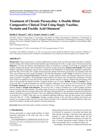
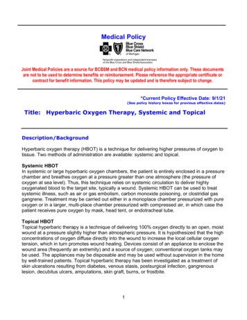
4/8/2021Case #1 40 yo Hispanic F w/ h/o HTN,anemia Joint pain and swelling in handswith morning stiffness Facial rash and body rashes Alopecia, oral ulcers No smoking Fam hx: No autoimmune diseaseJaccoud’s arthropathyCase #1: Workup and diagnosis ANA 1:320 speckled Sm, SSA ribosomal P, chromatin dsDNA 48 RF 27, ‐CCP Cq1 7 (L) early complementdeficiency increases risk of SLE C3 39, C4 8 (L) CBC with ACD otherwise normal ESR, CRP normal UA, UPCR normal Skin biopsy: interface dermatitisDiagnosis:SLE ( ANA, Sm, SSA, ribosomalP, chromatin, dsDNA, RF) withhypocomplementemia, anemia,Jaccoud’s arthropathy, oral ulcers,alopecia, acute cutaneous lupus10
4/8/2021Cutaneous lupusAcute cutaneous lupusLocalized: Malar rash Distinguish from: Rosacea Seborrhea dermatitis DermatomyositisGeneralizedDifferent forms of cutaneous LEChronic cutaneous lupusDiscoid lupusLupus profundus(panniculitis)ChilblainTumid lupusOther cutaneousmanifestations Raynaud Vasculitis Livedoreticularis Urticaria OthersSubacute cutaneous lupuserythematosus (SCLE) Types Annular Papulosquamous Photosensitive 50% have SLE 70% with ANA, SSA, 30% SSBAlopeciaNon‐scarring alopecia Focal or diffuse Differential:Recognize alopecia in SLEScarring alopecia Inflammatory,infiltrative conditions More focal than diffuse Discoid lupus Traction alopecia Female pattern hair loss crown, frontal, hereditary Telogen effluvium Iron deficiency Hypothyroidism11
4/8/2021Case #1: TreatmentBelimumab used as add‐on therapy for SLEBelimumab Topical therapy for cutaneous lupus Monoclonal ab against BLyS Topical corticosteroids SQ and IV For active seropositive SLE Topical calcineurin inhibitors Best for dsDNA, low complements, Immunomodulatorskin, MSK manifestations Hydroxychloroquine Adverse effects Infection Clinical course: Injection site/infusion reaction Inadequate control on plaquenil Intolerant to azathioprine,mycophenolate Started on belimumab Complicated by infection Diarrhea, nauseaHeadachePsych: depression, suicidal ideationCytopeniasPMLInfection evaluation, management, preventionVaccinations Recommended vaccines onWBC immunosuppressive therapy:ESR Yearly influenzaCRP ‐‐/ Prevnar13C3, C4, CH50 ‐‐/ Pneumovax23dsDNA‐‐ HAV and HBVSLE flare can be triggered by infections HPV ShingrixManagement during infection Hold immunosuppressive therapies until Avoid live vaccines Vaccines may be moreinfection resolveseffective when given before Ok to continue:starting ne if chronicInfectionSLE flare12
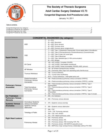
4/8/2021Case # 2 40 yo AAF with h/ocervical lymphadenopathy(biopsy negative formalignancy) whopresented with SOB andleg swelling SOB worse with lyingdown Smoker Exam BP 115/80, HR 101, RR 20,96% RA periorbital edema, diffuseanasarca, ascites Distant heart sounds Rash on trunk, extremities,hands and feetEKG: sinus tach, low voltageChest pain, SOB in SLECardiac manifestations Pericarditis Myocarditis Libman‐Sacks endocarditis Coronary arteritis Arrythmia CAD/MI Accelerated atherosclerosis 2 fold increase risk of CAD,CVA, PADDifferential for cardiacand pulmonarymanifestations in SLEPulmonary manifestations Pleuritis Parenchymal lung disease Pneumonitis Diffuse alveolar hemorrhge Interstitial lung disease Pulmonary vascular disease Pulmonary hypertension Pulmonary embolism Shrinking lung syndrome13
4/8/2021Sent to hospital WBC 3.02 (L), abs lymph 0.4 (L) concernfor SLE flare Hb 8.3 (L), MCV 85 Plt 175 less likely hemolytic Troponin normal less likely MI,myocarditis Cr 0.84 (baseline; BMP normal) UA trace proteins, no casts less likely GN LFT normal except Albumin 1.5 (L) Protein loss leading to anasarca Chest XR: cardiomegaly, left pleural effusion Serositis from SLE TTE: large, circumferential pericardialeffusion with early signs of tamponade serositis from SLE (No endocarditis)Case #2Serologies ANA 1:1280 speckled Sm, RNP, SSA, SSB dsDNA 70 (H) Low C3 39, C4 8 IgG 384 (L), IgA 59 (L), IgMnormal. ESR 91 (H), CRP normal UPCR 0.484 Stool alpha1antitrypsin 310 (H)Serologies suggestive of SLECT a/p: bowel walledema Concern for protein‐losing enteropathy Pericardiocentesis and paracentesis Exclude infection, malignancy Colonoscopy excluded alternativecausesDiagnosis:SLE ( ANA, Sm, RNP, SSA, SSB, dsDNA, low complements,leukopenia, lymphopenia, anemia)with acute cutaneous lupus,pericarditis, pleuritis and proteinlosing enteropathy14
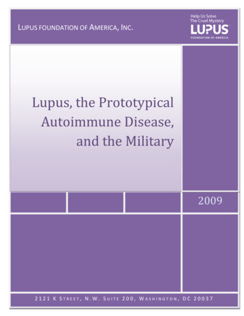
4/8/2021Case #2: ManagementTreatment: IVMP for rapid control Followed by PO taper Hydroxychloroquine IVIG for hypogammaglobulinemia Cyclophosphamide For induction therapy forsevere organ threateningdisease IV vs POCyclophosphamide effectiveinduction therapy for severe SLEbut has significant toxicitiesCyclophosphamide Used as Induction therapy for severe organ damage Toxicity increases with cumulative dose Transition to alternative agent for maintenance Adverse effects: Hemorrhagic cystitis, transitional cell carcinoma Cytopenia: Leukopenia/neutropenia Monitored closely and adjust dose GI upset, mucositis, stomatitis Alopecia Gonadal failure, teratogenic Fertility discussion with obgyn prior toinitiationCase #340 yo Caucaisan F w/ h/o Factor V Leiden and h/oDVT who presented with blurry vision, found tohave retinal hemorrhages by ophthalmology andadmitted to hospital for hypertensive emergencyChest XR: Smallbilateral pleuraleffusions, mildbibasilarairspace disease,markedcardiomegaly BP 215/126 WBC 2.42 (ALC 0.6), Hb 6.8, plt 112 Retic 2.79%, haptoglobin 30, LDH 265 Peripheral smear schistocytes hemolytic anemiaPancytopenia, serositis, hemolyticanemia, AKI, multisystem organinvolvement suspicious for SLE ANA 1:320 speckled ADAMTS13 activity 51% (normal 68%) SSA, SSB, dsDNA 380 UA: RBC, proteins. No casts. APS negative, negative DAT Cr 2.4 (baseline 1) C3 38, C4 8 (L) TTP/HUS, TMA, GN LFT normal except albumin 2 Normal ESR, CRP UPCR 4.2g/24hr nephrotic syndrome, LN15
4/8/2021Hematologic manifestations in SLE Leukopenia/lymphopenia Anemia Hemolytic anemia Anemia of chronic disease Thrombocytopenia ITP, TTP Thrombotic mbolism check for APSCytopenias: disease vs medications vsalternative causesHemolytic anemia Autoimmune TTP/HUS DIC APS Other: valves, malignanthypertension, PNHISN/RPS 2003 Classification of LNLupus nephritis Suspect in SLE with AKI, proteinuria, hematuria, activeurinary sediment, hypertension 50% of SLE, high morbidity and mortality More common and more severe in Black and Hispanic Role of kidney biopsy Establish diagnosis Evaluate for other causes Results determine treatment Indication for biopsy Increase Cr without clear cause Proteinuria 0.5g/24hr withactive urinary sedimentHahn BH. Arthr Care Res 2013ClassHistologic classificationClass IMinimal mesangial LNClass IIMesangial proliferative LNClass III Focal LN ( 50% of glomeruli)Class IV Diffuse ( 50% glomeruli)Diffuse segmental or globalClass VMembranous LNClass VI Advanced sclerosing LN ( 90%sclerosed glomeruli globally)Proliferative lupus nephritis (class III and IV)MaintenanceInduction Mycophenolate High dose steroids Azathioprine Cyclophosphamide Calcineurin Mycophenolate*inhibitors* African Americans respond better to MMF than CYCinduction for lupus nephritis16
4/8/2021Case #3: Diagnosis and managementRenal biopsy: Diffuse proliferative lupus nephritis(Class IV) Active thrombotic microangiopathy Moderate interstitial fibrosis andtubular atrophyDiagnosis: SLE ( SSA, SSB, dsDNA, lowcomplements), with pancytopenia,serositis Class IV nephritis Thrombotic microangiopathy (aHUS)Treatment: IVMP 1gx3 days, followed by PO prednisone For rapid control of LN and hemolysis Prophylaxis: Bactrim, PPI, calcium/vit D Screen for hepatitis B/C, TB Hydroxychloroquine Mycophenolate For LN after cell counts recover Anticoagulation with heparin transition tocoumadin For TMA Eculizumab for aHUSAntimetabolitesFor inflammatory lung disease, lupusnephritis, other deep organ involvement First line therapyMycophenolate * Inhibits purine synthesis PO 2‐3g/day in BID dosing Common adverse effects InfectionGI upsetCytopeniasElevated LFTsPPI may reduceabsorption Teratogenic OCP may be less effectiveFor cutaneous lupus, joints Used after topical and anti‐malarialsAzathioprine * Purine analog PO 2‐2.5mg/kg/day Common adverse effects InfectionGI upsetCytopeniaElevated LFTsHeadaches Avoid in poor TPMTmetabolizers Safe in pregnancy*Increase risk ofmalignancyRecommend: Age‐appropriate cancerscreening High vigilance HPV vaccineMycophenolate and azathioprine arecommonly used in SLE as first linetherapy for deep organ involvementbut may carry an increased risk ofmalignancy17
4/8/2021Case 4 31 yo Asian F w/ h/o SLE diagnosedin 2008 ( ANA 1:2560 speckled, dsDNA, Sm, RNP, RF, low C3/C4)with Raynaud, cutaneous ulcers andvasculitis with digital gangrene s/pamputation h/o non‐adherence to medicationsand lost to follow up Presented with confusion andworsening cutaneous vasculitisExam: cushingoid, malar rash, distalvasculitic purpura, livedo reticularis,Raynaud, flat affect, hyper‐reflexia in thelower extremities, up going toes bilaterallyCase #4: Workup Admitted and found to havebilateral basal ganglia strokes WBC 2.5 (ALC 0.8) Hb 10.9, plt 209ESR 40, CRP 10.3 (H)dsDNA 35 (H)C3 59, C4 10 (L)Lupus anticoagulant positiveB2GPI IgG 196, IgM 24Cardiolipin IgG 48, IgM normalUA, UPCR normalCSF with mildly elevated proteinsand WBC, oligoclonal bands Ruled out infectious etiologies18
4/8/2021Neuropsychiatric lupusDepression, anxietyPsychosisCognitive dysfunctionAseptic meningitisCerebritis/cerebral vasculitisCVA/TIASeizuresDemyelinating syndromesTransverse myelitisNeuromyelitis opticaPeripheral neuropathyCranial nerve palsyAutonomic disorderHeadacheNeuropsychiatric evaluationMRI of brain or spineCSF studiesEEGAutoantibodies: dsDNA, NMDA, NMO, ribosomal P, APSEMG/NCVNCVAutonomic testingCase #4: diagnosis and managementDiagnosisSLE with cutaneous vasculitis andencephalopathy, ischemic CVA associatedwith neuropsychiatric lupus and secondaryantiphospholipid syndrome (APS)Treatment IVMP followed by PO prednisone Cyclophosphamide Hydroxychloroquine Aspirin and anticoagulation Neurocognitive rehab Wound care Physical therapy Close follow up to ensure adherenceThromboembolic risk increased in SLE 2x risk of ischemic CVA in SLE aPL increases risk of thrombosis 40% SLE patients have aPL Evaluate and treat modifiable risk factors Lifestyle changesHypertensionHyperlipidemiaSmoking cessationAvoid estrogen‐containingcontraceptives Treatment Low dose aspirin Warfarin preferred over DOACs HydroxychloroquineSLE and aPL/APS increases risk of thromboembolism19
4/8/2021Summary Clinical manifestations of SLE Cutaneous lupusCardiopulmonary manifetsationsLupus nephritisHematologic abnormalitiesNeuropsychiatric lupusThromboembolism Increased risk of CAD, CVA, PAD aPL/APS increases risk Immunologic findings ANA and ENAs Complements Inflammatory markers Treatments Antimalarials Glucocorticoids Mycophenolate, azathioprine,cyclophosphamide, belimumab Complications InfectionsCardiovascular diseaseOsteoporosisMalignancyTake home points SLE can present with a wide range of clinical manifestation and diagnosis shouldbe made by an experienced physician based on clinical presentation excludingalternative diagnosis and supported by serologic findings Positive ANA is common and not sufficient to establish a diagnosis Treatment of SLE depends on areas being affected, disease activity and severity.Treatments may be associated with various toxicities that need to be monitoredclosely. Hydroxychloroquine improves outcomes in SLE SLE patients have increased risk of infections (if on immunosuppressivetherapy), cardiovascular disease, thromboembolism, renal disease,osteoporosis, and malignancy Patient should be counseled on immunizations and infectious management, evaluated andtreated for cardiovascular and thromboembolic risk factors, screened for lupus nephritis,evaluated for bone health, counseled on contraception with the use of teratogenicmedications, and follow appropriate cancer screenings.20
4/8/2021References Okon LG, Werth VP. Cutaneous lupus erythematosus: diagnosis andtreatment. Best Pract Res Clin Rheumatol. 8 Kuhn A, Bonsmann G, Anders HJ, Herzer P, Tenbrock K, Schneider M. TheDiagnosis and Treatment of Systemic Lupus Erythematosus. Dtsch Arztebl Int.2015;112(25):423‐432. doi:10.3238/arztebl.2015.0423 Hahn, B. H., McMahon, M. A., Wilkinson, A., Wallace, W. D., Daikh, D. I.,Fitzgerald, J. D., Karpouzas, G. A., Merrill, J. T., Wallace, D. J., Yazdany, J.,Ramsey‐Goldman, R., Singh, K., Khalighi, M., Choi, S. I., Gogia, M., Kafaja, S.,Kamgar, M., Lau, C., Martin, W. J., Parikh, S., American College ofRheumatology (2012). American College of Rheumatology guidelines forscreening, treatment, and management of lupus nephritis. Arthritis care &research, 64(6), 797–808. https://doi.org/10.1002/acr.21664ANA references Abeles AM, Abeles M. The clinical utility of a positive antinuclear antibody test result. Am J Med.2013 Apr;126(4):342‐8. doi: 10.1016/j.amjmed.2012.09.014. Epub 2013 Feb 8. PMID: 23395534. Grygiel‐Górniak B, Rogacka N, Puszczewicz M. Antinuclear antibodies in healthy people and non‐rheumatic diseases ‐ diagnostic and clinical implications. Reumatologia. 2018;56(4):243‐248.doi:10.5114/reum.2018.77976 Solomon DH, Kavanaugh AJ, Schur PH; American College of Rheumatology Ad Hoc Committee onImmunologic Testing Guidelines. Evidence‐based guidelines for the use of immunologic tests:antinuclear antibody testing. Arthritis Rheum. 2002 Aug;47(4):434‐44. doi: 10.1002/art.10561.PMID: 12209492. Satoh M, Chan EK, Ho LA, et al. Prevalence and sociodemographic correlates of antinuclearantibodies in the United States. Arthritis Rheum. 2012;64(7):2319‐2327. doi:10.1002/art.34380 Wandstrat AE, Carr‐Johnson F, Branch V, Gray H, Fairhurst AM, Reimold A, Karp D, Wakeland EK,Olsen NJ. Autoantibody profiling to identify individuals at risk for systemic lupus erythematosus. JAutoimmun. 2006 Nov;27(3):153‐60. doi: 10.1016/j.jaut.2006.09.001. Epub 2006 Oct 17. PMID:17052888.21
4/8/2021Neuropsychiatric references de Amorim LC, Maia FM, Rodrigues CE. Stroke in systemic lupuserythematosus and antiphospholipid syndrome: risk factors, clinicalmanifestations, neuroimaging, and treatment. Lupus. 2017 Apr;26(5):529‐536. doi: 10.1177/0961203316688784. PMID: 28394226. Jafri K, Patterson SL, Lanata C. Central Nervous System Manifestations ofSystemic Lupus Erythematosus. Rheum Dis Clin North Am. 2017Nov;43(4):531‐545. doi: 10.1016/j.rdc.2017.06.003. Epub 2017 Aug 23.PMID: 29061240. Ricarte IF, Dutra LA, Abrantes FF, Toso FF, Barsottini OGP, Silva GS, de SouzaAWS, Andrade D. Neurologic manifestations of antiphospholipid syndrome.Lupus. 2018 Aug;27(9):1404‐1414. doi: 10.1177/0961203318776110. Epub2018 May 17. PMID: 29768970.22
Antibodies alone are not sufficient to make diagnosis Other forms of lupus and lupus‐related disorders Cutaneous lupus Neonatal lupus Mixed connective tissue disease Antiphospholipid syndrome Sjogren's syndrome Undifferentiated connective tissue Overlap syndrome Drug‐induced lupus Hydralazine