Transcription
Fetal Pig Dissection
What do you thinkhumans have incommon with da.com/categories/pig-in-mud-cartoon
Humans and Pigs may becloser than you think! Both are mammals We share commonbody systems The anatomy ofthe pig is close tothat of humans The fetal pigs willtell us more aboutour own bodiesand give us a wayto w.biologycorner.com/pig/fetal pig02.jpg
SAFETY FIRST! NEVER point sharp Wash hand at theobjects at yourself orothers Always cut/makeincisions away formyourself and others No horseplay in thelab Properly mountspecimens to thedissection panend of each classperiod Don’t remove anyspecimens from theclassroom Properly dispose ofany materials Clean up area
Descriptive words are used to describe “where” onan animal. Like using North, South, East, or West forlocations on a map.
Dorsal-- toward the back Ventral -- toward the front/belly Separatedby the frontal planeV
Cranial-- toward the head Rostral -- toward the nose/beak Caudal -- toward the tail Separated by the transverse plane
Medial– directed toward themidline (sagittal plane) Lateral -- directed away from themidline (sagittal plane)Sagittal Plane
Proximal-- located close to thesagittal line of the body. Distal -- located away from thesagittal line of the body
External Anatomy Skin Nose Tongue Eyelids External Ear Digits Umbilical Cord 2 umbilical arteries Umbilical vein Teats Anus Identify the Sex Male – Scrotal Sac (ventral to anus)and Urogenital Opening Female – Urogenital opening (ventralto anus) and genital papilla
DEMO SLIDE BOX 23 Commercial slide. Eye, monkeyAlternative human acelensScleraCorneairis
Let’s take a Closer Look! Skin Skin Umbilical cord Tongue Fetal and Placental membranes Eye Fingertip
Digestive Tract(Gastrointestinal Tract)Goal: To get nutrients into the body via the breakdown offood into smaller molecules and to excrete waste Mouth Teeth Tongue Parotid Gland Sublingual Gland Mandibular Gland EpiglottisUpper GI Esophagus Liver Stomach GallbladderLower GI Small intestineLarger IntestineRectumAnus Pancreas Spleenhttp://www.clker.com/clipart-15593.html
Mouth Teeth: Helps aid inchewing of good Tongue: Muscle covered inmucous membranes withareas used for tasting.Papillae are the smallbumps on the tongue(taste buds) Epiglottis: Flexible flap atthe larynx. Acts as aswitch to allow air into thelarynx and food into lpig mouth2.jpg
Glands of the Mouth Parotid Gland :Largest of the salivaryglands located anteriorand inferior to the ear. Sublingual Gland:One of the salivaryglands; located underthe floor of the mouth.Mandibular Gland:One of the salivaryglands inferior to themandible.
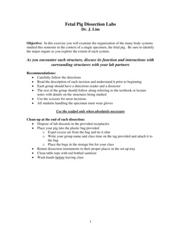
Slide #22 (BV3-113a1) – Tongue, bovine.Top of tongueskeletalmuscleBottomepithelium
Demo Slide #186 (SP-1-108). Parotid salivary gland,sheepstriated ductinterlobularductNervebundlesepithelium
DEMO SLIDE BOX 183 (BV-1-85A)- Mandibular salivary gland, cow.White fatStriated duct
Upper GI Esophagus: Long thin muscular tube, connects pharynx to thestomach. Moves bolus (wad of food)via peristaltic contractions. Posterior tothe trachea Liver: functions in digestion,metabolism, and storage of nutrients.Bile is produced which helps emulsifyfats Stomach: Food storage and digestion Gallbladder: Holds bile until needed to digest fatty acids Pancreas: Excretes enzymes to breakdown proteins, lipids, andcarbohydrates. Also produces insulin Spleen: Brown, flat, oval organ thatfilters and stores blood Lower GISmall intestine: Absorbs 90% ofthe nutrients from the food we eat.Very long, but SMALL in diameter Duodenum: receives chyme (Partiallydigested food) from stomach. Enzymesform the pancreas, liver, and gallbladdermix here for chemical digestion Jejunum: middle section Ileum: where remaining nutrients areabsorbedLarge intestine: Absorbs water andvitamins while converting digested foodinto feces. Short but LARGE in diameterRectum: Where feces wait to beexpelledAnus:Feces is expelled
GI TRACTLarynxTrachea
talpig.html
https://animalplanet.wikispaces.com/Snyder-Crago Mouth Anatomy
Slide #71 (Pf5-73/205). Esophagus and trachea,pig.stratified squamousepitheliummucous membrane
152VilliDuodenumSmooth MuscleFatCells
148
153
Let’s take a Closer Look! Submandibular gland Esophagus Spleen Liver and Spleen Bile Duct and portal vein Gallbladder Pancreas Ileum Duodenum Liver
Respiratory TractGoal: To remove carbon dioxide from the blood and replace itwith oxygen Works in conjunction with the circulatory system Organs: Upper Tract Nose with nares Pharynx Larynx Lower Tract Trachea Lungs which contain the bronchi, bronchioles, and alveoli Diaphragm
Nose Air enters the respiratorysystem through the pairednares or nostrils Two nasal passages:separated by bone Used for smell Hair and mucousmembranes clean outparticles Carbon dioxide is expiredthrough the tudy/biology/virtual-pig/respiratory-system
Pharynx and Epiglottis Nasopharynx Above the soft palate Leads from the nose to the trachea for breathing Laryngopharynx Upper border of the glottis Helps guide food and air Epiglottis Small flap of cartilage Lies above the pharynx Allows for air to flow into larynx and trachea Directs food into the esophagus
cch/genbios/fetalpig.html
Larynx Large hard structure attached to the trachea Contains the vocal cords As air passes through the vocal cords vibrate andproduce y/fetal%20pig/Mouth and neck region 4.jpg
Trachea Windpipe Connects larynx to lungs Contains cartilage rings that prevent tracheafrom collapsingExtends into the thoraciccavityBranches into bronchiAllows air into the lungsAnterior to theesophagushttps://www.google.com/search?q trachea fetal pig&espv 2&biw 972&bih 854&source lnms&tbm isch&sa X&ei qAQ4VKTyO4 o8AHl4oC4Ag&ved 0CAYQ AUoAQ#facrc &imgdii M%3A&imgrc 52Fflashcards%252F2198244%252Fjpg%252Fpulmonary trunk1352727288605.jpg%3Bhttp
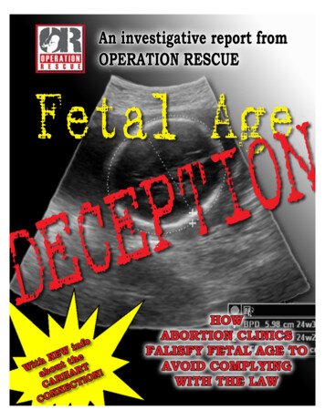
DEMO SLIDE BOX 81 – Trachea and elastic artery,“C” shaped rings of hyaline cartilage,pig.Lymph nodeelasticarterynervecompanion veinmusclePlasma cells
Lungs Branching of the trachea to bronchi to lungsLocated on either side of the heartThe bronchi divide into bronchiolesBronchioles branch into alveolar sacsAlveolar sacs are bunches of alveoliAlveoli hold air and tightly bound inblood vessels; allows for gas exchange
Lungs continued Sectioned into lobes Right lung is larger (4 lobes) cranial, medial, caudal, and accessory lobes Left lung is smaller because of space takenup by the heart (2 lobes) apical and caudal lobe
c-cavity
Slide #72(SP-1-81). Lung, sheep.smaller intrapulmonary bronchi.plates of hyalinecartilage.arteryveinglandsalveolar sacs
Slide #72(SP-1-81). Lung, nchiole
Diaphragm Below the lungsThin muscular sheet of tissueOnly in mammalsExpands to allow air in (diaphragm contracts)Compresses to expel air (diaphragm relaxes)Separates the chest and abdominal tory/fetal%20pig/Respiratory system 5.jpg
c-cavity
Let’s take a Closer Look! Cross section of the Trachea Nasal Tract Lungs Esophagus and Trachea Larynx Lung Larynx, Esophagus, and Glands
Circulatory SystemAllows blood, nutrients, oxygen and other gases, andhormones to flow throughout the body Mammals (including humans) Double loop system 4 chambered heart Organs Heart Lungs Arteries ils.php/id/1113
Think of it like a highway Blood: Transports cells just like abus transports people Heart: Controls the flow of blood like atraffic control light Blood Vessels: lead the flow of bloodthrough the body like roadslead
Heart Muscle used to pump blood 4 chambers in mammals Right Atria Right Ventricle Left Atria Left Ventricle Atria pump blood to ventricles Ventricles pump blood out of heart and into circulatorysystem Valves prevent backflow of blood
It’s more fun to sing
226
Heart withValve
**REMEMBER IT IS ALWAYS THE LEFTAND RIGHT OF THE Lables.html
Path of the Blood Oxygenated blood from the lungs enters from the pulmonary veins into the left atria, pumped through the mitral valve, andinto the left ventricle.Then it is pumped through the aortic valve where it leavesthrough the Aorta to the be taken to the rest of the bodyOnce circulated around the body the deoxygenated bloodenters through the Superior Vena Cava from the top of bodyand Inferior Vena Cava from the bottom of body all into theright atria.It is then pumped through the tricuspid valve into the rightventriclePumped through the pulmonary valve into the pulmonaryartery to the lungs to be oxygenated.Once oxygenated it will return through the pulmonary artery.
Lungs Provide oxygen to the blood and removecarbon dioxide from the blood Exchange of gases takes place in thealveolar sacshttp://en.wikipedia.org/wiki/Pulmonary circulation
Blood Liquid that circulates in the blood vessels,transporting oxygen, carbon dioxide, waste, andhormones around the body. Blood contains White Blood Cells (leukocytes)- fight against infections Red blood cells (erythrocytes) – transport oxygen Platelets (thrombocytes)- assist in clotting Plasma- liquid that suspends proteins and the solidcomponents of blood
Blood Vessels Veins- carry blood to the heart Arteries- carry blood away from the heart Capillaries – small pathway thatconnects arteries to veins Only one blood cell ic/deck/5473108
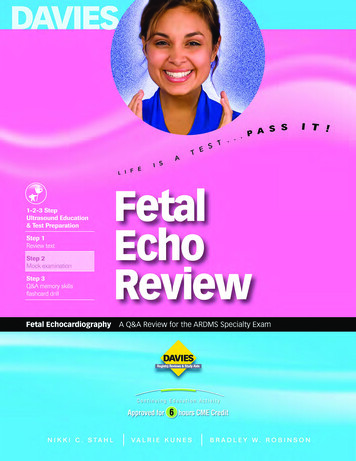
Slide #16 (Pmi 7A). Blood vessels, pigmediaendothelium(which is simplesquamousepithelium
BloodCellsLabeled
Let’s Take a Closer Look Peripheral Blood Smear Red Bone Marrow Smear Heart, Epicardium Heart, Endocardium Blood 1 Blood 2 Blood 3 Bone Marrow Heart with Valve Heart Muscle croscope-images/138-microscopes-lg.jpg
Reproductive SystemExpands the population of a certain species Male andfemale reproductivetracts are different Males-producetestosterone and sperm Females- produceoocytes
Male Repro Tract Testes – produce sperm Penis - deposits semen into female reproductive tract. Expelsurine from the body. Cremaster muscle –pulls the testescloser to the body to keepwarm Scrotum houses the testes Bulbourethral Gland gives off seminalfluid to urethra
Spermatic cord -contains Vasdeferens,spermatic arteryand vein, lymphaticvessels, and leadsto the epididymis Epididymis- storessperm Seminiferoustubules – spermproduced Vas deferenstransports sperm tourethra Urethra – receives seminal secretions form the testes. Drains excretory products fromthe bladder Prostate- secretes fluid that carries sperm
Female Repro Tract Ovaries- Produces oocytes (eggs) Oviduct- receives mature oocytes atovulation; site of fertilization Uterine Horns- Site of implantation andembryonic development Vagina- receives penis during copulation;serves as part of birth canal Urogenital Sinus- chamber in which thevagina and urethra meet
DEMO SLIDE BOX #208 – Cervix, sow.complex folds of the luminal surface of the cervix.
Slide #183 (PG-1-84). Mammary gland, sow.lactiferous ductducts
Let’s Take a Closer Look Ovary 1 Ovary 2 Ovary 3 Uterus Vagina Testis Seminal Vesicle Prostate Testis 2http://nerdbranding.com/product/microscope/
Urinary SystemEliminates wastes from the body, regulates bloodvolume and pressure, controls levels of electrolytesand metabolites, and regulates blood pHAdrenalGland Organs Adrenal GlandsKidney Kidneys Ureter Bladder ystem-pig-2/h4nsyh4nsUreter
Adrenal Glands Sit on top of the kidneys 2 total 2 Parts Adrenal Cortex- outer part thatproduces hormones vital to life,such as cortisol (metabolism) andaldosterone (blood pressure) Adrenal Medulla- inner part thatproduces nonessential hormones,such as adrenaline ychblog/4/
Kidneys Two kidneys Remove waste and excess waterfrom the blood Keep proper balance of salts andacids Nephrons are functional unit Filter the blood Water and waste removed Blood, waste and water enterkidney through the Renal Artery Blood cleaned of waste andexcess water leave kidneythrough Renal kidneyswork/tabid/590/Default.aspx
Kidney
Ureter, Bladder, and Urethra Ureter One from each kidney Carries urine fromkidney to bladder Strong muscular tube Bladder Hollow organ Expands as it is filled Urethra Takes urine from the bladder to be eliminated
Ureter
Urinary Bladder
Let’s Take a Closer Look Kidney 1 Kidney 2 Ureter Bladder Ureter 2 Adrenal gland Kidney 3http://www.biologyjunction.com/microscope lab1.htm
Who would havethought a pig wouldbe so closely relatedto a human?!!https://www.google.com/search?q pig and farmer friend cartoon&espv 2&biw 1197&bih 753&source lnms&tbm isch&sa X&ei 5khRVOqeA4upgwS7n4HQBw&ved 0CAYQ AUoAQ#tbm isch&q human in mud cartoon&imgdii
References http://peer.tamu.edu/ biology/virtual-pig/respiratory .edu/ntress/Bio1 Lab Manual New/fetal pig respiratory 20laboratory/fetal%20pig/Respiratory system 5.jpghttp://bioserv.fiu.edu/ walterm/human online/labs/fetal pig/fetal pig anatomy.htmhttp://www.shsu.edu/ agr www/documents/FetalPigDissectionLab ductive-system
The End
Humans and Pigs may be closer than you think! Both are mammals We share common body systems The anatomy of the pig is close to that of humans The fetal pigs will tell us more about our own bodies and give us a way