Transcription
AANEM PRACTICE TOPICEVIDENCE-BASED GUIDELINE: NEUROMUSCULAR ULTRASOUND FORTHE DIAGNOSIS OF CARPAL TUNNEL SYNDROMEMICHAEL S. CARTWRIGHT, MD,1 LISA D. HOBSON-WEBB, MD,2 ANDREA J. BOON, MD,3,4 KATHARINE E. ALTER, MD,5,6CHRISTOPHER H. HUNT, MD,7 VICTOR H. FLORES, MD,8 ROBERT A. WERNER, MD,9 STEVEN J. SHOOK, MD,10T. DARRELL THOMAS, MD,11 SCOTT J. PRIMACK, DO,12 and FRANCIS O. WALKER, MD11Department of Neurology, Wake Forest School of Medicine, Winston-Salem, North Carolina, USA2Department of Medicine, Division of Neurology, Duke University, Durham, North Carolina, USA3Department of Physical Medicine and Rehabilitation, Mayo Clinic, Rochester, Minnesota, USA4Department of Neurology, Mayo Clinic, Rochester, Minnesota, USA5Rehabilitation Medicine Department, National Institutes of Health, Bethesda, Maryland, USA6Mount Washington Pediatric Hospital, Baltimore, Maryland, USA7Department of Radiology, Mayo Clinic, Rochester, Minnesota, USA8Physical Medicine Associates, Arlington, Texas, USA9University of Michigan Health System, Ann Arbor, Michigan, USA10Neuromuscular Center, Cleveland Clinic, Cleveland, Ohio, USA11Knoxville Neurology Specialists, Knoxville, Tennessee, USA12Colorado Rehabilitation and Occupational Medicine, Aurora, Colorado, USAAccepted 12 March 2012ABSTRACT: Introduction: The purpose of this study was todevelop an evidence-based guideline for the use of neuromuscular ultrasound in the diagnosis of carpal tunnel syndrome(CTS). Methods: Two questions were asked: (1) What is the accuracy of median nerve cross-sectional area enlargement asmeasured with ultrasound for the diagnosis of CTS? (2) Whatadded value, if any, does neuromuscular ultrasound provideover electrodiagnostic studies alone for the diagnosis of CTS?A systematic review was performed, and studies were classifiedaccording to American Academy of Neurology criteria for ratingarticles of diagnostic accuracy (question 1) and for screeningarticles (question 2). Results: Neuromuscular ultrasound measurement of median nerve cross-sectional area at the wrist isaccurate and may be offered as a diagnostic test for CTS(Level A). Neuromuscular ultrasound probably adds value toelectrodiagnostic studies when diagnosing CTS and should beconsidered in screening for structural abnormalities at the wristin those with CTS (Level B).Muscle Nerve 46: 287–293, 2012Carpal tunnel syndrome (CTS) is a combinationof signs and symptoms resulting from mononeuropathy of the median nerve as it passes through therigid carpal tunnel in the wrist.1 It is a common condition that affects 2.7% of the general populationand results in health-care costs exceeding 500 million annually in the USA.2,3 CTS is typically diagnosed by history and physical examination, and electrodiagnostic studies (nerve conduction studies andAbbreviations: AAN, American Academy of Neurology; AANEM, American Association of Neuromuscular and Electrodiagnostic Medicine; CTS,carpal tunnel syndrome; NCS, nerve conduction studiesKey words: carpal tunnel syndrome; median nerve; mononeuropathy; nerveconduction studies; ultrasoundDisclosures: M.S.C. receives funding from the NIH/NINDS forneuromuscular ultrasound research and royalties from Elsevier for sales ofthe textbook Neuromuscular Ultrasound. F.O.W. receives royalties fromElsevier for sales of the Neuromuscular Ultrasound.Approved by the AANEM Board of Directors on January 23, 2012.Correspondence to: C. French, AANEM, 2621 Superior Drive NW,Rochester, MN 55901; e-mail: cfrench@aanen.orgC 2012 American Association of Neuromuscular and Electrodiagnostic Medicine.VPublished online 20 March 2012 in Wiley(wileyonlinelibrary.com). DOI 10.1002/mus.23389AAEM Practice TopicOnlineLibrarysometimes electromyography) are used to confirmthe presence of a median mononeuropathy. Electrodiagnostic studies have limitations; they are uncomfortable and do not directly assess the anatomy ofthe median nerve and its surrounding structures.Over the past 20 years, neuromuscular ultrasound has been introduced into electrodiagnosticlaboratories as a complement to nerve conductionstudies and electromyography for the diagnosis ofa variety of nerve and muscle conditions.4 CTS isthe condition most commonly studied with neuromuscular ultrasound, and individuals with CTShave displayed ultrasonographic evidence of focalenlargement of the median nerve at the wrist.5 Inaddition, neuromuscular ultrasound can identifycauses of median mononeuropathy at the wristand structural anomalies that could not bedetected with electrodiagnostic studies alone, suchas compressive cysts, tumors, and vessels.5This evidence-based guideline was designed toaddress 2 critical questions regarding the use ofneuromuscular ultrasound for the diagnosis ofCTS. First, what is the accuracy of median nervecross-sectional area enlargement, as measured withultrasound, for the diagnosis of CTS? Second, whatadded value, if any, does neuromuscular ultrasound provide over electrodiagnostic studies alonefor the diagnosis of CTS?DESCRIPTION OF THE ANALYTIC PROCESSThe American Association of Neuromuscular andElectrodiagnostic Medicine (AANEM) convened anexpert panel of physicians specializing in neurology, physical medicine and rehabilitation, and radiology, selected to represent a broad range of expertise related to neuromuscular ultrasound andCTS. Some panel participants reported usingMUSCLE & NERVEAugust 2012287
neuromuscular ultrasound frequently for clinicaland research purposes, and others reported neverusing the technology. All panel participants hadexpertise in the clinical and electrodiagnosticassessment of CTS.In May 2011, PubMed was used to search Medline to identify all potential abstracts. The searchterms ‘‘carpal tunnel syndrome OR median nerveOR median neuropathy’’ were combined with theterms ‘‘ultrasound OR ultrasonography OR sonogram OR sonography.’’ This produced 724 articlesfrom 1990 to May 2011. This was narrowed to 641articles by including ‘‘English-only’’ and ‘‘humanonly’’ studies. The titles of those articles werereviewed for relevance, which yielded 240 articles,and each abstract was then reviewed by at least 2investigators. This resulted in 121 articles for fullmanuscript review. After each article was reviewedin its entirety by 2 investigators, 67 were identifiedas relevant for this guideline. In order to be considered relevant, the article had to describe theuse of ultrasound to image the wrist in individualssuspected of having CTS.The 67 relevant articles were rated by at least 2investigators according to criteria set by the American Academy of Neurology (AAN).6 Articles pertaining to the accuracy of median nerve cross-sectional area measurements for the diagnosis of CTS(45 articles) were assessed using the AAN criteriafor rating an article on diagnostic accuracy, andarticles pertaining to neuromuscular ultrasound asa screening tool to identify anatomic explanationsfor CTS were assessed using AAN criteria for ratinga screening article (23 articles). One article wasassessed for both diagnostic accuracy and as ascreening article. Both rating systems are includedin the Appendices.Studies with the highest levels of evidence(Class I and II) are discussed in the text and summarized in the evidence tables. At each step in theprocess, disagreements were arbitrated by a thirdinvestigator.ANALYSIS OF THE EVIDENCEAccuracy of Neuromuscular Ultrasound.Forty-fiverelevant articles pertaining to the accuracy of neuromuscular ultrasound in the diagnosis of CTSwere identified. Four were graded as Class I and 2as Class II. Of the 27 that were Class III, 18received this rating because they had spectrumbias (in all cases healthy volunteers served as thecontrol group); 6 received this rating because theultrasonographer was not blinded to clinical orother diagnostic information; and 3 had both spectrum bias and lack of blinding.7–33 Spectrum biasoccurs when cases and controls are potentially atopposite ends of the disease spectrum, which may288AAEM Practice Topicartificially enhance the diagnostic accuracy of atest. For example, the articles with spectrum biasreviewed in this study used healthy volunteers ascontrols rather than a control population morerepresentative of individuals referred for possibleCTS in a typical clinical practice. If not explicitlystated in the article that blinding of the ultrasonographer occurred, the corresponding author wase-mailed to clarify this issue, and if no responsewas received it was categorized as having not beenblinded. In general, the Class III articles demonstrated high diagnostic accuracy of ultrasound forthe diagnosis of CTS, similar to the Class I and IIarticles. Of the 12 articles graded as Class IV, 8received this rating because they did not reportenough measures of diagnostic accuracy (sensitivityand specificity, or a likelihood ratio) and 4because the control group selection was notacceptable.34–45 In all 4 cases in which the controlgroup selection was unacceptable, the controlgroup included individuals with symptoms consistent with CTS but normal nerve conduction studies.Although it is important to study individuals withthis type of conflicting clinical data (incongruentsymptoms and nerve conduction studies), it is theauthors’ opinion that it is problematic to use suchindividuals as a control group when assessing theaccuracy of a diagnostic test.Four articles were identified that met Class Ilevel of evidence (Table 1), meaning that the studies were prospective (cohort studies), wereblinded, were free of spectrum bias, used appropriate reference standards, and included measures ofdiagnostic accuracy.46–49 Three of the studiesdefined individuals as having CTS if they had bothconsistent clinical symptoms and abnormal nerveconduction studies, whereas Altinok et al. definedCTS as those with a consistent clinical presentationand improvement in symptoms after 3 months ofnon-surgical treatment. Three studies used wristscontralateral to the wrist with CTS as the controls(as long as the participant was asymptomatic onthat side and the nerve conduction studies werenormal on that side),46,47 and Altinok et al.recruited controls from outpatients presenting tothe same clinic for causes unrelated to CTS. Thepresence of different case definitions for CTS isnot problematic. In fact, in a condition such asCTS, in which the ‘‘gold standard’’ for diagnosis isdebated, it is beneficial in evidence-based guidelines to have studies in which the reference standards for diagnosis were established using differentmethods. Similarly, it is beneficial to have differentcontrol groups, and the authors thought the 2 different control groups (contralateral unaffectedhands and individuals referred to the clinic fornon-CTS indications) were clinically appropriateMUSCLE & NERVEAugust 2012
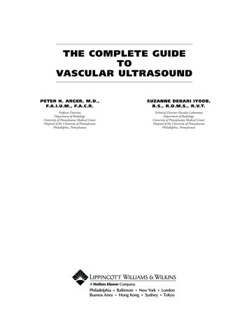
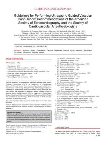
Table 1. Class I level of evidence studies* of the accuracy of neuromuscular ultrasound for the diagnosis of carpal tunnel syndrome.YearFirst authorReference standardNumberwith CTSNumberwithout CTSSensitivitySpecificityAccuracyArea cut-off2004T. Altinok200420052010S.M. WongH.-R. ZiswilerA. MohammadiClinical þ improvementand NCSClinical þ NCSClinical þ NCSClinical þ .5%92.5%72.7%87.0%98.0%78.9%NA79.3%83.4%97.2%9 mm29 mm210 mm210 mm28.5 mm2NA, not available (data presented in the article did not include this value or permit calculation of this value); NCS, nerve conduction studies.*To meet Class I level of evidence these studies are prospective, are blinded, are free of spectrum bias, have appropriate reference standards, and includemeasures of diagnostic accuracy.and minimized spectrum bias. The use of contralateral unaffected wrists also raised the question ofwhether there was statistical independencebetween the control wrists and the wrists with CTS.This cannot be definitively answered without theoriginal data. However, if they were not independent, that would make the control and affectedwrists more similar, which would likely result in anunderestimation of the diagnostic accuracy of thetest.The four Class I studies used slightly differentmedian nerve cross-sectional area cut-offs to diagnose CTS (ranging from 8.5 to 10 mm2), and allstudies used the direct tracing method to measuremedian nerve area. Ziswiler et al. measured themedian nerve at the site of maximal enlargementat the wrist, and the other 3 studies measured thenerve at, or just proximal to, the level of the pisiform bone. Our guideline development panel considered studies with diagnostic accuracy 70% tobe acceptable and supportive of neuromuscularultrasound for the diagnosis of CTS (accuracy ¼sensitivity prevalence þ specificity (1 prevalence)). The sensitivity of median nerve cross-sectional area for the diagnosis of CTS ranged from65% to 97%, specificity from 72.7% to 98%, andaccuracy from 79% to 97%. Interestingly, Altinoket al. also analyzed their data using nerve conduction studies in addition to clinical diagnosis, andthis more strict case definition of CTS resulted inan increase in the sensitivity of neuromuscularultrasound from 65% to 100%.Two Class II articles were identified (Table 2),and they met the same criteria as the Class Iarticles, except they involved retrospective data collection (case–control studies).50,51 Nakamichi andTachibana used only clinical criteria to classify participants as CTS or controls, and the control subjects were recruited from a health fair. The mediannerve cross-sectional area was calculated by tracingthe nerve at 3 sites within the carpal tunnel andtaking the mean of those measurements, with 12mm2 selected as the cut-off for the diagnosis ofCTS. The study by Klauser et al. is unique amongthe 6 Class I and II articles in that it only examined those with bifid median nerves within the carpal tunnel. The area of each portion of the nervewas traced and then added together, and a singlecut-off of 12 mm2 was used. They also reportedimproved accuracy when the difference betweenthe median nerve area measured at the wrist andproximal forearm was calculated, with a differenceof 4 mm2 or greater used to diagnose CTS.Conclusion. Based on consistent Class I andClass II evidence, neuromuscular ultrasound measurement of median nerve cross-sectional area atthe wrist is established as accurate for the diagnosisof CTS.Recommendation: If available, neuromuscularultrasound measurement of median nerve crosssectional area at the wrist may be offered as anaccurate diagnostic test for CTS (Level A).Clinical Context. As is the case with all ultrasonographic imaging, neuromuscular ultrasound ofthe median nerve at the wrist should be performedand interpreted by clinicians experienced with thetechnique. Scanning protocols and reference values for median nerve cross-sectional area at theTable 2. Class II level of evidence studies* of the accuracy of neuromuscular ultrasound for the diagnosis of carpal tunnel syndrome.YearFirst authorReferencestandardNumberwith CTSNumberwithout CTS20022011†K. NakamichiA.S. KlauserClinicalClinical þ NCS4145340828SensitivitySpecificityAccuracyArea 9%12 mm212 mm2D4 mm2‡*To meet Class II level of evidence these studies are retrospective, are blinded, are free of spectrum bias, and include measures of diagnostic accuracy.†This study only evaluated those with bifid median nerves.‡This is the difference in median nerve area at the wrist compared to the forearm.AAEM Practice TopicMUSCLE & NERVEAugust 2012289
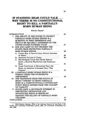
Table 3. Class II level of evidence studies* of the added value of neuromuscular ultrasound in the diagnosis of carpal tunnel syndrome.YearFirst authorNumberwith CTS1993K. Nakamichi2020002008E. IannicelliI.K. Bayrak2943202011L. Padua35Percentage and type of abnormal structures25% with unilateral CTS (by clinical and NCS criteria) have occultganglia in the carpal tunnel2% with CTS have a bifid median nerve13% with CTS have a bifid median nerve6% with CTS have a persistent median artery17% with CTS have a finding that changes therapeutic approach9% have a bifid median nerve9% have a persistent median artery6% have tenosynovitis3% have accessory muscles*To meet Class II level of evidence these studies draw from a statistical and non-referral clinic-based sample of patients, evaluate all CTS patients prior tosurgery, and conduct neuromuscular ultrasound on all study participants.wrist should be established by each laboratoryprior to using neuromuscular ultrasound for thediagnosis of CTS.Added Value of Neuromuscular Ultrasound. Twentythree articles were identified that potentiallydemonstrated the added value of neuromuscularultrasound as a diagnostic tool when used in combination with electrodiagnostic studies. Four weregraded as Class II (Table 3), and the rest wereclassified as Class IV (all of these were case reportsand case series). None were graded as Class I,because no studies drew from a population-basedsample of patients. None were Class III, because allthe studies that were not case reports or case seriesmet Class II criteria.The 4 articles that met Class II criteriadescribed studies that drew from a statistical andnon-referral clinic-based sample of patients, evaluated all CTS patients prior to surgery, andincluded neuromuscular ultrasound on all studyparticipants.7,52–54 Three of these articles describedthe detection of bifid median nerves at the wrist,and 2–13% of those with CTS had a bifidnerve.7,53,54 Two studies described the detection ofpersistent median arteries, which occurred in 9–13% of those with CTS.7,54 The study by Paduaet al. also described the detection of tenosynovitis(6%) and accessory muscles within the wrist (3%)in those with CTS. The study by Nakamichi andTachibana was unique in that it assessed theaffected wrists in those with unilateral CTS (meaning the contralateral side was normal by clinicaland nerve conduction criteria). Neuromuscularultrasound detected occult ganglia causing medianmononeuropathy in 25% of the CTS-affected wristsin this population.All 19 of the Class IV articles were case reportsor case series in which neuromuscular ultrasoundwas used to identify abnormal structures causing290AAEM Practice Topicmedian mononeuropathy at the wrist. These structures included traumatic neuromas, Schwannomas,lipofibromatous hamartomas, ganglion cysts,thrombosed persistent median arteries, an abscess,and compressive gouty tophus.55–73Conclusion. Based on Class II evidence, neuromuscular ultrasound of the wrist probably addsvalue to electrodiagnostic studies when assessingCTS as it can detect structural abnormalities.Recommendation: If available, neuromuscularultrasound should be considered to screen forstructural abnormalities at the wrist in those withCTS (Level B).Clinical Context. Screening for structuralabnormalities at the wrist that cause CTS is likelyto be of higher yield in those with atypical CTS.This was demonstrated by Nakamichi and Tachibana, who found a high rate of occult ganglia onlyin those with unilateral CTS. This is an atypicalpresentation, as most patients have bilateral CTS(defined by symptoms, nerve conduction studies,or both).52 Other atypical presentations of CTSinclude sudden onset and onset in the setting oftrauma. Although ultrasound can identify structural abnormalities, it is possible these abnormalities may not always be the underlying cause of themedian mononeuropathy. In addition, the prevalence of such abnormalities in the general population is not known, so the sensitivity and specificityof ultrasound for the identification of these structures cannot be calculated based on currently available data. The wide prevalence range for bifid median nerves (2–13%) may be secondary toultrasound device resolution (earlier studies identified fewer bifid nerves), ultrasound technique andsite of imaging within the wrist, or patient population. The presence of structures such as persistentmedian arteries and accessory muscles is clearly oftherapeutic interest, as it may alter the choice ofinterventional approach (either injection orMUSCLE & NERVEAugust 2012
surgery). Knowledge of a bifid median nerve andother anatomic variants is also of interest in planning the treatment of CTS,53 and identification ofsuch variants prior to invasive intervention caneven assist later in the assessment of failed intervention. In addition, the presence of a bifid median nerve may be an independent risk factor forthe development of CTS.7CLINICAL CONTEXT SUMMARY FOR ALL EVIDENCEA single neuromuscular ultrasound evaluation ofthe wrist in those with CTS allows for assessment ofboth median nerve cross-sectional area and thepresence of structural abnormalities, and this complements well the information obtained during anelectrodiagnostic study (which is the gold standardfor diagnosis of CTS). Some variability exists in thedevices, scanning protocols, and reference rangesfor the diagnosis of CTS when using neuromuscularultrasound, but this is to be expected. As a comparison, similar variability exists in electrodiagnostictechniques. It is anticipated that with continued experience with neuromuscular ultrasound techniques, more uniformity will occur as consensus develops regarding optimal use of the technology. Itshould also be noted that many studies have proposed other neuromuscular ultrasound parametersthat can be used to assist in the diagnosis of CTS,but these were not assessed in this guideline. Theseinclude median nerve flattening ratios; measures ofmedian nerve mobility, echogenicity, and vascularity; and measures of flexor retinaculum bowing.RECOMMENDATIONS FOR FUTURE RESEARCH1 A standardized protocol for using neuromuscularultrasound in the diagnosis of CTS should be developed. This should include definition of the optimalsite of median nerve cross-sectional area measurement, standardization of nerve outlining technique,and further refinement of reference values.2 Further research and evidence-based guidelinesshould assess the usefulness of other neuromuscular ultrasound parameters for the diagnosis ofCTS, such as median nerve flattening, mobility,echogenicity, vascularity, and bowing of theflexor retinaculum.3 Large population-based studies that enroll consecutive patients with CTS should be performed toassess for all structural abnormalities that may becausative or change therapeutic approach, whichwill help further determine the added benefit ofneuromuscular ultrasound in the diagnosis of CTS.4 Finally, large studies should be performed todetermine whether neuromuscular ultrasoundchanges treatment strategies and outcomes inthose with CTS when compared with those inwhich CTS is established using only electrodiagAAEM Practice Topicnostic studies. This type of study should alsoallow for cost–benefit analyses of neuromuscularultrasound in the diagnosis of CTS.DISCLAIMERThis statement has been provided as an educational service of the AANEM. It is based on anassessment of current scientific and clinical information. It is not intended to include all possibleproper methods of care of a particular neurological problem or all legitimate criteria for choosingto use a specific procedure. Neither is it intendedto exclude any reasonable alternative methodology.The AANEM recognizes that specific patient caredecisions are the prerogative of the patient andthe physician caring for the patient, based on allof the circumstances involved. The clinical contextsection is made available in order to place the evidence-based guidelines into perspective with current practice habits and challenges. No formalpractice recommendation should be inferred.The authors thank Gary Gronseth, MD, for his gracious assistanceand shared expertise regarding the grading of evidence, which wascritical for the creation of this evidence-based guideline.APPENDIX 1: AAN CLASSIFICATION OF THE EVIDENCEFOR RATING OF A DIAGNOSTIC ARTICLE6Class I: A cohort study with prospective data collection of a broad spectrum of persons with the suspected condition, using an acceptable referencestandard for case definition. The diagnostic test isobjective or performed and interpreted withoutknowledge of the patient’s clinical status. Study resultsallow calculation of measures of diagnostic accuracy.Class II: A case–control study of a broad spectrum of persons with the condition established byan acceptable reference standard compared with abroad spectrum of controls, or a cohort study witha broad spectrum of persons with the suspectedcondition where the data were collected retrospectively. The diagnostic test is objective or performedand interpreted without knowledge of disease status. Study results allow calculation of measures ofdiagnostic accuracy.Class III: A case–control study or cohort studywhere either persons with the condition or controls are of a narrow spectrum. The condition isestablished by an acceptable reference standard.The reference standard and diagnostic test areobjective or performed and interpreted by different observers. Study results allow calculation ofmeasures of diagnostic accuracy.Class IV: Studies not meeting Class I, II, or IIIcriteria, including consensus, expert opinion, or acase report.MUSCLE & NERVEAugust 2012291
APPENDIX 2: AAN CLASSIFICATION OF THE EVIDENCEFOR RATING OF A SCREENING ARTICLE6Class I: A statistical, population-based sample ofpatients studied at a uniform point in time (usuallyearly) during the course of the condition. Allpatients undergo the intervention of interest. Theoutcome, if not objective, is determined in an evaluation that is masked to the patients’ clinicalpresentations.Class II: A statistical, non-referral clinic-basedsample of patients studied at a uniform point intime (usually early) during the course of the condition. Most patients undergo the intervention of interest. The outcome, if not objective, is determined in an evaluation that is masked to thepatients’ clinical presentations.Class III: A sample of patients studied duringthe course of the condition. Some patientsundergo the intervention of interest. The outcome, if not objective, is determined in an evaluation by someone other than the treating physician.Class IV: Studies not meeting Class I, II, or IIIcriteria, including consensus, expert opinion, or acase report.APPENDIX 3: CLASSIFICATION OFRECOMMENDATIONSThe four possible recommendation classificationsinclude: A ¼ Established as effective, ineffective or harmful (or established as useful/predictive or notuseful/predictive) for the given condition inspecified population. (Level A rating requires atleast 2 consistent Class I studies.) [In exceptionalcases, 1 convincing Class I study may suffice foran ‘‘A’’ recommendation if: (1) all criteria aremet; and (2) the magnitude of effect is large(relative rate improved outcome 5 and thelower limit of the confidence interval is 2.] B ¼ Probably effective, ineffective, or harmful(or probably useful/predictive or not useful/predictive) for the given condition in the specifiedpopulation. (Level B rating requires at least 1Class I study or 2 consistent Class II studies.) C ¼ Possibly effective, ineffective, or harmful (orpossibly useful/predictive or not useful/predictive) for the given condition in the specifiedpopulation. (Level C rating requires at least 1Class II study or 2 consistent Class III studies.) U ¼ Data inadequate or conflicting; given current knowledge, treatment (test, predictor) isunproven.REFERENCES1. Gelberman RH, Hergenroeder PT, Hargens AR, Lundborg GN, Akeson WH. The carpal tunnel syndrome. A study of carpal canal pressures. J Bone Joint Surg Am 1981;63:380–383.292AAEM Practice Topic2. Atroshi I, Gummesson C, Johnsson R, Ornstein E, Ranstam J, RosenI. Prevalence of carpal tunnel syndrome in a general population.JAMA 1999;282:153–158.3. Stevens JC, Sun S, Beard CM, O’Fallon WM, Kurland LT. Carpal tunnel syndrome in Rochester, Minnesota, 1961 to 1980. Neurology1988;38:134–138.4. Walker FO, Cartwright MS, Wiesler ER, Caress J. Ultrasound of nerveand muscle. Clin Neurophysiol 2004;115:495–507.5. Beekman R, Visser LH. Sonography in the diagnosis of carpal tunnelsyndrome: a critical review of the literature. Muscle Nerve 2003;27:26–33.6. American Academy of Neurology. Clinical practice guideline processmanual. St. Paul, MN: American Academy of Neurology; 2011.7. Bayrak IK, Bayrak AO, Kale M, Turker H, Diren B. Bifid mediannerve in patients with carpal tunnel syndrome. J Ultrasound Med2008;27:1129–1136.8. Duncan I, Sullivan P, Lomas F. Sonography in the diagnosis of carpal tunnel syndrome. AJR Am J Roentgenol 1999;173:681–684.9. El Miedany YM, Aty SA, Ashour S. Ultrasonography versus nerve conduction study in patients with carpal tunnel syndrome: substantiveor complementary tests? Rheumatology (Oxford) 2004;43:887–895.10. Iannicelli E, Almberger M, Chianta GA, Salvini V, Rossi G, MonacelliG, et al. High resolution ultrasonography in the diagnosis of the carpal tunnel syndrome. Radiol Med 2005;110:623–629.11. Kele H, Verheggen R, Bittermann HJ, Reimers CD. The potentialvalue of ultrasonography in the evaluation of carpal tunnel syndrome. Neurology 2003;61:389–391.12. Kotevoglu N, Gulbahce-Saglam S. Ultrasound imaging in the diagnosis of carpal tunnel syndrome and its relevance to clinical evaluation.Joint Bone Spine 2005;72:142–145.13. Koyuncuoglu HR, Kutluhan S, Yesildag A, Oyar O, Guler K, OzdenA. The value of ultrasonographic measurement in carpal tunnel syndrome in patients with negative electrodiagnostic tests. Eur J Radiol2005;56:365–369.14. Sarria L, Cabada T, Cozcolluela R, Martinez-Berganza T, Garcia S.Carpal tunnel syndrome: usefulness of sonography. Eur Radiol 2000;10:1920–1925.15. Wiesler ER, Chloros GD, Cartwright MS, Smith BP, Rushing J,Walker FO. The use of diagnostic ultrasound in carpal tunnel syndrome. J Hand Surg Am 2006;31:726–732.16. Wong SM, Griffith JF, Hui AC, Tang A, Wong KS. Discriminatorysonographic criteria for the diagnosis of carpal tunnel syndrome. Arthritis Rheum 2002;46:1914–1921.17. Yesildag A, Kutluhan S, Sengul N, Koyuncuoglu HR,
A systematic review was performed, and studies were classified according to American Academy of Neurology criteria for rating articles of diagnostic accuracy (question 1) and for screening articles (question 2). Results: Neuromuscular ultrasound mea-surement of median nerve cross-sectional area at the wrist is