Transcription
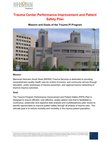
Trauma Center Performance Improvement and PatientSafety PlanMission and Goals of the Trauma PI ProgramMission:Memorial Hermann South West (MHSW) Trauma Services is dedicated to providingcomprehensive quality health care for victims of trauma, and community service througheducation, public awareness of trauma prevention, and regional trauma networking toimprove trauma outcomes.Goal:The Trauma Program Performance Improvement and Patient Safety (PIPS) Plan isdesigned to ensure efficient, cost effective, quality patient care that is facilitated bycontinuous, systematic and objective data analysis and multidisciplinary peer review toidentify opportunities to improve patient safety through all phases of trauma care. Theultimate goal is to reduce mortality and morbidity in the trauma patient population.This document is privileged and confidential Quality Committee or Peer Review work product under Hospital Committee Privilege contained in the TEXAS HEALTH ANDSAFETY CODE §§ 161.031 & 161.032, or Medical Peer Review under the Medical Practice Act, TEXAS OCCUPATIONS CODE, § § 151.001 et. seq & 160.007.; and theMedical Peer Review immunity provided by federal law, the Health Care Quality Improvement Act, 42. U.S.C. 11101, et. seq.1
I. Trauma Center PI ProgramA. Credentialing For Call Panel ParticipationAll physicians who participate in the care of injured patients will be credentialedaccording to the Medical Staff Bylaws. The Trauma Medical Director has the authorityto set additional criteria, and to recommend changes to the trauma call panel based onperformance review.B. Patient PopulationTrauma patients are defined by the inclusion criteria contained in Appendix A.C. Administrative StructurePerformance improvement consists of ongoing evaluation of all facets of trauma careprovided to the trauma patient. The Trauma Medical Director and Trauma ProgramDirector provide ongoing and systematic monitoring of care provided by medical,nursing, and ancillary personnel. Performance Improvement review consists of theutilization of state pre-selected performance improvement “audit filters” and additionalhospital and regional indicators. In addition, a process of tracking complications,systems issues, provider issues, and adverse events is determined. The TraumaProgram Director will report all issues and opportunities for improvement to the TraumaMedical Director for determination of the need for further review via the Trauma PeerReview Committee, Trauma Multi-Disciplinary Committee, Section Chair Committee,Joint Quality Review Committee, or the Medical Executive Committee. Documentationof resolution of identified issues (loop closure) is the responsibility of the TraumaMedical Director and the Trauma Program Director.The use of indicators to measure, evaluate, and improve performance is an importantcomponent of the Trauma Performance Improvement Plan. Examples of suggestedindicators are contained in Appendix B.D. Data Collection and AnalysisConcurrent and retrospective data is collected and entered in Trauma Base. Datadefinitions are consistent with those of the American College of Surgeons (ACS)National Trauma Data Standard: Data Dictionary.Data sources for the collection of this information include: Hospital Medical RecordPre-hospital Patient Care Report (run sheets)Referring Hospital RecordThis document is privileged and confidential Quality Committee or Peer Review work product under Hospital Committee Privilege contained in the TEXAS HEALTH ANDSAFETY CODE §§ 161.031 & 161.032, or Medical Peer Review under the Medical Practice Act, TEXAS OCCUPATIONS CODE, § § 151.001 et. seq & 160.007.; and theMedical Peer Review immunity provided by federal law, the Health Care Quality Improvement Act, 42. U.S.C. 11101, et. seq.2
Medical Examiner ReportsE. Performance Improvement Process1. Primary ReviewThe Trauma Program Director or designee will do the initial case review ofall trauma patients. Appropriate clinical care without provider or systemissues identified will need no further review.2. Second Level of ReviewOpportunities for improvement in the system or provider and sentinelevents are referred to the Trauma Medical Director (TMD). The TraumaMedical Director and the Trauma Program Director will perform the secondlevel of review. Further analysis of the case and issue(s) identified will occur.Those cases in which a simple action plan, such as trending of the issue,targeted education, provider counseling or discussion is the only correctiveaction identified need not proceed to the next level of review. Deaths,significant adverse events and cases involving more than one service orprovider with opportunities for improvement should be elevated to the ThirdLevel of Review.Trauma PI issues will be documented in “Trauma Base”. This program tracksall patient care issues, serves as a reference for all PI activity, and assuresproper documentation and loop closure by tracking all aspects of the casereview to include: Clinical summary, Trauma Medical Director review, Judgment of committee, Corrective actions, Re-evaluation and loop closure date. Referral to Section Chair Committee, Joint Quality Review Committee,or the Medical Executive Committee for further review and PI withfeedback to Trauma Services within defined time limits.3. Third Level of ReviewTertiary Review will occur at the committee level. Cases for tertiary reviewmay be referred to the Trauma Peer Review Committee, Trauma MultiDisciplinary Committee, Section Chair Committee, Joint Quality ReviewCommittee, or the Medical Executive Committee.4. Purpose of the MeetingsThis document is privileged and confidential Quality Committee or Peer Review work product under Hospital Committee Privilege contained in the TEXAS HEALTH ANDSAFETY CODE §§ 161.031 & 161.032, or Medical Peer Review under the Medical Practice Act, TEXAS OCCUPATIONS CODE, § § 151.001 et. seq & 160.007.; and theMedical Peer Review immunity provided by federal law, the Health Care Quality Improvement Act, 42. U.S.C. 11101, et. seq.3
a) Process Improvement-issues identified in the review that deal with thesystem of care in the facility are appropriate to discuss in this venue.These include, but are not limited to, issues such as:i.Creation and/or Clarification of Trauma Activation Criteriaii.Creation of pathways and protocolsiii.Process for reviewing interdepartmental activities related totraumaiv.Determination of additional requirements for service on thetrauma call panelv.Review key metric values related to trauma (e.g. call volume,transfer referrals, etc.)These issues deal more with the system of care and not an individualprovider. It is important to have representation from all hospital andpre-hospital stakeholders (representatives) at this meetingb) Provider Peer Review-issues identified in the review that deal withspecific cases and provider issues that arise. These include, but arenot limited to, issues such as :i.Timeliness of response to a high level activationii.Appropriateness of evaluation and treatmentiii.Appropriateness of admission or transferiv.Trauma Deathc) A judgment will be rendered by the committee with regards to theappropriateness of the issue referred for further review and on allmortality being reviewed according to the following metrics: Survival with Opportunity for Improvement(OFI) in the care Unanticipated Mortality with OFI Anticipated Mortality with OFI Mortality without OFIFurther recommendations for performance improvement based on tertiaryreview will be made to the relevant hospital committees who, with the traumaprogram, are responsible for resolution of identified issues or loop closure.5. Performance Improvement Action PlanAll corrective action planning and implementation will be overseen by theTrauma Medical Director and Trauma Program Director. Possible correctiveactions may include, but are not limited to, the following: EducationTrending of IssuePolicy or Guideline Development/RevisionCounselingReferralThis document is privileged and confidential Quality Committee or Peer Review work product under Hospital Committee Privilege contained in the TEXAS HEALTH ANDSAFETY CODE §§ 161.031 & 161.032, or Medical Peer Review under the Medical Practice Act, TEXAS OCCUPATIONS CODE, § § 151.001 et. seq & 160.007.; and theMedical Peer Review immunity provided by federal law, the Health Care Quality Improvement Act, 42. U.S.C. 11101, et. seq.4
Peer ReviewFocused AuditResource EnhancementPrivilege Action6. Loop Closure and Re-EvaluationAn essential component in Performance Improvement is demonstratingthat a corrective action has the desired effect. The outcome of any actionplan will be monitored for expected change and re-evaluated accordingly sothat the PI loop can be closed. No issue will be considered as “closed” untilthe re-evaluation process has been complete and it demonstrates a measureof performance that has been deemed acceptable. Documentation shouldinclude the following aspects of follow-up and re-evaluation: Time Frame for Re-evaluationDocumentation of FindingsResults of Re-monitoring7. Integration into the Hospital Performance ImprovementTrauma Performance Improvement issue reports are prepared insummary format of problem identification and resolution. These reports arethen integrated into the Hospital Quality Department through reporting ofcommittee meeting minutes.This document is privileged and confidential Quality Committee or Peer Review work product under Hospital Committee Privilege contained in the TEXAS HEALTH ANDSAFETY CODE §§ 161.031 & 161.032, or Medical Peer Review under the Medical Practice Act, TEXAS OCCUPATIONS CODE, § § 151.001 et. seq & 160.007.; and theMedical Peer Review immunity provided by federal law, the Health Care Quality Improvement Act, 42. U.S.C. 11101, et. seq.5
MHSW GuidelinesTable of ContentsCervical Spine Clearance in Obtunded Patients.7Cervical Spine Evaluation .8Evaluation of Genitourinary Trauma .10Post Splenectomy Vaccination .15Resuscitative Endovascular Balloon Occlusion of the Aorta .17Management of Rib Fractures .20Screening for Blunt Cerebrovascular Injury.24Specialty Service Consultations.28Stress Ulcer Prophylaxis .34STICU Ventilator Weaning and Extubation Protocol .38Venous Thromboembolism Prophylaxis .41Trauma Team Activation, Roles, Responsibilities, and Resuscitation.44This document is privileged and confidential Quality Committee or Peer Review work product under Hospital Committee Privilege contained in the TEXAS HEALTH ANDSAFETY CODE §§ 161.031 & 161.032, or Medical Peer Review under the Medical Practice Act, TEXAS OCCUPATIONS CODE, § § 151.001 et. seq & 160.007.; and theMedical Peer Review immunity provided by federal law, the Health Care Quality Improvement Act, 42. U.S.C. 11101, et. seq.6
Memorial Hermann Southwest HospitalClinical GuidelineGUIDELINE TITLE:Cervical Spine Clearance in Obtunded PatientsPUBLICATION DATE:VERSION:01/15/181GUIDELINE PURPOSE:To identify a method of clearing the cervical spine in the obtunded trauma patient to preventdecubitus ulcers, and not miss any clinically significant injuries.SCOPE:This applies to Trauma Services at Memorial Hermann Southwest Hospital.DEFINITION(S):PARAMETERS:The C collar can be removed in the unconscious or obtunded patients once the followingcriteria have been met: The radiologist has dictated a final report of the CT scan of the cervical spine This final report has no cervical spine fracture or acute abnormality A tertiary survey has been completed and documentedThe C collar should remain in place and the spine service consulted if ANY of the followingcriteria are present: Any signs of focal neurologic deficit on physical exam Any acute abnormal findings on CT scan of the cervical spine Please change to Miami J collar within 12 hoursReferences1. Hennessy D, Widder S, Zygun D, Hurlbert RJ, Burrowes P, Kortbeek JB. Cervical spineclearance in obtunded blunt trauma patients: a prospective study. J Trauma.2010;68(3):576–582. doi:10.1097/TA.0b013e3181cf7e55.2. Raza M, Elkhodair S, Zaheer A, Yousaf S. Safe cervical spine clearance in adult obtundedblunt trauma patients on the basis of a normal multidetector CT scan--a meta-analysisand cohort study. Injury. 2013;44(11):1589–1595. doi:10.1016/j.injury.2013.06.005.3. Ackland H, Cooper J, Malham G, Kossmann T. Factors predicting cervicalcollar-related decubitus ulceration in major trauma patients. Spine.2007;32(4): 423-428.This document is privileged and confidential Quality Committee or Peer Review work product under Hospital Committee Privilege contained in the TEXAS HEALTH ANDSAFETY CODE §§ 161.031 & 161.032, or Medical Peer Review under the Medical Practice Act, TEXAS OCCUPATIONS CODE, § § 151.001 et. seq & 160.007.; and theMedical Peer Review immunity provided by federal law, the Health Care Quality Improvement Act, 42. U.S.C. 11101, et. seq.7
Memorial Hermann Southwest HospitalClinical GuidelineGUIDELINE TITLE:Cervical Spine EvaluationPUBLICATION DATE:VERSION:01/15/181GUIDELINE PURPOSE:To optimize the cervical spine evaluation in adult trauma patientsSCOPE:This applies to Trauma Services at Memorial Hermann Southwest Hospital.DEFINITION(S):PARAMETERS:1) Criteria for radiographic (CT C-Spine) evaluation of cervical spine on a patient arriving toEC: Age 65Paresthesias in extremities/neurologic deficitsAltered mental status/intoxicationDistracting injury2) The patient must be awake, alert, and not distracted in order to properly examine thecervical spine. If unable to do this, proceed to radiographic evaluation. If the patient isalert and cooperative and exhibits no midline bony tenderness to palpation, next,passively rotate the patients head to right and left. If there is absence of midline cervicaltenderness, the patient is to lift their head off the bed and touch their chin to their chest.If able to perform all these maneuvers, the collar can be removed.If a patient is obtunded/persistently altered, the c-collar can be removed if anattending radiologist has posted a final negative acute read of a CT C-Spine. Thecollar should remain in place if ANY of the following are present: any signs ofneurologic deficit on exam, or abnormalities on CT scan. If any abnormality ispresent on CT C-Spine, and the Philadelphia collar has been on 12 hours, order aMiami-J and proceed to MRI C-Spine without contrast and/or spine consult.This document is privileged and confidential Quality Committee or Peer Review work product under Hospital Committee Privilege contained in the TEXAS HEALTH ANDSAFETY CODE §§ 161.031 & 161.032, or Medical Peer Review under the Medical Practice Act, TEXAS OCCUPATIONS CODE, § § 151.001 et. seq & 160.007.; and theMedical Peer Review immunity provided by federal law, the Health Care Quality Improvement Act, 42. U.S.C. 11101, et. seq.8
Appendix AThis document is privileged and confidential Quality Committee or Peer Review work product under Hospital Committee Privilege contained in the TEXAS HEALTH ANDSAFETY CODE §§ 161.031 & 161.032, or Medical Peer Review under the Medical Practice Act, TEXAS OCCUPATIONS CODE, § § 151.001 et. seq & 160.007.; and theMedical Peer Review immunity provided by federal law, the Health Care Quality Improvement Act, 42. U.S.C. 11101, et. seq.9
Memorial Hermann Southwest HospitalClinical GuidelineGUIDELINE TITLE:Evaluation of Genitourinary TraumaPUBLICATION DATE: 01/15/18VERSION:1GUIDELINE PURPOSE:To guide the work up of genitourinary traumaSCOPE:The urinary tract may be damaged by a variety of blunt and penetrating mechanisms. Thepresence of gross hematuria in the trauma patient mandates evaluation for genitourinaryinjury. This includes evaluation of the kidneys, bladder, and urethra. The purpose of thisguideline is to provide guidance for the evaluation of genitourinary trauma.DEFINITION(S): Gross hematuriao Blood in the urine that can be seen as a change in the color of the urine. Microscopic hematuriao Urine that appears normal in color but has tested positive for blood onmicroscopic examination.PARAMETERS:EVALUATION FOR RENAL INJURY:1. Evaluation for the presence of blunt solid organ injury (including renal injury) isinitially dictated by the hemodynamic status of the patient.a. In hemodynamically stable patients with gross hematuria, abdominal computedtomography with intravenous contrast and immediate and delayed imaging is theradiologic gold standard for the evaluation of renal parenchymal injury andshould be performed in hemodynamically stable patients with gross hematuria1, 2.b. Hemodynamically unstable patients with gross hematuria should proceed to theoperating room for exploratory laparotomy, especially if additional intraabdominal injuries are suspected. A one shot IVP can be considered intraoperatively to evaluate the functional status of the kidneys1, 2. Use of IVP fordetermination of renal function should only be utilized in hemodynamically stablepatients that have been adequately resuscitated. When ordering CT contrast studies, it is the ordering physician’sdiscretion whether or not to wait for the creatinine results.2. The presence of microscopic hematuria does not mandate performance of CT toevaluate for renal injuries. However, CT imaging to rule out renal injury should beThis document is privileged and confidential Quality Committee or Peer Review work product under Hospital Committee Privilege contained in the TEXAS HEALTH ANDSAFETY CODE §§ 161.031 & 161.032, or Medical Peer Review under the Medical Practice Act, TEXAS OCCUPATIONS CODE, § § 151.001 et. seq & 160.007.; and theMedical Peer Review immunity provided by federal law, the Health Care Quality Improvement Act, 42. U.S.C. 11101, et. seq.10
undertaken in patients with major associated injuries, flank ecchymosis, and/or rapiddeceleration injuries.3. If there is no mechanism to suggest intra-abdominal injury then no further diagnosticstudies are needed.EVALUATION FOR URETERAL INJURIES:There are no classic clinical symptoms and signs of ureteral injury.1. Injury to the ureters should be suspected in all cases of penetrating abdominal injury,and in cases of blunt deceleration trauma in which the kidney and renal pelvis can betorn away from the ureter.2. Abdominal and pelvic CT imaging with IV contrast with both immediate and delayedimaging is the recommended diagnostic study for evaluation of ureteral trauma.3. If CT scan cannot be performed, a one shot intravenous pyelogram (IVP) can beperformed. If the patient is undergoing laparotomy, direct visualization of the uretersshould be performed to evaluate for injury. The technique consists of a bolusintravenous injection of 2 ml/kg radiographic contrast (Omnipaque 350) followed by asingle plain film taken after 10 minutes. This study provides important information fordecision-making in the critical time of urgent laparotomy, and documents thepresence of a functioning contralateral kidney.EVALUATION FOR BLADDER INJURIES:Bladder injuries can be divided into extra peritoneal (60%), and intraperitoneal (30%).Simultaneous extra peritoneal and intraperitoneal injuries occur in 10% of all traumaticbladder injuries3. About 70–97% of patients with bladder rupture from blunt trauma haveassociated pelvic fractures4. The two most common sign and symptoms are gross hematuria(82%–100%) and abdominal tenderness (62%) 4. Other findings may include the inability tovoid, bruises over the suprapubic region, and abdominal distension. Extravasation of urinemay result in swelling in the perineum, scrotum, thighs, and anterior abdominal wall.1. The combination of pelvic fracture and gross hematuria constitutes an absoluteindication for immediate cystography in blunt trauma patients2–4.2. All patients with gross hematuria and a pelvic ring fracture should undergo radiologicexamination of the bladder.a. Conventional cystography is the preferred screening method for the evaluationof both intraperitoneal and extra peritoneal bladder injury.b. CT Cystography with installation of 350 ml of contrast agent into the bladder isalso an accepted diagnostic study for the evaluation of bladder injury.3. The presence of microscopic hematuria is only a relative indication of injury. Inpatients with microscopic hematuria, imaging should be reserved for those withanterior rami fractures (straddle fracture) or severe pelvic ring disruption.4. The presence of pelvic fluid in patients with pelvic fractures other than acetabularfractures should prompt cystography to evaluate for bladder injury.This document is privileged and confidential Quality Committee or Peer Review work product under Hospital Committee Privilege contained in the TEXAS HEALTH ANDSAFETY CODE §§ 161.031 & 161.032, or Medical Peer Review under the Medical Practice Act, TEXAS OCCUPATIONS CODE, § § 151.001 et. seq & 160.007.; and theMedical Peer Review immunity provided by federal law, the Health Care Quality Improvement Act, 42. U.S.C. 11101, et. seq.11
5. Microscopic hematuria with isolated acetabular fracture or minimally displaced pelvicring fractures is not an indication for cystography.EVALUATION FOR URETHRAL TRAUMA IN THE MALEBlood at the meatus is present in 37–93% of patients with posterior urethral injury and at least75% of patients with anterior urethral trauma2. The presence of blood at the meatus shouldpreclude any attempts at urethral instrumentation, until the entire urethra is adequatelyimaged.1. Retrograde urethrogram (RUG) is considered to be the gold standard diagnostic test forthe evaluation of urethral injury. Evaluation for urethral injuries is recommended for thefollowing patients:a. Presence of blood at the urethral meatusb. “High-riding” prostate on rectal examinationc. Gross hematuriad. Penetrating trauma to the penis or perineume. Displaced fracture of the anterior pelvic ring ( 10 mm displacement)f. Inability to void in the setting of pelvic traumag. Unable to pass urethral catheter2. In the event that a Foley catheter has been inserted prior to urethral evaluation (in apatient with concern for urethral trauma) a pericatheter retrograde urethrogram shouldbe performed in a non- emergent fashion to identify a potential missed urethral injury.a. This is done by injecting contrast via a 3 French catheter or angiocatheter held inthe fossa navicularis to distend the urethra and prevent contrast leak from themeatus.RETROGRADE URETHROGRAM (RUG) INSTRUCTIONS:Where to Perform: RUG is optimally performed in a fluoroscopy room In urgent situations, RUG may be performed in the trauma room using digitalradiography (DR) equipmento A member of the ER and/or trauma team will place the urethral catheter andinject the contrasto One or more members of the Emergency Radiology team will be present to assistwith timing the radiographic exposures and real-time interpretation of theimagesProcedure: West modification of Sandler procedure1. The external meatus is prepared in a standard sterile fashion.2. Use an 8-F pediatric Foley catheter.3. The catheter, with both the irrigating syringe and inflating (saline solution) syringeattached, should be flushed before use.4. Apply a very thin coat of water soluble lubricant to the tip and balloon of the catheter.Very thin means a barely visible coating – less than 0.1 ml.This document is privileged and confidential Quality Committee or Peer Review work product under Hospital Committee Privilege contained in the TEXAS HEALTH ANDSAFETY CODE §§ 161.031 & 161.032, or Medical Peer Review under the Medical Practice Act, TEXAS OCCUPATIONS CODE, § § 151.001 et. seq & 160.007.; and theMedical Peer Review immunity provided by federal law, the Health Care Quality Improvement Act, 42. U.S.C. 11101, et. seq.12
5. Insert the catheter approximately 2.0 – 2.5 cm into the penis so that the balloon portionof the catheter is seated in the fossa navicularis of the penile urethra. The balloonshould be aligned with the corona of the glans penis.6. The patient should be reassured about the discomfort that is experienced duringballoon inflation.7. The balloon is inflated with 0.5 - 1.0 mL of saline solution while the port is held with thefree hand to partially inflate the balloon. Watch the patient grimace to judge when tostop inflating. A properly inflated catheter should remain in place when gentle tractionis applied to gently stretch the penis.8. If possible, the patient is rolled in a supine 45 oblique position. The penis should begently pulled laterally over the proximal thigh using moderate traction on the catheter.9. 5 ml of Omnipaque-300 is injected and the first radiograph is made. This first radiographlow volume radiograph may depict massive urethral disruption.10. If no contrast extravasation is seen on the initial radiograph, an additional 15–25 mL ofOmnipaque-300 is injected so that the anterior urethra is filled. Commonly, spasm of the external urethral sphincter will be encountered,which prevents filling of the deep bulbar, membranous, and prostaticurethras. Slow, gentle pressure is usually needed to overcome this resistance. The pressure on the injection syringe often noticeably diminishes as theexternal sphincter relaxes and the expressions on the patient’s face changes. When these events occur, the physician performing the injection says “shoot”for the second radiograph.11. Timing of the second radiograph is important. The technologist should start the x-ray tube rotor when the higher volumeinjection commences and push the exposure button when the injectingphysician says “shoot.”12. If the second radiograph shows neither contrast extravasation nor filling of theposterior urethra, a third radiograph may be made during the injection of an additional25 ml of Omnipaque-300 while the patient is told to “bear down” and try to forciblyurinate against the contrast stream. This maneuver sometimes relaxes the recalcitrant external sphincter. Maximum 50 ml Omnipaque-300.13. If the posterior urethra is still not filled after 50 ml injection, consider getting a voidingradiograph after the urinary bladder has been filled with contrast from the CT. For this radiograph, the technologist starts the rotor when the patient beginsto urinate into a urinal. The technologist then asks the patient to squeeze his penis to interrupt theurine stream and shoots radiograph as the urine stream stops.This document is privileged and confidential Quality Committee or Peer Review work product under Hospital Committee Privilege contained in the TEXAS HEALTH ANDSAFETY CODE §§ 161.031 & 161.032, or Medical Peer Review under the Medical Practice Act, TEXAS OCCUPATIONS CODE, § § 151.001 et. seq & 160.007.; and theMedical Peer Review immunity provided by federal law, the Health Care Quality Improvement Act, 42. U.S.C. 11101, et. seq.13
REFERENCES:1. Morey AF, Brandes S, Dugi DD, et al. Urotrauma: AUA guideline. J Urol. 2014;192(2):327335. doi:10.1016/j.juro.2014.05.004.2. Lynch TH, Martínez-Piñeiro L, Plas E, et al. EAU guidelines on urological trauma. Eur Urol.2005;47(1):1-15. doi:10.1016/j.eururo.2004.07.028.3. Brandes S, Borrelli J. Pelvic fracture and associated urologic injuries. World J Surg.2001;25(12):1578-1587.4. Morey AF, Iverson AJ, Swan A, et al. Bladder rupture after blunt trauma: guidelines fordiagnostic imaging. J Trauma. 2001;51(4):683-686.5. Kawashima A, Sandler CM, Wasserman NF, LeRoy AJ, King BF Jr, Goldman SM.6. Imaging of urethral disease: a pictorial review. Radiographics. 2004 Oct;24 Suppl 1:S195216. Review. PubMed PMID: 15486241.7. lity safety/app criteria/pdf/ExpertPanelonUrolog px8. view#a1This document is privileged and confidential Quality Committee or Peer Review work product under Hospital Committee Privilege contained in the TEXAS HEALTH ANDSAFETY CODE §§ 161.031 & 161.032, or Medical Peer Review under the Medical Practice Act, TEXAS OCCUPATIONS CODE, § § 151.001 et. seq & 160.007.; and theMedical Peer Review immunity provided by federal law, the Health Care Quality Improvement Act, 42. U.S.C. 11101, et. seq.14
Memorial Hermann Southwest HospitalClinical GuidelineGUIDELINE TITLE:Post Splenectomy VaccinationPUBLICATION DATE:VERSION:01/15/181GUIDELINE PURPOSE:To delineate timing of post-splenectomy vaccinesSCOPE:This applies to Trauma Services at Memorial Hermann Southwest Hospital.DEFINITION(S):Injured patients will be categorized and given an activation level based on criteria in thisguideline and roles and responsibilities of each trauma team member will be defined.PARAMETERS: All patients status post-splenectomy All patients with 50% perfus
Director provide ongoing and systematic monitoring of care provided by medical, nursing, and ancillary personnel. Performance Improvement review consists of the utilization of state pre-selected performance improvement "audit filters" and additional hospital and regional indicators. In addition, a process of tracking complications,
