Transcription
Veterinary World, EISSN: 2231-0916Available at SEARCH ARTICLEOpen AccessAnticancer activity of milk fat rich in conjugated linoleic acid againstEhrlich ascites carcinoma cells in female Swiss albino miceAbdelrahman M. Abd El-Gawad1, Diea G. Abo El-Hassan2 , Ahmed M. Aboul-Enein3 , Sherein S. Abdelgayed4 ,Salwa A. Aly5 , Gamal Esmat6 , Amr A. Mostafa3, Mohamed H. Bakr1, Rida A. Ali1 and Mahmoud A. Ayoub71. Department of Animal Production, Faculty of Agriculture, Cairo University, Giza, Egypt; 2. Department of Medicine andInfectious Diseases, Faculty of Veterinary Medicine, Cairo University, Giza, Egypt; 3. Department of Biochemistry, Facultyof Agriculture, Cairo University, Giza, Egypt; 4. Department of Pathology, Faculty of Veterinary Medicine, Cairo University,Giza, Egypt; 5. Department of Food Hygiene, Faculty of Veterinary Medicine, Cairo University, Giza, Egypt; 6. Departmentof Hepatogastroenterology and Infectious Diseases, Faculty of Medicine, Cairo University, Giza, Egypt; 7. Department ofClinical and Chemical Pathology, National Cancer Institute, Cairo University, Giza, Egypt.Corresponding author: Diea G. Abo El-Hassan, e-mail: dieaabo@cu.edu.egCo-authors: AMA: amabdelgawad2007@gmail.com, AhMA: aboul.enein1@agr.cu.edu.eg,SSA: sherein.abdelgayed@vet.cu.edu.eg, SAA: salwaaly@cu.edu.eg, GE: gesmat@cu.edu.eg, AAM: dean@agr.cu.edu.eg,MHB: mhmbakr@agr.cu.edu.eg, RAA: rida100@gmail.com, MAA: ayoub.9177@yahoo.comReceived: 21-09-2020, Accepted: 02-02-2021, Published online: 20-03-2021doi: www.doi.org/10.14202/vetworld.2021.696-708 How to cite this article: Abd El-Gawad AM, Abo El-Hassan DG,Aboul-Enein AM, Abdelgayed SS, Aly SA, Esmat G, Mostafa AA, Bakr MH, Ali RA, Ayoub MA (2021) Anticancer activity ofmilk fat rich in conjugated linoleic acid against Ehrlich ascites carcinoma cells in female Swiss albino mice, VeterinaryWorld, 14(3): 696-708.AbstractBackground and Aim: The major conjugated linoleic acid (CLA) isomers have anticancer effect, especially breast cancercells, inhibits cell growth and induces cell death. Also, CLA has several health benefits in vivo, including antiatherogenesis,antiobesity, and modulation of immune function. The present study aimed to assess the safety and anticancer effects of milkfat CLA against in vivo Ehrlich ascites carcinoma (EAC) in female Swiss albino mice. This was based on acute toxicitystudy, detection of the tumor growth, life span of EAC bearing hosts, and simultaneous alterations in the hematological,biochemical, and histopathological profiles.Materials and Methods: One hundred and fifty adult female mice were equally divided into five groups. Groups (1-2)were normal controls, and Groups (3-5) were tumor transplanted mice (TTM) inoculated intraperitoneally with EAC cells(2 106/0.2 mL). Group (3) was (TTM positive control). Group (4) TTM fed orally on balanced diet supplemented withmilk fat CLA (40 mg CLA/kg body weight). Group (5) TTM fed orally on balanced diet supplemented with the samelevel of CLA 28 days before tumor cells inoculation. Blood samples and specimens from liver and kidney were collectedfrom each group. The effect of milk fat CLA on the growth of tumor, life span of TTM, and simultaneous alterations in thehematological, biochemical, and histopathological profiles were examined.Results: For CLA treated TTM, significant decrease in tumor weight, ascetic volume, viable Ehrlich cells accompaniedwith increase in life span were observed. Hematological and biochemical profiles reverted to more or less normal levels andhistopathology showed minimal effects.Conclusion: The present study proved the safety and anticancer efficiency of milk fat CLA and provides a scientific basisfor its medicinal use as anticancer attributable to the additive or synergistic effects of its isomers.Keywords: anticancer activity, conjugated linoleic acid, Ehrlich ascites carcinoma, % increase in life span, mean survivaltime, tumor transplanted mice.IntroductionCancer is a category of malignant illnesses thatare caused by rapid and uncontrolled formation ofabnormal cells which may lump forming tumor orproliferate and imitate abnormal growth at other sitesin the body [1]. It is a devastating disease with tremendous negative implications for the personal, healthcare economics, and social levels [2]. Statistically,it is the second major cause of human death afterCopyright: Abd El-Gawad, et al. Open Access. This article isdistributed under the terms of the Creative Commons Attribution4.0 International License (http://creativecommons.org/licenses/by/4.0/), which permits unrestricted use, distribution, andreproduction in any medium, provided you give appropriate creditto the original author(s) and the source, provide a link to theCreative Commons license, and indicate if changes were made.The Creative Commons Public Domain Dedication waiver ) applies to the datamade available in this article, unless otherwise stated.Veterinary World, EISSN: 2231-0916 cardiovascular diseases worldwide and is responsible for the deaths of 9.6 million person in 2018 [3].About 14.1 million cancer cases were diagnosed allover the world in 2012; of these, 52.5% were in menand 47.5% in women, and will rise up to 21.7 million by 2030 [4]. It causes about 13% of all humandeaths and according to the American Cancer Societyaround 7.6 million people die every year from cancer[5]. Several methods are used for the treatment of cancer, such as chemotherapy, radiotherapy, and surgery,the chemotherapy is now considered as an efficientmethod for treatment. Success of cancer chemotherapy is limited by drugs that have hepatotoxic, nephrotoxic, cardiotoxic, myelosuppressive, multidrugresistance, and other side effects [6] and have becomeserious medical problems [7]. Recent strategies forcancer prevention depend on the modification of696
Available at festyle such as diet, some dietary elements such asconjugated linoleic acid (CLA) can minimize the cancer risk [8]. Therefore, many extensive studies havebeen devoted recently to search for natural preventiveand therapeutic approaches with antitumor, antioxidant, and anti-inflammatory potential can treat various kinds of diseases with less side effects [9]. Milkand its products are considered as important sourcesof energy and bioactive substances positively associated with human health [10].Tumor cell line, Ehrlich ascites carcinoma(EAC); is an undifferentiated carcinoma, not havea tumor-specific transplantation antigen, of 100%malignancy, highly transplantable, very rapid proliferative, and shorter lifespan [11]. It is mostly used toassess the anticancer activity of different agents [12].This cell line is more sensitive to chemotherapy andused in determining whether the tumor is responding to therapy or not and has similarity with humantumors due to its undifferentiation and rapid growthrate nature [13].More than 28 different geometrical and positional isomers of linoleic acid conjugated with a double bond system are defined as CLA. Many biologicalimpacts of CLA until now are imputed to two majorisomers, namely, cis-9 (c9), trans-11 (t11) and trans10 (t10), cis-12 (c12) constitute about 90% and 10%of the total CLA isomers. These isomers are synthesized through a biohydrogenation process by the ruminal bacteria in ruminants, or through bioconversion inmammary gland [14]. The isomers of CLA are activebiological molecules, have protective effects againstseveral diseases include atherosclerosis, obesity, osteoporosis, diabetes, cancer, and certain chronic inflammatory afflictions [15]. Anti-carcinogenic effects havebeen observed in all cancer types, with doses varyingbetween 55 mg and 3.5 g CLA/day [16]. Milk and itsproducts represent main source of CLA in the humandiet, with nearly 70% of the daily requirement, andan average intake of 650 mg/day. That intake value isinsufficient for achieving beneficial effects on humanhealth. Various technological alternatives in the fieldof food technology are exploring ways to produce milkand dairy product rich in CLA [17]. Several studies onhuman nutrition recorded that daily safe requirementof CLA ranged between 3 and 6 g, although someresearches mentioned that administration more than3.4 g CLA/day has no beneficial action [18].Loss of endogenous estrogen production aftermenopause increases the risk of osteoporosis, cardiovascular diseases, and obesity [19]. Estrogen surrogate therapy effectively reduces and/or preventsthese health issues in postmenopausal women [20].At the same time, exogenous estrogen administration increases the risk of endometrial hyperplasia andbreast cancer [21], so there is great interest in naturalalternatives to estrogen therapy to avoid postmenopausal hazard on women’s health. In postmenopausalwomen, CLA can inhibit the estrogen receptor (ER)Veterinary World, EISSN: 2231-0916 signaling in human endometrial and breast cancercells [22], acting as an estrogen antagonist throughthe inhibition of ER alpha (ERα)-mediated responses.Bocca et al. [23] reported the strong anticancer actionof CLA through the ER signaling inhibition. Amaruet al. [24] concluded that CLA effectively reducesbreast cancer risk by inhibiting breast tumor initiation,promotion, and progression. Concerning colorectal cancer, CLA intake succeeded in achieving 30%reduction and showed significant role in preventingtesticular cancer [25].From this viewpoint, this study was performedto assess the efficacy of milk fat rich in CLA on modulating cancer induced by Ehrlich cells in femaleSwiss albino mice to enhance a natural tumor therapy. To achieve this goal, several parameters includecell growth inhibition, volume of the ascetic fluid, andmean survival time (MST) of tumor transplanted mice(TTM); in addition, restoration of the hematological,biochemical, and histopathological alterations wasstudied.Materials and MethodsEthical approvalThis study protocol was approved by theInstitutional Animal Care and Use Committee,the Ethics Committee of the Faculty of VeterinaryMedicine, Cairo University, Giza, Egypt (ApprovalNo. VetCU10102019088).Study period and locationThis study was conducted from September2019 to March 2020 at the Laboratory of Facultyof Agriculture and Veterinary Medicine, CairoUniversity, Egypt.Milk fat rich in CLAThe milk fat rich in CLA was produced fromHolstein-Friesian Cows fed on diet contained protected fat, high in unsaturated fatty acids. The CLApercentage of the produced milk fat was 2% of thefatty acids, while the percentage of CLA in normalcow milk fat ranged between 0.3% and 0.4%. Thecow milk fat rich in CLA was supplemented to thenormal balanced diet of mice (10% w/w) and used infeeding the experimental female mice orally at a levelof 50 mg (containing 1 mg CLA) per mice, 40 mgCLA/kg body weight (BW), daily.Experimental animalsOne hundred and fifty adult female Swiss albinomice (23-25 g) from the Laboratory Animal Farm inHelwan, Egypt, were housed in plastic mesh cagesunder strict standard hygienic measures. Female miceselected for this study because EAC is grown anddivided in female peritoneal mice and not in male,due this tumor is working on female hormones. Themice were adapted to the laboratory circumstance, 28days before the experiment onset. The mice were fedon a balanced ration with a good source of water adlibitum, during the acclimatization and experimental697
Available at riods and were exposed to 12 h of light/ dark cycleduring the study.Acute toxicity studyThe acute oral toxicity investigation of the milkfat enriched CLA was performed in six adult femaleSwiss albino mice, two mice per treatment dose, atthree increasing oral doses 50, 100, and 200 mg ofthe tested milk fat rich in CLA equivalent 40, 80, and160 mg CLA/kg BW, respectively. Following treatment, the mice were observed for 48 h for any mortality or behavioral changes [26]. Adding different concentrations of milk fat to ration did not produce anymortality in any of the tested dose levels, this encouraged us to perform further assessments using the doselevel of 50 mg milk fat (40 mg CLA/kg BW).Ascitic tumor inductionThe tumor cell line, EAC, was kindly obtainedfrom the National Cancer Institute, Cairo, Egypt,and propagated intraperitonially (i/p) in Swiss albinomice [27]. Viable tumor cells were detected usingTrypan blue stain and counted by hemocytometer.The ascitic fluid was diluted in normal saline to obtaintumor cell suspension of 10 106 cells/mL. From thisstock suspension, 0.2 mL (2 106 cells/mice) was inoculated i/p to induce ascitic tumor.used for estimating the MST and percent increase inlife span (% ILS). BW of mice, ascitic fluid volume,hematological parameters, red blood cells (RBCs),hemoglobin (Hb), white blood cells (WBCs), differential count, and biochemical parameters was evaluated.Evaluation of the anticancer activityThe anticancer activity of milk fat CLA wasassessed through definitive parameters include theMST, % ILS, tumor weight and volume, and restoringof the hematological, biochemical, and histopathological alterations [28].Life spanSurvival time for TTM and effect of milk fat CLAon tumor growth was observed by MST and % ILS [29].MSTTwenty mice in each group were monitored by5 weeks. Survival days of each animal from the day oftumor induction were counted, and spontaneous deathof mice was considered the endpoint of experiments.The calculation of MST was calculated according tothe following equation:MST survival time (days) of ach micein a groupTotal number of miceExperimental designOne hundred and fifty adult female Swiss albino% ILSmice, about 25 g each, were equally divided into fiveThe effect of milk fat CLA on % ILS of the anigroups for anticancer activity evaluations. Group 1 (G1)mals was calculated by comparing the survival timewas kept as normal control; feed on normal balancedfor treated group with that of control group using theration without tumor cell induction. Group 2 (G2) feedfollowing equation:on normal balanced ration supplemented with 50 mgmilk fat, 1 mg CLA/mice, (40 mg CLA/kg BW) byMST of treated group oral route daily without tumor cell inoculation. GroupMST of control group 100% ILS3 (G3) was positive TTM, inoculated i/p byEACMST of control groupcells (2 106 cells/mouse) and fed on normal balancedration. Group 4 (G4) was inoculated (i/p) by EACcells (2 106 cells/mouse), after 24 h of inoculation,Tumor weight and tumor volumethe mice were treated with 50 mg milk fat, containedFor the determination of tumor weight, ten mice1 mg CLA/mice (40 mg CLA/kg BW) by oral routefrom each group were sacrificed on day 16 of inocdaily. Group 5 (G5) was inoculated (i/p) by EAC cellsulation; the weight was recorded before and after(2 106 cells/mouse), fed with 50 mg milk fat, 1 mgthe collection of the ascitic fluid. The difference inCLA/mice, (40 mg CLA/kg BW) by oral route daily,weight before and after gives the tumor weight and is28 days pre inoculation. The oral administration of theexpressed in grams. Tumor volume was determinedtreatments was done in the form of paste, representingby measuring the ascitic fluid volume using a gradu10% of the normal mice ration, daily. After 16 days ofated centrifuge tube [30].tumor induction, ten mice of each group were fastedCounting of viable tumor cellsfor 18 h, then anesthetized with diethyl ether and sacriThe ascitic fluid was collected using WBCsficed. The ascitic fluid was collected and tumor growthdilutingpipette and diluted up to 100 times with phoswas assessed. Percent of tumor growth inhibition wasphate-bufferedsaline. The viable cell counting was percalculated by comparing the tumor cells count in theformedin64squareson Neubauer’s chamber using aascitic fluid of treated groups and the TTM positivedropoftheprepareddilution.Trypan blue stain (0.4%control group (G3). Tumor cell growth in peritonealinnormalsaline)wasusedtoassess the viable cells,fluids of the normal control group (G1) was taken asviable cells did not take the color of the dyes [30].100% cell growth to assess the anticancer action of themilk fat CLA.Blood samplingFor histopathological examination, liver and kidFrom the sacrificed mice, 2 mL of blood wereneys were harvested. The twenty remaining mice weretaken per mouse and placed on previously twoVeterinary World, EISSN: 2231-0916 698
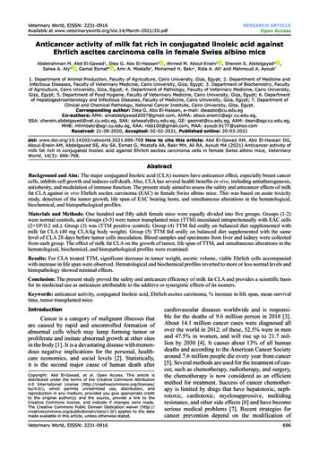
Available at entified microtubes; one with ethylenediaminetetra-acetic acid (EDTA) 10% as anticoagulant usedto measure hematological parameters, and the secondone without anticoagulant for separation of serum wasused to measure the biochemical parameters [31].Hematological parametersBlood parameters were performed according toSantos et al. [32]. Blood on EDTA was used for RBCcounts, estimation of Hb levels, and WBC counts.Differential count of WBCs was determined usingblood smears stained with Giemsa stain.Biochemical parametersBiomarkers of liver function include serum alanine aminotransferase (ALT), aspartate aminotransferase (AST), and alkaline phosphatase (ALP) activitiesin addition to serum creatinine, a biochemical analysis for renal function evaluation, were measured andinterpreted, according to Tietz [33]. Total protein wasmeasured by means of Biuret reagent and albumin wasdetermined using the modified Bromocresol green,according to Moreira et al. [34]. Globulins were measured by subtracting of albumin from total serum proteins and albumin/globulin (A/G) ratio was calculatedfrom dividing albumin value on globulin value for eachsample. Liver and renal function tests were performedto assess functioning of the liver and kidneys aftermilk fat CLA administration. Total antioxidant capacity (TAC) was estimated, according to Lalithadeviet al. [35]. The previous biochemical parameters weremeasured colorimetrically using bio-diagnostic kits.Histopathological examinationThe treated mice and their controls were anesthetized with diethyl ether and immediately sacrificed,quickly dissected; liver and kidney were removed andfixed in Bouin’s fluid. After 24 h, tissues were rinsed 3times in 70% ethanol, dehydrated using a graded ethanol series and then embedded in paraffin wax. Paraffinthick slices, 5 µm, were stained with hematoxylinand eosin and examined under light microscope andphotographed using a digital microscope (OlympusBX50, Japan) [36].Acute toxicity of milk fat CLAMilk fat CLA was safe at doses as high as3 mg/mouse daily (160 mg CLA/kg BW) by oral route.The behavior of the mice was monitored, every 4 h,for a period of 48 h. Milk fat CLA supplemented dietdid not induce mortality, behavioral changes, locomotor ataxia, diarrhea, or weight loss in mice during the48-h observation period. Furthermore, feed and waterintake did not differ among the groups of study. Dilzerand Park [15] reported similar findings.Anticancer activityEffect on MST and % ILSEffect of milk fat CLA on life span, MST and %ILS, of TTM was investigated (Table-1 and Figure-1).In TTM positive control group (G3), the MST was16.5 days while it significantly (p 0.05) increased to23.5 and 25.2 days in G4 and G5 fed 1 mg milk fat CLA(40 mg CLA/kg BW.) daily 28 days post- and pre-tumor cells inoculation, respectively. The administrationof CLA at a dose of 40 mg/kg BW, orally significantly(p 0.05) increased the % ILS values to 42.4% and52.7% in the treated groups (G4) and (G5), respectively, when compared with the TTM, positive controlgroup (G3). The results of survival analysis revealedTable-1: Effect of daily oral administration of milk fat richin CLA on MST and % ILS in EAC inoculated mice.GroupsThe values expressed as mean SE, TTM Tumortransplanted mice with EAC cells, *Significant at (p 0.05)as compared with positive control group (Group 3, TTM).CLA Conjugated linoleic acid, MST Mean survival time,EAC Ehrlich ascites carcinoma, % ILS Percent increasein life span, TTM Tumor transplanted mice, MST Meansurvival timeCancer remains a major health problem causing mortality despite novel findings and several newanticancer drugs, its incidence is increasing with anannual rate of 1.2% [37]. Improvement in cancertreating strategies will result in prolonged survival ofpatients; however, there is a growing need for additional means of cancer management in both palliativeand curative treatments [38].Veterinary World, EISSN: 2231-0916 60MST5052.7%ILS42.440DaysResults and DiscussionMST/days % ILSGroup 3 Positive control (TTM)16.5 0.19‑Group 4* TTM feed on 50 mg milk23.5 0.26* 42.42*fat/mouse (1 mg CLA) dailypost‑inoculation.Group 5* TTM feed on 50 mg milk25.2 0.35* 52.73*fat/mouse (1 mg CLA) daily28 days pre‑inoculation.Statistical analysisThe experimental results were expressed as themean standard error of the mean. The significantdifference between the groups was statistically determined by one-way analysis of variance. Data wereaccepted as statistically significant considering thedifference at p 0.05.Treatment302023.525.216.51000G 3 (Cancer Control)G 4 (CLA treated)G 5 (CLA treated 28 daysbefor induction)Figure-1: Mean survival time/day and percent increase inlife span of tumor transplanted mice treated with milk fatrich in conjugated linoleic acid.699
Available at at MST and ILS% were significantly increased inmilk fat CLA treated mice (Groups 4 and 5).Effect on tumor weight and tumor volumeMilk fat CLA supplement leads to significant(p 0.05) reduction in the weight and volume of tumorin TTM treated groups (Table-2 and Figure-2a).Tumor weight of TTM control group (G3) was foundto be 5.8 g and that of milk fat CLA treated mice G4and G5 at a dose of 40 mg CLA/kg BW, was 3.7 and3.1 g with weight growth inhibition 36.2 and 46.6%,respectively. The ascitic fluid volume of the TTM control group was 7 mL, and it was significantly (p 0.05)reduced to 4.3 and 3.2 mL with a volume inhibition37.1 and 54.3% in treated mice groups G4 and G5,respectively.Effect on viable cell count (cells 06/mL)As shown in Table-3 and Figure-2b, the meanviable cell count of TTM positive control group (G3)was 330 0.2 and significantly (p 0.05) decreasedto 216 1 and 152 9 in treated mice G4 and G5, atdose of 40 mg/kg BW, respectively. The viable cellreduction in both treated groups was 65.5% and46.1% while tumor cell inhibition was 34.5% and53.9%, respectively. The antitumor nature of milk fatCLA was evidenced by the significant inhibition intumor weight and ascitic volume and the significantreduction in viable tumor cell count, in milk fat CLAtreated mice as compared to the TTM positive controlgroup (G3).EAC is one of the commonly used experimental breast tumor that derived from spontaneous mouseadenocarcinoma that characterized by ascites [28]. Incancer, the increased volume of ascitic fluid indicatestumor growth; hence, decrease in ascitic fluid volumeand arresting the tumor growth increase the life spanof animals [39]. The obtained results in vivo supportedthis hypothesis as i/p inoculation of EAC cells intomice significantly increased the number of life EACcells and subsequent ascites. Oral administration of50 mg milk fat rich in CLA per mice (40 mg CLA/kglive BW) to TTM treated groups (G4 and G5) significantly (p 0.05) enhanced the MST and increased thelife span of the inducted mice (Figure-1), significantlydecreased tumor weight and volume (Figure-2a),number and percent of life EAC cells (Figure-2b) aswell as WBCs count (Figure-3c), as compared to theTTM positive control (G3). Reddy et al. [38] reportedthat a 25% increase in the life span of ascites bearinganimals is a significant indication of drug activity. Theobservations in the present experiment indicated theeffectiveness of milk fat CLA on EAC cells and itsrole in the delay of cell division, thereby suggestingthe reduction in EAC volume and increased survivaltime in mice. This meets the steadfast criteria, prolongation of life span and decrease in WBC, for judgingthe potency of any anticancer agent [30,38,39].Hematological parametersThe results in Table-4, Figures-3 and 4 showedthat; on the 16th day after tumor cells inoculation,the hematological parameters of TTM positive control group (G3) were significantly (p 0.05) altered,relative to the normal group (G1). The RBCs count(Figure-3a) and Hb (Figure-3b) were significantly(p 0.05) decreased to 2.2 106 cells/mm3 and 8.3 g%,respectively, while WBCs count (Figure-3c) was significantly (p 0.05) increased to 14.2 103 cells/mm3.Table-2: Effect of daily oral administration of milk fat rich in CLA on tumor growth in EAC inoculated mice.GroupsGroup 1Group 2Group 3Group 4Group 5TreatmentMean bodyweightsNormal control (mice feed on balanced rationwithout inoculation)Mice feed on 50 mg milk fat/mouse (1 mg CLA)daily for 28 days without inoculationPositive control (TTM) feed on balanced rationTTM feed on 50 mg milk fat/mouse (1 mg CLA)daily post‑inoculation.TTM feed on 50 mg milk fat/mouse (1 mg CLA)daily 28 days pre‑inoculation.Tumor growthW (g)WI (%)V ( mL)VI (%)24.9 0.3‑‑‑‑25.0 0.3‑‑‑‑30.7 0.128.6 0.15.8 0.1*3.7 0.2**100.036.2**7.0 0.4*4.4 0.2**100.037.1**28.1 0.13.1 0.2**46.6**3.2 0.1**54.3**The values expressed as mean SE, W Weight, WI Weight inhibition, V Volume, VI Volume inhibition, *Significantat P 0.05 as compared with normal control group (Group 1), **Significant at P 0.05 as compared with EACgroup (Group 3). CLA Conjugated linoleic acid, EAC Ehrlich ascites carcinoma, TTM Tumor transplanted miceTable-3: Effect of daily oral administration of milk fat rich in CLA on viable cell count.GroupsGroup 3Group 4Group 5Viable cell count ( 106)Viable cell count (Numberof tumor cells)Viable cell count% (Tumor cell growth %)Viable cells inhibition (I%) (Tumorcells inhibition %)330 2216 1*152 9*100.065.5*46.1*100.034.5*53.9*The values expressed as mean SE. *Significant at (p 0.05) as compared with positive control group (Group 3, TTM).CLA Conjugated linoleic acid, TTM Tumor transplanted miceVeterinary World, EISSN: 2231-0916 700
Available at 00G3G45013.511.61246.134.553.90Viable cell count %Viable cell count ( - x 106 )Viable cells inhibition(I %)Figure-2: Tumor growth rate in tumor transplanted micetreated with milk fat rich in conjugated linoleic acid:(a) Weight and volume inhibition percent, (b) viable cellcount and percentage of viable cell inhibition.In addition, the differential count of WBCs showedsignificant decrease (31.1%) in the percentage oflymphocytes (Figure-4a) while the percentage of neutrophils (Figure-4b) and monocytes (Figure-4c) significantly increased (55.0% and 2.0%), respectively.Significantly (p 0.05) restoring all the hematologicalalterations, in the treated groups (G4) and (G5) to nearnormal values, after oral administration of milk fatCLA at the same time period.Anemia in TTM or during chemotherapy protocols, mainly due to reduction in RBC or hemoglobinpercentage as a result of iron deficiency or myelosuppression [40]. Restoration of hemoglobin content andmaintenance for the nearly normal values of RBCs andWBCs count of the treated mice groups (G4 and G5)after 16 days of tumor induction clearly indicatesthat milk fat CLA was able to reverse the alterationin the hematological parameters consequent to tumorinoculation and possesses a protective action on thehemopoietic system, suggesting its anticancer naturewithout inducing myelotoxicity.Lymphocyte count significantly increased in theTTM treated groups (G4 and G5) after administration of milk fat CLA demonstrating that, CLA has thepotentials of improving the cellular immune systemand its effectiveness in treatment of disease conditionscaused by lymphocytopenia in mammals [41].Biochemical parametersLiver functionIn respect to liver functions, transaminasesand ALP levels were determined in the sera of theVeterinary World, EISSN: 2231-0916 Hb (g%)65.5Viable cell count14.51410002.22G5Experimental Groups2001503.4303002503.94G5Treated Groups1012.88.38642b0G1G2G3G4G5Experimental Group1614.21412WBCs CountRate of viable cell Growth and Inhibitionb00a4.936.2 37.1RBCs CountRate of Inhibition6volume inhibition % 54.3weight inhibition %10.31087.66.66.1G1G26420cG3G4G5Experimental GroupsFigure-3: Effect of daily oral administration of milk fat richin conjugated linoleic acid on: (a) Red blood cells count,(b) hemoglobin content, and (c) white blood cells count.tested mice groups. Results, as shown in Table-5 andFigure-5, proved that supplementation of diet withmilk fat CLA with level of 40 mg CLA/kg live BW didnot affect transaminase, ALT and AST enzyme, activities in the sera of the tested normal animal group (G2).Induction of cancer into animals (G3), severely affecttransaminases activity in its sera. The activities ofboth ALT (Figure-5a) and AST (Figure-5b) were significantly (p 0.05) elevated from 17.87 to 22.78 IU/Land from 15.89 to 25.61 IU/L, respectively. WhenTTM fed on diet supplemented with 40 mg CLA/kglive BW (Group G4), the elevation in transaminaseactivities significantly (p 0.05) lowered than that inTTM (G3). The enzyme activity levels reached nearlythe normal levels as compared to Groups G1 and G2but the levels were slightly still high (19.40 1.3 and701
Available at ble-4: Effect of daily oral administration of milk fat rich in CLA on hematological parameters in EAC inoculated Mice.GroupsGroupGroupGroupGroupGroup12345RBC count(106cells/mm3)Hb (g %)WBC ocytes(%)5.2 0.14.9 0.12.2 0.1*3.4 0.1**3.9 0.2**14.5 0.713.5 0.38.3 0.2*11.6 0.2**12.8 0.3**6.6 0.16.1 0.114.2 0.2*10.3 0.20**7.6 0.15**69 0.974 0.231.1 1.0*48.9 2.1**59.9 1.5**25.5 0.826.2 0.455 1.9*38.2 0.9**27.1 0.7**1 0.31 0.52 0.6*1.2 0.3**1.1 0.1**The values expressed as mean SE, *Significant
The milk fat rich in CLA was produced from Holstein-Friesian Cows fed on diet contained pro-tected fat, high in unsaturated fatty acids. The CLA percentage of the produced milk fat was 2% of the fatty acids, while the percentage of CLA in normal cow milk fat ranged between 0.3% and 0.4%. The cow milk fat rich in CLA was supplemented to the