Transcription
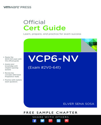
Chapter One: IntroductionANATOMICAL POSITIONAND TERMS OFDIRECTIONWhen studying the human body it isimportant to place the body inanatomical position. Anatomicalposition is described as the bodyfacing you, feet placed together andflat on the floor. The head is helderect, arms straight by the side withpalms facing forward. All referencesto the body are made as if the body isin this position so when you describesomething as being above somethingelse it is always with respect to thebody being in anatomical position.a.a.e.)The relative position of the parts ofthe human body has specific terms.Superior means above whileinferior means below. Medial refersto being close to the midline whilelateral means to the side. Anterioror ventral is to the front whileposterior or dorsal is to the back.Superficial is near the surface whiledeep means to the core of the body.When working with the limbs,proximal means closer to the trunkwhile distal is to the ends of theextremities. Write the directionalterms in the spaces provided andcolor in the arrows in reference tothese terms. Note that these termsare somewhat different for fourlegged animals.h.).(1.lII('---:-----':----;- . k . - -1)I, \VeIf.b.b.g. ----- V?1h. .I. \!l« Answer Key: a. Superior, b. Inferior,c. Lateral, d. Medial, e. Proximal,f. Distal, g. Anatomical position,h Posterior, i. Anterior, j. Dorsal,k. Ventral) I.
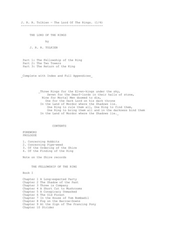
Chapter OneIntroductionIANATOMICAL PLANES OFTHE BODYMany specimens in anatomy aresectioned so that the interior of theorgan or region can be examined. Itis important that the direction of thecut is known so that the properorientation of the specimen isknown. A heart looks very differentif it is cut along its length as opposedto horizontally. A horizontal cut isknown as a transverse section or across section. A cut that divides thebody or an organ into anterior andposterior parts is a coronal sectionor frontal section. One that dividesthe structure into left and right partsis a sagittal section. If the body isdivided directly down the middle thesection is known as a midsagittalsection. A midsagittal section isusually reserved for dividing thebody into to equal left and rightparts. If an organ (such as the eye) issectioned into two equal parts suchthat there is a left and right half thenthis is known as a median section.Label the illustrations and color inthe appropriate planes.c.a.Answer Key: a. Frontal (coronal) plane,b. Transverse (cross-section) plane,c. Median (midsagittal) planeb.c.mKAPeLAN(I·-Ical3
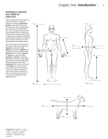
Chapter OneIntroductionHIERARCHY OF THE BODYThe human body can be studied at different levels. Organs such as thestomach can be grouped into organ systems (digestive system) or can bestudied on a smaller scale like the cellular level. The ranking of theselevels is called a hierarchy. The smallest organizational unit is the atom.Individual atoms are grouped into larger structures called molecules.I KAPLA .meulCaIThese in turn make up organelles, which are part of a larger, morecomplicated systems called cells. Cells are the structural and functionalunits of life. Cells are clustered into tissues. Organs are discreet unitsmade up of two or more tissues and organs are grouped into organsystems that compose the organism. Label the levels of the hierarchyand color each item a different color. a.b.og.-----c. - - - - -d.Answer Key: a. Organism (human), b. Organ system (respiratory system) c. Organ (lung), d. Tissue (epithelium), e. Organelle (cilia), f. Molecule, g.Atom, h. cells5
Chapter OneIntroductionI me dlea- IKAPLAlfREGIONS OF THEABDOMENIn anatomy the abdomen is dividedinto nine regions. Write the names ofthe regions in the spaces indicated.Color both the left and righthypochondriac regions in light blue.Hypochondriac means "below thecartilage." The common use of theword (someone who thinks they aresick all the time) reflects the Greekorigin of the word as the ancientGreeks considered the region to bethe center of sadness. Inferior to thehypochondriac regions are thelumbar or lateral abdominalregions. These are commonly knownas the "love handles." Use yellow forthese regions. Below the lumbarregions are the inguinal or iliacregions. You should color in theseregions with the same shade ofgreen. In the middle of theabdomen is the umbilical region.Color this region in red. Above thisis the epigastric region (epi aboveand gastric stomach). Color thisregion in purple. Below theumbilical region is the hypogastricregion (hypo below). Color thisregion in a darker blue.In clinical settings a quadrantapproach is used. Write the names ofthe regions (right upper quadrant,left upper quadrant, right lowerquadrant, left lower quadrant) inthe spaces provided. Color eachquadrant a different color.Answer Key: a Right hypochondriac,b. Right lumbar (lateral abdominal),c. Umbilical, d. Right Inguinal or iliac,e. Epigastric, f. Left hypochondriac,g. Left lumbar (lateral abdominal),h. Left inguinal or iliac, i. Hypogastric,) Left upper quadrant, k. Right upperquadrant, I. Left lower quadrant,m. Right lower quadranta.b.c.d.g.-----7
Chapter OneIIntroductionORGAN SYSTEMSThe human body is either studied by regions or by organs systems. Thisbook uses the organ system approach in which individual organs (suchas bones) are grouped into the larger organ system (for example, theskeletal system). Typically eleven organ systems are described. Theskeletal system consists of all of the bones of the body. Examples are thefemur and the humerus. The nervous system consists of the nerves,spinal cord, and brain while the lymphatic system consists of lymphglands, conducting tubes called lymphatics, and organs such as thespleen. The term immune system is more of a functional classificationIIAPLAN· ·me dlea9and will not be treated as a separate system here. The muscular systemconsists of individual skeletal muscles as organs such as the pectoralismajor and deltoid. Label the organ systems underneath each illustrationand label the selected organs by using the terms available. When youfinish, select different colors for each organ system and color them in.Organ SystemOrganOrganSkeletal systemNervous systemLymphatic systemMuscular systemFemurNervesLymph glandsPectoralis majorHumerusSpinal ---1.-----1.Answer Key:J. - - - - - - - - -ffi .a. Humerus,b. Femur, c. Skeletal,d. Brain, e. Spinalcord, f. Nerves,g. Nervous,h. Spleen, i. Lymphnodes, j. Lymphatic,k. Deltoid,I Pectoralis major,m. Muscular
Chapter OneIntroductionORGAN SYSTEMS (CONTINUED)The skin and other structures are in the integumentary system and thedigestive system involves the breakdown and absorption of food withorgans such as the esophagus and stomach. The endocrine system ismade of the glands that secrete hormones such as the thyroid gland andthe adrenal glands. The respiratory system involves the transfer ofoxygen and carbon dioxide between the air and the blood. Therespiratory system consists of organs such as the trachea and lungs.ImKAPeLANd'-IcalLabel the organ systems underneath each illustration and label theselected organs by using the terms available. When you finish, selectdifferent colors for each organ system and color them in.Organ SystemOrganOrganIntegumentary systemDigestive systemEndocrine systemRespiratory systemSkinEsophagusThyroid glandTracheaStomachAdrenal glandsLungsa.--------J"', .,-,.,"1b.- ,\ \I(\e.Q---)fjp). --- /,\ '. I\,I'I'\\h.-------.//\/k.Answer Key: a. Skin, b. Integumentary, c. Esophagus, d. Stomach, e. Digestive, f. Thyroid gland, g. Adrenal gland, h. Endocrine, i, Trachea, j. Lung, k. Respiratory11
Chapter OneIntroductionORGAN SYSTEMS (COI\ITINUED)The heart and associated blood vessels compose the cardiovascularsystem which circulates blood throughout the body. The urinary systemfilters, stores, and conducts some wastes from the body. The bladder andurethra are part of the urinary system. The testes and ovaries are partof the reproductive system and this system perpetuates the species. Thedifferentiation of male and female systems makes this organ systemunique among the other systems. These eleven organs systems can beremembered by the memory clue LN Cries Drum. Each letter representsOrgan SystemOrganOrganCardiovascular systemUrinary systemReproductive systemHeartBladderTestesBlood vesselsUrethraOvariesb.----J C\::c.h.Answer Key: a. Heart, b. Bloodvessels, c. Cardiovascular, d. Bladder,e. Urethra, f. Urinary, g. Ovary, h. Testis,I. Reproductive1.d' -.eallA PeLmAN13the first letter of a name of an organ system. Label the organ systemsunderneath each illustration and label the selected organs by using theterms available. When you finish, select different colors for each organsystem and color them in.-a.I-J
Chapter OneIntroductionI KAPLA .meulCaIBODY REGIONS(AI\ITERIOR)There are specific anatomical termsfor regions of the body. These areasor regions frequently have Greek orLatin names because early westernstudies in anatomy occurred inGreece and Rome. During theRenaissance, European scholarsstudied anatomy and applied theancient names to the structures.Label the various regions of the bodyand fill in their names. You can use astandard anatomy text or follow thekey at the bottom of the page. A listof terms and their common namesfollows for the anterior side of thebody. Color in the regions of thebody.cranial (head)facial (face)cervical (neck)deltoid (shoulder)pectoral (chest)sternal (center of chest)brachial (arm)antebrachial (forearm)manual (hand)digital (fingers)abdominal (belly)inguinal (groin)coxal (hip)femoral (thigh)genicular (knee)crural (leg)pedal (foot)digital (toes)qr- )a. - - - - - - - - -\ ··: lf.e-;;))1).:§ b '. .::::. ::-.: r":::::;::::. d. - - - - - -"',""--:''\. . .(. '] ( -.' .!. . \. y e.1\L\.·····f.----: ./.·V.). .g. -.0(.f\/·t.\. ." ./ -/ 1. -\ \J1.m.n.O.J .TpAnswer Key: a. Cranial (head), b. Facial(face), c. Cervical (neck), d. Deltoid(shoulder), e. Sternal (center of chest),f Pedoral (chest), g. Brachial (arm),h. Abdominal (belly), i. Antebrachial(forearm), j. Coxal (hip), k. Manual(hand), I. Digital (fingers), m. Inguinal,n. Femoral (thigh), o. Genicular (knee),p. Crural (leg), q. Pedal (foot), r. Digital(toes)} ;{---jr'' Lq\ . r. - - - - - - -15
Chapter OneIntroductionIlAP LANd' me leaIBODY REGIONS(POSTERIOR)For the posterior view of the bodyfill in the terms and color the regionsof the body. The anatomical namesare given first with the commonnames in parentheses.cephalic (head)nuchal (neck)scapular (shoulder blade)vertebral (backbone)lumbar (love handles)brachial (arm)olecranon (elbow)antebrachial (forearm)gluteal (buttocks)femoral (thigh)popliteal (back of knee)sural (calf)calcaneal (heel)----i---J. - - - - - - - - - -.'/--11}\.Answer Key: a.Cephalic (head),b. Nuchal (neck), c. Scapular (shoulderblade), d. Brachial (arm), e. Vertebral(backbone), f. Olecranon (elbow),g. Lumbar(love handles),h. Antebrachial (forearm), i. Gluteal(buttocks), j. Femoral (thigh),k. Popliteal (back of knee), I. Sural(calf), m. Calcaneal (heel) m. - - - - - - - - - -17
Chapter OneIntroductionBODY CAVITIESThe organs of the body are frequently found in body cavities. The bodyis divided into two main cavities, the dorsal body cavity and the ventralbody cavity. The dorsal body cavity consists of the cranial cavity, whichhouses the brain and the spinal canal, which surrounds the spinal cord.The ventral body cavity contains the upper thoracic cavity, which issubdivided into the pleural cavities, housing the lungs, and theIKAPLAIf Ime d lea19mediastinum. The mediastinum contains the heart in the pericardialcavity, the major vessels near the heart, nerves, and the esophagus.Below the thoracic cavity is the abdominopelvic cavity, which containsthe upper abdominal cavity, housing the digestive organs, and theinferior pelvic cavity, which holds the uterus and rectum in females orjust the rectum in males. Label the specific and major cavities of thebody and color them with different colors.b.------c. - - - - - - - - -Answer Key: a. Dorsal body cavity, b. Cranial cavity, c. Spinalcanal, d. Ventral body cavity, e. Thoracic cavity, f. Mediastinum, g Pericardial cavity, h. Pleural cavity,I. Abdominopelvic cavity, j. Abdominal cavity, k. Pelvic cavity
Chapter Two: Cells, Tissues, and IntegumentOVERVIEW OF CELL AND CELL MEMBRANECells consist of an enclosing plasma membrane, an inner cytoplasmwith numerous organelles, and other cellular structures. The fluidportion of the cell is called the cytosol. Color the cytosol in last after youcolor the rest of the cellular structures. One of the major structures inthe cell is the nucleus. It is the genetic center of the cell and consists oftluid karyoplasm, chromatin (containing DNA), and the nucleolus.Color these features and label them on the illustration.The cytoskeleton consists of microtubules, intermediate filaments andmicrofilaments. It is involved in maintaining cell shape, fixingorganelles, and directing some cellular activity.21Golgi apparatus assembles complex biomolecules and transports themout of the cell. Proteins are made in the cell by ribosomes. If theribosomes are found by themselves in the cytoplasm, they are called freeribosomes. If they are attached to the rough endoplasmic reticulum,they are called bound ribosomes. The smooth endoplasmic reticulummanufactures lipids and helps in breaking down toxic materials in thecell. Other structures in the cell are vesicles (sacs that hold liquids).Phagocytic vesicles ingest material into the cell. Lysosomes containdigestive enzymes while peroxisomes degrade hydrogen peroxide in thecell. After you label and color the organelles make sure to go back andshade in the cytosol. Centrioles are microtubules grouped together andare involved in cell division.Label the organelles of the cell and use a different color for each one. Themitochondria are the energy-producing structures of the cell while thea.p.o.ll.m.1.The plasma membrane is composed of a phospholipid bilayer. Colorthe phosphate molecules on the outside and inside of the membraneone color and the lipid layer another color. Cholesterol molecules occurin the membrane and, depending on their concentration, can make themembrane stiff or more fluid. Proteins that are found on the outside ofthe membrane are called peripheral proteins while proteins that passthrough the membrane are called integral proteins. Frequently thesemake up gates or channels that allow material to pass through themembrane. Attached to proteins on the cell membrane are carbohydratechains. These provide cellular identity. Label and color the cellmembrane structures.r.q.------v.Answer Key: a. Golgi apparatus, b. Lysosome, c. Peroxisome, d. Phagocytic vesicle, e. Nucleus, f. Nucleolus, g. Chromatin, h. Karyoplasm, '1. Cytoskeleton,J. Centrioles, k. Plasma membrane, I. Cytoplasm, m. Rough endoplasmic reticulum, n. Smooth endoplasmic reticulum, o. Mitochondrion, p. Free ribosomes,q. Phospholipid bilayer, r.Integral protem, s.Carbohydrate chain, t. Peripheral protein, u. Phosphate molecule, v. lipid layer, w. Cholesterol molecule
Chapter TwoCells, Tissues, and IntegumentSIMPLE EPITHELIAThere are four types of tissues inhumans and these make up all of theorgans and binding material in thebody. Epithelial tissue makes uplinings of the body. In many cases,where there is exposure (outside,such as the skin, or inside, such as inblood vessels), epithelium is thetissue found. It is named accordingto its layers (typically simple orstratified) and the shape of cells(such as cuboidal). Simplesquamous epithelium is a singlelayer of flattened cells. Simplecuboidal epithelium is also a singlelayer of cells but the cells are in theshape of cubes. Simple columnarepithelium is a single layer of longcolumnar cells. Label and color theseepithelial types and pay attention tothe basement membrane, thenoncellular layer that attaches theepithelium to lower layers. It shouldbe colored red. Color the nuclei inpurple, the cytoplasm blue, andlabel the cells.Top viewSide viewa.b.Pseudostratified ciliated columnarepithelium is in a single layer of cellshut it looks stratified on firstappearance. Not all of the cells reachthe surface of the tissue. All of thecells reach the basement membrane.Label and color the nuclei,basement membrane, cellmembrane and the cilia in thistissue.c.d. - - - - - - - - -Answer Key: a. Simple squamousepithelium, b. Simple cuboidalepithelium, c. Simple columnarg.epithelium, d.Cilia, e. Cell membrane,t. Nuclei, g. Basement membrane,h. Pseudostratlfled ciliated columnarepitheliumh.IUPLANd··me leaI23
Chapter TwoCells, Tissues, and IntegumentSTRATIFIED EPITHELIAThere are two common epithelialtissues that are many-layered.Stratified squamous epithelium ismany layers of flattened cells. Labeland color the basement membranered, color the cytoplasm blue, andthe nuclei purple. There are twomajor types of stratified squamousepithelium. Keratinized epitheliumis found on the skin and istoughened by the protein keratin.Non-keratinized stratified squamousepithelium is found in the oral cavityand vagina and is a mucousmembrane.Another main type of layeredepithelial tissue is transitionalepithelium. This is tissue that linespart of the urinary tract includingthe bladder. When the bladder isempty, the cells bunch up on oneanother and the tissue is thick. Whenthe bladder is full, the cells stretchout into a few layers. Label the celltypes for each picture and color thestructures in the same way as inprevious illustrations.a.StretchedRelaxedb. - - - - - - - - - - - Answer Key: a. Stratified squamousepithelium, b. Transitional epitheliumIKAPLAdlf.me leaI25
Chapter TwoCells, Tissues, and IntegumentIKAPLA!.meulCaIGLANDSThere are several types of glands inthe human body. Some of theseglands secrete their products intotubes or ducts. These are known asexocrine glands. Other glandssecrete their products into the spacesbetween cells where they are pickedup by the blood or lymph system.These are the endocrine glands.Endocrine glands secrete hormonesthat have an impact on target tissuesof the body.Glands can be unicellular ormulticellular. Glands that consist ofjust one cell are called goblet cells.They secrete mucus, which is alubricant. There are many types ofmulticellular glands. They areclassified by how they secrete theirproducts. Some glands secreteproducts from vesicles pinched offfrom the cell. These are calledmerocine glands. In these glands nocellular material is lost in thesecretion of material. An example ofa merocrine gland is a sweat gland.Some cells squeeze parts of the celloff to secrete cellular products.These are known as apocrine glands.The lactiferous glands that producemilk are apocrine glands. Somesecretions occur by the entire cellrupturing. These are calledholocrine glands. Oil glands of theskin are holocrine glands. Label theglands and color them in on thefigure.b.a.c.e.-----Answer Key: a. Exocrine gland,b Endocrine gland, c. Goblet cell,d. Merocrineglands, e. Vesicles,f. Apocrine glands, g. Holocrine glandsd.f.g. ------27
Chapter TwoCells, Tissues, and IntegumentIlAP LANd' me leaCOI\II\IECTIVE TISSUEConnective tissue is a varied groupof associated tissues, all of which arederived from an embryonic tissueknown as mesenchyme. Connectivetissue not only has cells, as do all ofthe other tissues, but it also hasfibers and a large amount ofbackground substance calledmatrix. There are many specifictissues that belong to connectivetissue. Loose connective tissue isfound wrapping around organs orunder the epidermis and it iscomposed of collagenous, elastic,and reticular fibers, a liquid matrixand numerous cells, many of whichhave an immune function. Denseregular connective tissue has a fewcells called fibrocytes and a smallamount of matrix with most of thetissue composed of a regulararrangement of collagenous fibers.This specific tissue makes up tendonsand ligaments. If the fibers are not inan orderly arrangement, then thetissue is called dense irregularconnective tissue. This tissue isfound in places like the white ofthe eye.a.b.c.d.e.c.f.c. C . \ '------J .' »:g.I ,, ,-, / Answer Key: a. Matrix, b. I"ibrocyte,c. Collagenous fiber, d. Elastic fiber,e. Loose connective tissue, f. Denseregular connective tissue, g. Dense'Irregular connectivetissue . ) .I29
IChapter TwoCells, Tissues, and IntegumentCONNECTIVE TISSUE(CONTINUED)Elastic connective tissue containselastic fibers and is found in areasthat recoil when stretched such as inthe walls of arteries. Reticularconnective tissue consists ofreticular fibers that form an internalsupport in soft organs such as theliver and spleen. Adipose tissueconsists of specialized fat-storingcells called adipocytes. Label andcolor the components of theseconnective tissues.c.d.e.Answer Key: a. Collagenous fibers,b. Elastic fibers, c. Elastic connectivetissue, d. Reticular fibers, e. Reticularconnective tissue, f. Adipose tissuef.IAPLAlf Ime dlea31
Chapter TwoCells, Tissues, and IntegumentCARTILAGEThere are three types of cartilage inconnective tissue. The mostcommon kind of cartilage is hyalinecartilage. It contains a semisolidmatrix, collagenous fibers, andchondrocytes (cartilage cells). Theend of the nose is pliable due tohyaline cartilage. Fibrocartilage islike hyaline cartilage, having thesame components, but there aremore collagenous fibers infibrocartilage. It is found in areaswhere there is more stress, such asthe joint between the bones of thethigh and leg. Elastic cartilage has amatrix, chondrocvtes, and elasticfibers. These fibers make thecartilage more bendable than hyalinecartilage. Label and color the cellsand fibers of cartilage and use a lightcolor to shade the matrix such as apale pink or blue.a. - - - - - - - - - -c.e.Answer Key: a. Matrix, b. Chondrocytes,c. Hyalinecartilage, d. Collagenousfibers, e Fibrocartilage, f. Elastic fibers,g. ElastiC cartilagef.-------g.IKAPLAll"d-me leaI:n
Chapter TwoCells, Tissues, and IntegumentBONE AND BLOODBone is a connective tissue. The cellsare the osteocytes and the fibers arecollagenous fibers enclosed in a hardmatrix of bone salts. You will not seethe fibers in the illustration becausethey are covered by the dense matrix.Label and color the osteocytes andmatrix of bone.Blood is another kind of connectivetissue. The matrix in blood is theplasma and the cells areerythrocytes (red blood cells) andleukocytes (white blood cells).IKAPLAIf Ime d leaa.b.I, .Platelets are small flat disks in theblood that aid in clotting.c. - - - - - - - - - - - - - - - - - - - - - - - -d.o e.0 O 0oGoDo !I00 oO \V. 0oCb'cP\\)OOC6o (JOc; OO\JOC]Jf.h.Answer Key: a. MatriX, b. Osteocyte.c. Bone, d. Erythrocyte, e. Platelet,f. Leukocytes, g. Plasma, h. Bloodg. - - - - - - - - -35
Chapter TwoCells, Tissues, and IntegumentMUSCLE AND NERVOUS TISSUEMuscular tissue is composed of specialized cells involved in contraction.Skeletal muscle makes up body muscles and represents around 40percent of the body mass. Skeletal muscle is striated and the fusion ofindividual cells produces longer, mature cells that are multinucleate.These nuclei are found on the edges of the cells. Skeletal muscle can beconsciously controlled and is called voluntary muscle. Label and color thestriations of the skeletal muscle cells, the nuclei, and individual cells.Cardiac muscle is also striated but the striations are not as obvious as inskeletal muscle. This muscle is found in the heart and is involuntary. Itdoes not involve conscious control. Cardiac muscle typically has onlyone centrally located nucleus per cell, and the cells themselves arebranched. They attach to other cells by intercalated discs, which allowcommunication between cells for the conduction of impulses during thecardiac cycle. Label and color these features on the illustration.a.b.Irleame CKAPLAISmooth muscle is not striated and it is involuntary. The cells are slenderand have one nucleus located in the center of the cell. It is widelydistributed in the body, making up, among other things, part of thedigestive system, reproductive system, and integumentary system.Smooth muscle is found in glands and other areas not under consciouscontrol. Label and color the nucleus and cell of smooth muscle.Nervous tissue consists of the neuron and associated glial cells.Neurons have numerous branched extensions called dendrites, a centralnerve cell body (soma) that houses the nucleus, and a long extensioncalled an axon. The glial cells, also known as neuroglia, have manyfunctions. Some of these are supportive of the neuron and some mayinvolve processing of neural information. Label and color the parts ofthe neuron and the glial cells.b. '\\ } / ;:CF-;C C : ':':C ': e.-------c.d.f.b.c.1.g.37h.Answer Key: a. Striations, b. Nuclei, c. Cell, d. Skeletal muscle, e. Intercalated disc, f. Cardiacmuscle, g. Smooth muscle, h. Nervous tissue, i. Nerve cell body,j. Glial cells (Neuroglia), k. Dendrites, I. Nucleus, m. Axon
Chapter TwoCells, Tissues, and IntegumentIKAPLAlfdmeleaI39INTEGUMENTARY SYSTEMThe most superficial layer of the skinis the epidermis. Color the five layersof the epidermis. The deepest layer isthe stratum basale and there arespecific cells called melanocytes thatsecrete the brown pigment melanin.Color the majority of the stratumbasale pink but color themelanocytes brown. Color thestratum spinosum a light blue. Thestratum granulosum has purplegranules in it so color that layerusing purple dots. The stratumlucidum (found only in thick skin)is a thin, light colored layer so yellowor white are good colors for thistissue. Color the superficial stratumcorneum orange.The overview of the skin containsmany layers. Color the epidermis ared-orange. The dermis consists oftwo layers, an upper papillary layer,which should be colored in a lightpink, and a deeper reticular layer,which should be colored a darkerpink. There are sweat glands that arefound in the dermis that can becolored purple. You should color thehypodermis (not a part of theintegument) yellow because of theamount of fat found there. Twotypes of touch receptors can easily beseen in microscopic sections. Theseare the Meissner corpuscles and thePacinian corpuscles.f.------1.Fl ,1-----1.Answer Key: a. Stratum corneum,b. Stratum IUCIdum, c. Stratumgranulosum, d. Stratum spinosum,e. Stratum basale, f. Melanocyte,g. Epidermis, h. Papillary layer,I. Reticular layer, J. Dermis,k. Hypodermis, L Sweat gland,m. Pacinian corpuscle,n. Meissnercorpusclem.--------g.-----).---
Chapter TwoCells, Tissues, and IntegumentHAIR AND NAILSHair consists of several parts. The hair originates from the dermalpapilla and the deepest part of the hair is known as the hair bulb. Thehair is pushed superficially and forms the hair root (the part of the hairenclosed in the skin). Once the hair erupts from the skin it is known asthe hair shaft. Color the three sections of hair different shades of blue.The hair is enclosed by the hair follicle, which should be colored purple.IKAPLA .meulcaIAssociated with the hair are the arrector pili muscle, which is made ofsmooth muscle and is colored pink, and an oil-secreting gland known asthe sebaceous gland, which should be colored yellow.Fingernails and toenails are considered accessory structures of theintegument. Color the diagram labeling the nail plate, the free edge, thenail fold, the lunula, eponychium (cuticle), nail root, hyponychiumand the nail bed.a.1.k.J.1. - - - - - - - - -h.41m.n.o.Answer Key: a. Bulb, b. Follicle, c. Root, d. Shaft,e. Sebaceous gland, f. Arrector pili. g. Pacinian corpuscle, h. Nail plate, i. Nail fold, J. Lunula, k. Eponychium,I. Nail root, m. Free edge, n. Hyponychium, o. Nail matrix (Nail bed)
Chapter Three: Skeletal SystemFRONTAL ASPECT OF THE SKULLThe skull is a complex structure. There are 8 cranial bones and 14 facialbones in the skull. From the anterior view most of the facial bones can beseen and some of the cranial bones are visible too. The bone that makesup the forehead and extends beyond the eyebrows is the frontal bone.This bone forms the upper rim of the orbit, which is a socket thatencloses the eye. In the back of the orbit is the sphenoid bone and thelateral walls of the orbit are composed of the zygomatic bones. Thebridge of the nose consists of the paired nasal bones and just lateral toa.43them are the two maxillae. These bones hold the upper teeth. The lowerteeth are held by the mandible. Inside the nasal cavity two projectionscan be seen. These are the inferior nasal conchae. The wall that divides thenasal cavity is the nasal septum and it consists of two bones, the ethmoidbone and the vomer. Along the side of the skull are the temporal bones,located posterior to the zygomatic bones. Label the major bones of theskull and color them in. As you color in the skull try to use the same colorfor the same bone on different pages. This will help you associate thesame bone with various views from which it can be seen.d. ------e. - - - - - - - -f.g.-------h.Answer Key: a. Orbit, b. Frontal bone, c. Temporal bone, d. Sphenoid bone, e. Nasal bone, f. Zygomatic bone, g. Nasal septum, h. Maxilla, i. Mandible
Chapter ThreeSkeletal System IMany bones seen from the anterior view can also be seen from the lateralview. The frontal bone is joined to the parietal bones by the coronalsuture. The parietal bones span much of the cranium and articulate withthe occipital bone at the lambdoid suture. There is a posteriorextension of the occipital bone known as the external occipitalprotuberance. The exterior aspect of the temporal bone is seen from thelateral view and many of the significant features such as the mastoidprocess, external acoustic meatus, and styloid process are visible. On theside is the elongated zygomatic process. The temporal bone articulateswith other cranial bon
the human body has specific terms. Superior means above while inferior means below. Medial refers to being close to the midline while lateral means to the side. Anterior or ventral is to the front while posterior or dorsal is to the back. Superficial is near the surface while deep means to the core of the body. When working with the limbs,