Transcription
P A R TV IRENAL PHYSIOLOGY AND BODY FLUIDSC H A P T E R22Kidney FunctionGeorge A. Tanner, Ph.D.LEARNING OBJECTIVESUpon mastering the material in this chapter you should beable to: Summarize the functions of the kidneys. Define the renal clearance of a substance. Explain how glomerular filtration rate is measured, thenature of the glomerular filtrate, and the factors that affectfiltration rate. Describe how renal blood flow can be determined from theclearance of p-aminohippurate (PAH) and the hematocrit,and discuss the factors that influence renal blood flow. Calculate rates of net tubular reabsorption or secretion of asubstance, given the filtered and excreted amounts of thesubstance. For glucose, explain what is meant by tubular transportmaximum, threshold, and splay. Discuss the magnitude and mechanisms of solute and waterreabsorption in the proximal convoluted tubule, loop ofHenle, and distal nephron. Explain why sodium reabsorptionis a key operation in the kidneys.he kidneys play a dominant role in regulating the composition and volume of the extracellular fluid (ECF).They normally maintain a stable internal environmentby excreting in the urine appropriate amounts of many substances. These substances include not only waste productsand foreign compounds, but also many useful substances thatare present in excess because of eating, drinking, or metabolism. This chapter considers the basic renal processes thatdetermine the excretion of various substances.The kidneys perform a variety of important functions:T1. They regulate the osmotic pressure (osmolality) ofthe body fluids by excreting osmotically dilute orconcentrated urine.2. They regulate the concentrations of numerous ionsin blood plasma, including Na , K , Ca2 , Mg2 , Cl ,bicarbonate (HCO3 ), phosphate, and sulfate.3. They play an essential role in acid–base balance byexcreting H when there is excess acid or HCO3 whenthere is excess base.4. They regulate the volume of the ECF by controlling Na and water excretion. Describe the active tubular secretion of organic anions andorganic cations in the proximal tubule and passive transportvia nonionic diffusion.5. They help regulate arterial blood pressure by adjusting Na excretion and producing various substances(e.g., renin) that can affect blood pressure. Explain how arginine vasopressin increases collecting ductwater permeability. Discuss the countercurrent mechanisms responsible forproduction of osmotically concentrated urine. Explain howosmotically dilute urine is formed.6. They eliminate the waste products of metabolism,including urea (the main nitrogen-containing endproduct of protein metabolism in humans), uric acid(an end product of purine metabolism), and creatinine(an end product of muscle metabolism).7. They remove many drugs (e.g., penicillin) and foreignor toxic compounds.391
392PART VIRENAL PHYSIOLOGY AND BODY FLUIDSFUNCTIONAL RENAL ANATOMY8. They are the major sites of production of certainhormones, including erythropoietin (see Chapter 9)and 1,25-dihydroxy vitamin D3 (see Chapter 35).9. They degrade several polypeptide hormones, includinginsulin, glucagon, and parathyroid hormone.10. They synthesize ammonia, which plays a role in acid–base balance (see Chapter 24).11. They synthesize substances that affect renal bloodflow and Na excretion, including arachidonic acidderivatives (prostaglandins, thromboxane A2) andkallikrein (a proteolytic enzyme that results in theproduction of kinins).When the kidneys fail, a host of problems ensue. Dialysisand kidney transplantation are commonly used treatmentsfor advanced (end-stage) renal failure.Each kidney in an adult weighs about 150 g and is roughly thesize of one’s fist. If the kidney is sectioned (Fig. 22.1), tworegions are seen: an outer part, called the cortex, and aninner part, called the medulla. The cortex typically is reddishbrown and has a granulated appearance. All of the glomeruli,convoluted tubules, and cortical collecting ducts are locatedin the cortex. The medulla is lighter in color and has a striated appearance that results from the parallel arrangementof loops of Henle, medullary collecting ducts, and blood vessels of the medulla. The medulla can be further subdividedinto an outer medulla, which is closer to the cortex, and aninner medulla, which is farther from the cortex.The human kidney is organized into a series of lobes, usually 8 to 10 in number. Each lobe consists of a pyramid ofmedullary tissue, plus the cortical tissue overlying its baseCLINICAL FOCUS22.1Chronic kidney disease is usually progressiveDialysis can enable patients with otherwiseand may lead to renal failure. Common causesfatal renal disease to live useful and productiveDialysis andinclude diabetes mellitus, hypertension, inflamlives. Many physiological and psychological probTransplantationmation of the glomeruli (glomerulonephritis),lems persist, however, including bone disease,urinary reflux and infections (pyelonephritis),disorders of nerve function, hypertension, atherand polycystic kidney disease. Renal damageosclerotic vascular disease, and disturbances ofmay occur over many years and may be undesexual function. There is a constant risk of infectected until a considerable loss of functioning nephrons hastion and, with hemodialysis, clotting and hemorrhage. Dialysisoccurred. When GFR has declined to 5% of normal or less, thedoes not maintain normal growth and development in children.internal environment becomes so disturbed that patients usuAnemia (primarily resulting from deficient erythropoietin producally die within weeks or months if they are not dialyzed or protion by damaged kidneys) was once a problem but can now bevided with a functioning kidney transplant.treated with recombinant human erythropoietin.Most of the signs and symptoms of renal failure can beRenal transplantation is the only real cure for patientsrelieved by dialysis, the separation of smaller molecules fromwith end-stage renal failure. It may restore complete health andlarger molecules in solution by diffusion of the small moleculesfunction. In 2003, about 15,000 kidney transplantation operathrough a selectively permeable membrane. Two methods oftions were performed in the United States. At present, aboutdialysis are commonly used to treat patients with severe, irre95% of kidneys grafted from a living donor related to the recipversible (“end-stage”) renal failure.ient function for 1 year; about 90% of kidneys from cadaverIn continuous ambulatory peritoneal dialysis (CAPD), thedonors function for 1 year.peritoneal membrane, which lines the abdominal cavity, acts asSeveral problems complicate kidney transplantation. Thea dialyzing membrane. About 1 to 2 L of a sterile glucose/saltimmunological rejection of the kidney graft is a major chalsolution are introduced into the abdominal cavity, and smalllenge. The powerful drugs used to inhibit graft rejection commolecules (e.g., K and urea) diffuse into the introduced solution,promise immune defensive mechanisms so that unusual andwhich is then drained and discarded. The procedure is usuallydifficult-to-treat infections often develop. The limited supplydone several times every day.of donor organs is also a major, unsolved problem; thereHemodialysis is more efficient in terms of rapidly removingare many more patients who would benefit from a kidney transwastes. The patient’s blood is pumped through an artificial kidneyplantation than there are donors. The median waiting timemachine. The blood is separated from a balanced salt solution byfor a kidney transplantation is currently more than 900 days.a cellophanelike membrane, and small molecules can diffuseFinally, the cost of transplantation (or dialysis) is high. Fortunatelyacross this membrane. Excess fluid can be removed by applyingfor people in the United States, Medicare covers the costpressure to the blood and filtering it. Hemodialysis is usually doneof dialysis and transplantation, but these life-saving therthree times a week (4 to 6 hours per session) in a medical facilapies are beyond the reach of most people in developingity or at home.countries.
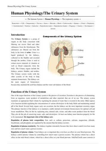
CHAPTER 22393the urinary bladder, which stores the urine until the bladder is emptied. The medial aspect of each kidney is indentedin a region called the hilum, where the ureter, blood vessels,nerves, and lymphatic vessels enter or leave the kidney.Cortical radial arteryand glomeruliArcuate arteryKIDNEY FUNCTIONInterlobararteryPyramidThe Nephron Is the Basic Unit of RenalStructure and FunctionOutermedullaRenalarteryInnermedullaEach human kidney contains about one million nephrons(Fig. 22.2), each of which consists of a renal corpuscle and arenal tubule. The renal corpuscle consists of a tuft of capillaries, the glomerulus, surrounded by Bowman’s capsule.The renal tubule is divided into several segments. The partof the tubule nearest the glomerulus is the proximal tubule.This is subdivided into a proximal convoluted tubule andproximal straight tubule. The straight portion heads towardthe medulla, away from the surface of the kidney. The loopof Henle includes the proximal straight tubule, thin limb,and thick ascending limb. Connecting tubules connect thenext segment, the short distal convoluted tubule, to the collecting duct system. Several nephrons drain into a corticalcollecting duct, which passes into an outer medullary collecting duct. In the inner medulla, inner medullary collecting ducts unite to form large papillary ducts.The collecting ducts perform the same types of functions as the renal tubules, so they are often considered tobe part of the nephron. The collecting ducts and nephronsdiffer, however, in embryological origin, and because thecollecting ducts form a branching system, there are manymore nephrons than collecting ducts. The entire renal tubuleand collecting duct system consists of a single layer of epithelial cells surrounding fluid (urine) in the tubule or duct lumen.PapillaHilumRenalveinPelvisMajor calyxRenalcapsuleSegmentalarteryMinor calyxCortexUreterFIGURE 22.1 The human kidney, sectioned vertically.(Modified from Smith HW. Principles of Renal Physiology. New York:Oxford University Press, 1956.)and covering its sides. The tip of the medullary pyramidforms a renal papilla. Each renal papilla drains its urine intoa minor calyx, The minor calices unite to form a majorcalyx, and the urine then flows into the renal pelvis.Peristaltic movements propel the urine down the ureters toFIGURE 22.2 Components of thenephron and the collecting duct system. Onthe left is a long-looped juxtamedullary nephron;on the right is a superficial cortical nephron.(Modified from Kriz W, Bankir L. A standardnomenclature for structures of the kidney.Am J Physiol tingductOuter imbInner medullaInnermedullarycollectingductAscendingthin limbPapillaryductProximalconvolutedtubuleRenal corpusclecontainingBowman’s capsuleand glomerulusProximal straighttubuleDescendingthin limb
394PART VIRENAL PHYSIOLOGY AND BODY FLUIDSCells in each segment have a characteristic histologicalappearance. Each segment has unique transport properties(discussed later).Not All Nephrons Are AlikeThree groups of nephrons are distinguished, based onthe location of their glomeruli in the cortex: superficial, midcortical, and juxtamedullary nephrons. The juxtamedullarynephrons, whose glomeruli lie in the cortex next to themedulla, comprise about one eighth of the total nephronpopulation. They differ in several ways from the othernephron types: they have a longer loop of Henle, longer thinlimb (both descending and ascending portions), lower renincontent, different tubular permeability and transport properties, and different type of postglomerular blood supply.Figure 22.2 shows superficial and juxtamedullary nephrons;note the long loop of the juxtamedullary ulusOutermedullaArcuateveinAscendingvasa rectaArcuatearteryDescendingvasa rectaInnermedullaThe Kidneys Have a Rich BloodSupply and InnervationEach kidney is typically supplied by a single renal artery,which branches into anterior and posterior divisions,which give rise to a total of five segmental arteries. Thesegmental arteries branch into interlobar arteries, whichpass toward the cortex between the kidney lobes (see Fig.22.1). At the junction of the cortex and medulla, the interlobar arteries branch to form arcuate arteries. These, inturn, give rise to smaller cortical radial arteries, whichpass through the cortex toward the surface of the kidney.Several short, wide, muscular afferent arterioles arisefrom the cortical radial arteries. Each afferent arteriole givesrise to a glomerulus. The glomerular capillaries are followedby an efferent arteriole. The efferent arteriole then dividesinto a second capillary network, the peritubular capillaries, which surround the kidney tubules. Venous vessels,in general, lie parallel to the arterial vessels and have similar names.The blood supply to the medulla is derived from the efferent arterioles of juxtamedullary glomeruli. These vesselsgive rise to two patterns of capillaries: peritubular capillaries, which are similar to those in the cortex, and vasa recta,which are straight, long capillaries (Fig. 22.3). Some vasarecta reach deep into the inner medulla. In the outer medulla,descending and ascending vasa recta are grouped in vascular bundles and are in close contact with each other. Thisarrangement greatly facilitates the exchange of substancesbetween blood flowing into and blood flowing out of themedulla.The kidneys are richly innervated by sympathetic nervefibers, which travel to the kidneys mainly in thoracic spinalnerves X, XI, and XII and lumbar spinal nerve I. Stimulationof sympathetic fibers causes constriction of renal blood vessels and a fall in renal blood flow. Sympathetic nerve fibersalso innervate tubular cells and may cause an increase in Na reabsorption by a direct action on these cells. In addition,stimulation of sympathetic nerves increases the release ofrenin by the kidneys. Afferent (sensory) renal nerves arestimulated by mechanical stretch or by various chemicals inthe renal barveinFrom renal arteryRenalpelvisTo renalveinFIGURE 22.3 The blood vessels in the kidney. Peritubularcapillaries are not shown. (Modified from Kriz W, Bankir L.A standard nomenclature for structures of the kidney.Am J Physiol 1988;254:F1–F8.)Renal lymphatic vessels drain the kidneys, but little isknown about their functions.The Juxtaglomerular Apparatus Is the Site ofRenin ProductionEach nephron forms a loop, and the thick ascending limbtouches the vascular pole of the glomerulus (see Fig. 22.2).At this site is the juxtaglomerular apparatus, a regioncomprising the macula densa, extraglomerular mesangialcells, and granular cells (Fig. 22.4). The macula densa (densespot) consists of densely crowded tubular epithelial cellson the side of the thick ascending limb that faces theglomerular tuft; these cells monitor the composition of thefluid in the tubule lumen at this point. The extraglomerularmesangial cells are continuous with mesangial cells of theglomerulus; they may transmit information from maculadensa cells to the granular cells. The granular cells aremodified vascular smooth muscle cells with an epithelioidappearance, located mainly in the afferent arterioles closeto the glomerulus. These cells synthesize and release renin,a proteolytic enzyme that results in angiotensin formation(see Chapter 23).
CHAPTER 22Macula leNerveExtraglomerularmesangial cellAfferentarterioleGlomerularcapillaryFIGURE 22.4 Histological appearance of the juxtaglomerularapparatus. A cross section through a thick ascending limb is on top, andpart of a glomerulus is below. The juxtaglomerular apparatus consistsof the macula densa, extraglomerular mesangial cells, and granularcells. (Modified from Taugner R, Hackenthal E. The JuxtaglomerularApparatus: Structure and Function. Berlin: Springer, 1989.)AN OVERVIEW OF KIDNEY FUNCTIONThree processes are involved in forming urine: glomerular filtration, tubular reabsorption, and tubular secretion(Fig. 22.5). Glomerular filtration involves the ultrafiltrationof plasma in the glomerulus. The filtrate collects in the urinary space of Bowman’s capsule and then flows downstream through the tubule lumen, where tubular activityalters its composition and volume. Tubular reabsorptioninvolves the transport of substances out of tubular urine.These substances are then returned to the capillary blood,which surrounds the kidney tubules. Reabsorbed substances include many important ions (e.g., Na , K , Ca2 ,Mg2 , Cl , HCO3 , phosphate), water, important metabolites(e.g., glucose, amino acids), and even some waste products(e.g., urea, uric acid). Tubular secretion involves the transport of substances into the tubular urine. For example, manyExcreted Filtered Re absorbed SecretedReabsorptionGlomerulusSecretion(1)The functional state of the kidneys can be evaluated usingseveral tests based on the renal clearance concept (seebelow). These tests measure the rates of glomerular filtration, renal blood flow, and tubular reabsorption or secretion of various substances. Some of these tests, such as themeasurement of glomerular filtration rate, are routinelyused to evaluate kidney function.Renal Clearance Equals Urinary Excretion RateDivided by Plasma ConcentrationA useful way of looking at kidney function is to think of thekidneys as clearing substances from the blood plasma.When a substance is excreted in the urine, a certain volumeof plasma is, in effect, freed (or cleared) of that substance.The renal clearance of a substance can be defined as thevolume of plasma from which that substance is completelyremoved (cleared) per unit time. The clearance formula isCx U x V Px(2)where X is the substance of interest, CX is the clearance ofsubstance X, UX is the urine concentration of substanceX, PX.is the plasma concentration of substanceX,andVistheurine.flow rate. The product of UX times V equals the excretion rateand has dimensions of amount per unit time (e.g., mg/min ormEq/day). The clearance of a substance can easily be determined by measuring the concentrations of a substance inurine and plasma and the urine flow rate (urine volume/timeof collection) and substituting these values into the clearance formula.Inulin Clearance Equals theGlomerular Filtration RateKidney tubuleFiltration395organic anions and cations are taken up by the tubular epithelium from the blood surrounding the tubules and added to thetubular urine. Some substances (e.g., H , ammonia) are produced in the tubular cells and secreted into the tubular urine.The terms reabsorption and secretion indicate movementout of and into tubular urine, respectively. Tubular transport(reabsorption, secretion) may be either active or passive,depending on the particular substance and other conditions.Excretion refers to elimination via the urine. In general,the amount excreted is expressed by the following equation:Bowman’scapsuleMesangial cellKIDNEY FUNCTIONExcretionPeritubular capillaryFIGURE 22.5 Processes involved in urine formation. Thishighly simplified drawing shows a nephron and its associated bloodvessels.An important measurement in the evaluation of kidney function is the glomerular filtration rate (GFR), the rate at whichplasma is filtered by the kidney glomeruli. If we had a substance that was cleared from the plasma only by glomerular filtration, it could be used to measure GFR.The ideal substance to measure GFR is inulin, a fructosepolymer with a molecular weight of about 5,000. Inulin issuitable for measuring GFR for the following reasons: It is freely filterable by the glomeruli. It is not reabsorbed or secreted by the kidney tubules.
396PART VIRENAL PHYSIOLOGY AND BODY FLUIDS It is not synthesized, destroyed, or stored in the kidneys. It is nontoxic. Its concentration in plasma and urine can be determinedby simple analysis.The principle behind the use of inulin is illustrated inFigure 22.6. The amount of inulin (IN) filtered per unit time,the filtered load, is equal to the product of the plasma[inulin] (PIN) timesGFR. The rate of inulin excretion is equal.to UIN times V . Because inulin is not reabsorbed, secreted,synthesized, destroyed, or stored by the kidney tubules,the filtered inulin load equals the rate of inulin excretion.The equation can be rearranged. by dividing by the plasma[inulin]. The expression UIN V /PIN is defined as the inulinclearance. Therefore, inulin clearance equals GFR.Normal values for inulin clearance or GFR (corrected to abody surface area of 1.73 m2) are 110 15 (SD) mL/min foryoung adult women and 125 15 mL/min for young adult men.In newborns, even when corrected for body surface area, GFRis low, about 20 mL/min per 1.73 m2 body surface area. Adultvalues (when corrected for body surface area) are attainedby the end of the first year of life. After the age of 45 to 50,GFR declines, and it is typically reduced by 30% to 40% byage 80.If GFR is 125 mL plasma/min, then the volume of plasma filtered in a day is 180 L (125 mL/min 1,440 min/day). Plasmavolume in a 70-kg young adult man is only about 3 L, so the kidneys filter the plasma some 60 times in a day. The glomerularfiltrate contains essential constituents (salts, water, metabolites), most of which are reabsorbed by the kidney tubules.The Endogenous Creatinine Clearance Is UsedClinically to Estimate Glomerular Filtration RateInulin clearance is the gold standard for measuring GFR andis used whenever highly accurate measurements of GFR aredesired. The clearance of iothalamate, an iodinated organiccompound, also provides a reliable measure of GFR. It is notcommon, however, to use these substances in the clinic. Theymust be infused intravenously and the bladder is usuallycatheterized, because short urine collection periods are used;these procedures are inconvenient. It would be simpler to useFiltered inulinPIN x GFRGFR Excreted inulinUIN x VUINV CINPINFIGURE 22.6 The principle behind the measurement ofglomerular filtration rate (GFR). PIN, plasma [inulin]; UIN, urine[inulin]; V̇, urine flow rate; CIN, inulin clearance.an endogenous substance (i.e., one native to the body) that isonly filtered, is excreted in the urine, and normally has a stable plasma value that can be accurately measured. There isno such known substance, but creatinine comes close.Creatinine is an end product of muscle metabolism, aderivative of muscle creatine phosphate. It is producedcontinuously in the body and is excreted in the urine. Longurine collection periods (e.g., a few hours) can be used,because creatinine concentrations in the plasma are normally stable and creatinine does not have to be infused; consequently, there is no need to catheterize the bladder. Plasmaand urine concentrations can be measured using a simple colorimetric method. The endogenous creatinine clearance iscalculated from the formulaCCREATININE U CREATININE V PCREATININE(3)There are two potential drawbacks to using creatinine to measure GFR. First, creatinine is not only filtered but secreted bythe human kidney. This elevates urinary excretion of creatinine, normally causing a 20% increase in the numerator of theclearance formula. The second drawback is related to errorsin measuring plasma [creatinine]. The colorimetric methodusually used also measures other plasma substances, such asglucose, leading to a 20% increase in the denominator of theclearance formula. Because both numerator and denominatorare 20% too high, the two errors cancel, so the endogenouscreatinine clearance fortuitously affords a good approximation of GFR when it is about normal. When GFR in an adult hasbeen reduced to about 20 mL/min because of renal disease,the endogenous creatinine clearance may overestimate theGFR by as much as 50%. This results from higher plasma creatinine levels and increased tubular secretion of creatinine.Drugs that inhibit tubular secretion of creatinine or elevatedplasma concentrations of chromogenic (color-producing)substances other than creatinine may cause the endogenouscreatinine clearance to underestimate GFR.Plasma Creatinine Concentration Can Be Usedas an Index of Glomerular Filtration RateBecause the kidneys continuously clear creatinine from theplasma by excreting it in the urine, the GFR and plasma [creatinine] are inversely related. Figure 22.7 shows the steadystate relationship between these variables—that is, whencreatinine production and excretion are equal. Halving theGFR from a normal value of 180 L/day to 90 L/day resultsin a doubling of plasma [creatinine] from a normal value of1 mg/dL to 2 mg/dL after a few days. A reduction in GFR from90 L/day to 45 L/day results in a greater increase in plasmacreatinine, from 2 to 4 mg/dL. Figure 22.7 shows that withlow GFR values, small absolute changes in GFR lead to muchgreater changes in plasma [creatinine] than occur at highGFR values.The inverse relationship between GFR and plasma [creatinine] allows the use of plasma or serum [creatinine] as anindex of GFR, provided certain cautions are kept in mind:1. It takes a certain amount of time for changes in GFR toproduce detectable changes in plasma [creatinine].
CHAPTER 22Steady state for creatinineProduced Filtered Excreted161.8 g/day 10 mg/L 180 L/day 1.8 g/day1.8 g/day 20 mg/L 90 L/day 1.8 g/day1.8 g/day 40 mg/L 45 L/day 1.8 g/day1.8 g/day 80 mg/L 22 L/day 1.8 g/dayPlasma [creatinine] (mg/dL)121.8 g/day 160 mg/L 11 L/day 1.8 g/day8KIDNEY FUNCTIONRBF RPF (1 Hematocrit )397( 4)The hematocrit is easily determined by centrifuging a bloodsample. Renal plasma flow is estimated by measuring theclearance of the organic anion p-aminohippurate (PAH),infused intravenously. PAH is filtered and also vigorouslysecreted, so it is nearly completely cleared from all of theplasma flowing through the kidneys. The renal clearanceof PAH, at low plasma PAH levels, approximates the renalplasma flow.The equation for calculating the true value of the renalplasma flow isRPF CPAH E PAH(5)4004590GFR (L/day)135180FIGURE 22.7 The inverse relationship between plasma[creatinine] and glomerular filtration rate (GFR). If GFR is decreasedby half, plasma [creatinine] is doubled when the production andexcretion of creatinine are in balance in a new steady state.where CPAH is the PAH clearance and EPAH is the extractionratio (see Chapter 15) for PAH—the difference between thearterial and renal venous plasma [PAH]s (PaPAH PrvPAH)divided by the arterial plasma [PAH] (PaPAH). The equation isderived as follows. In the steady state, the amounts of PAHper unit time entering and leaving the kidneys are equal. ThePAH is supplied to the kidneys in the arterial plasma andleaves the kidneys in urine and renal venous plasma, or:PAH entering kidneys PAH leaving kidneysRPF Pa PAH U PAH V RPF P rv PAH(6)Rearranging, we get:2. Plasma [creatinine] is also influenced by muscle mass. Ayoung, muscular man will have a higher plasma [creatinine] than an older woman with reduced muscle mass.3. Some drugs inhibit tubular secretion of creatinine, leading to a raised plasma [creatinine] even though GFRmay be unchanged.The relationship between plasma [creatinine] and GFR isone example of how a substance’s plasma concentration candepend on GFR. The same relationship is observed for several other substances whose excretion depends on GFR. Forexample, the plasma [urea] (or blood urea nitrogen [BUN])rises when GFR falls.Several empirical equations have been developed thatallow physicians to estimate GFR from serum creatinineconcentration. These equations often take into considerationsuch factors as age, gender, race, and body size. The equation: GFR (in mL/min per 1.73 m2) 186 (serum [creatinine]in mg/dL) 1.154 (age in years) 0.203 0.742 (if the subject isfemale) or 1.212 (if the subject is black) is recommended bythe National Kidney Disease Education Program, which provides a calculator on its Web site: www.nih.nkdep.gov.Para-Aminohippurate Clearance Nearly EqualsRenal Plasma FlowRenal blood flow (RBF) can be determined from measurements of renal plasma flow (RPF) and blood hematocrit, usingthe following equation:RPF U PAH V ( Pa PAH P rv PAH )(7 )If we divide the numerator and denominator of the rightside of the equation by PaPAH, the numerator becomes CPAHand the denominator becomes EPAH.If we assume extraction of PAH is 100% (EPAH 1.00), thenthe RPF equals the PAH clearance. When this assumption ismade, the renal plasma flow is usually called the effectiverenal plasma flow and the blood flow calculated is called theeffective renal blood flow. However, the extraction of PAH byhealthy kidneys at suitably low plasma PAH concentrations isnot 100% but averages about 91%, so the assumption of 100%extraction results in about a 10% underestimation of the truerenal plasma flow. To calculate the true renal plasma flow orblood flow, it is necessary to sample renal venous blood tomeasure its plasma [PAH], a procedure not often done.Net Tubular Reabsorption or Secretion of aSubstance Can Be Calculated From Filtered andExcreted AmountsThe rate at which the kidney tubules reabsorb a substancecan be calculated if we know how much is filtered and howmuch is excreted per unit time. If the filtered load of a substance exceeds the rate of excretion, the kidney tubulesmust have reabsorbed the substance. The equation isTreabsorbed Px GFR U x V where T is the tubular transport rate.(8)
398PART VIRENAL PHYSIOLOGY AND BODY FLUIDSThe rate at which the kidney tubules secrete a substanceis calculated from this equation:Tsec reted U x V Px GFR(9)Note that the quantity excre
in blood plasma, including Na , K , Ca 2 , Mg , Cl , bicarbonate (HCO 3 ), phosphate, and sulfate. 3. They play an essential role in acid–base balance by excreting H when there is excess acid or HCO 3 when there is excess base. 4. They regulate the volume of the ECF by