Transcription
Modeling cytoskeleton self assemblyDimitrios VavylonisDepartment of Physics, Lehigh UniversityOctober 31, 2008
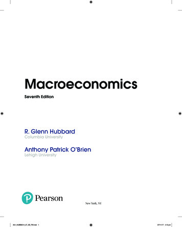
Cell organization How does the cell achieveinternal organization? How is it regulated? Can we model cell structure patterson.htmldynamics?
Thermal and viscous forces are verylarge on the scale of proteinsv protein 9 m/ sect 0t 3 psec 3 10-12 secin this time the protein travels0.027 nm protein size 4 nmThe result of this is diffusion
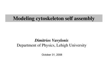
Modeling diffusion: random walk on a latticecomputer picks arandom direction (up/down/left/right)with equal probability at each step(a “Monte Carlo” method)“drunkard’s walk”
Brownian motion, continuedx(t)0 steps100 steps1000 stepsx(t) constant · (D t)1/2Diffusion coefficient D for a protein in the cell 4 μm2/sec5 μmt 0fraction of a secseveral sec
Polymerization of proteins to filamentsactinmonomers D D/2 D/3actin filament cross-links Æ rigid, stationary cytoskeletonKovar, Harris, Mahaffy, Higgs, PollardCell, 124 423 (2006)Alberts et al, MBOC
Microtubule filaments and cell organizationAlberts et al, MBOC
Search and capture of chromosomesby microtubules during mitosisdynamic instabilitydividing cell
GTP-cap model of dynamic instabilityGTP-tubulincapGDP-tubulinAlberts et al, MBOC
GTP-cap “toy model”k chydrolysis at blue/red interface, rate khydroGTP-tubulinGTP-tubulin monomersdiffuse in the cytoplasmat concentration ck-GDP-tubulinmicrotubule filamentkrescuekcatGillespie algorithm (a Monte Carlo method)1. pick an event at random according to rate constants(polymerization, depolymerization, hydrolysis)2. update configuration and time
Numerical simulations of the modellarge GTP-tubulin monomer concentrationsexample of lengthtrajectory at smallconcentrations:subunitssmall GTP-tubulin monomer concentrationsTime (sec)
Insights from simple modelother trajectoriessubunitsaverageelongation rate0time (sec)c*catastropheand rescueregionmonomerconcentration
Models of chromosome search and capture
Cytokinesis5 nm 50 μmPollard and Earnshaw, “Cell Biology” (2002)Ring assembly involves 50 or more protein components.How do protein molecules assemble in a ring?What is the mechanistic pathway?How is it coordinated and controlled?How does the ring exert the required force?actinmonomersactinfilament
Fission yeast is a a model organism for the study of cytokinesisnucleuscell tipcell tiptimecontractileringyeastsbuild rigid cell wallsaround them15 μm
Contractile ring assembles from 63 nodes in 10 minRlc1p-3GFPspinning disk confocal microscopyVavylonis, Wu, Hao, O’Shaughnessy,Pollard, Science 2008 40 myosin II (Myo2p) molecules/node 2 formin Cdc12p dimers/nodeWu and Pollard, Science 2005
radial projection0arc length2πRActin filament meshwork establishes connections among nodesred: nodes (Rlc1-RFP1)green: actin filaments (GFP-CHD)
Lateral contraction modelnodeWu, Sirotkin, Kovar, Lord, Beltzner, Kuhn, Pollard J. Cell Biol. 2006
Model with static connections condenses nodes into clumps!numerical simulationtimeÆ pattern and dynamics of connections is important
Nodes move in stochastic manner, making manystarts, stops and changes of direction15x time lapse10 minuteseach frame:average of 6typical node speedv 30 nm/sectypical durationτ 20 sec
Evidence for dynamic actin meshwork growing out of nodesGFP-CHD (binds actin filaments), Rlc1p-tdTomato (nodes)4x time lapse (0.3 s/frame)single confocal slice
Search, capture, pull and release modelactin filamentpolymerization 0.2 μ m/secactin filament capturerc 100 nmlifetime of connections 20 secF 4 pNtraction on filamentsbetween nodeslifetime of filamentsDynamic reestablishment of connections Æ plasticity of network 20 sec
Simulations with search, capture, pull and releasered: nodesgreen: actinSimulated radial projection030x time lapse,20min model reproduces many observed features2πRexperiment:
5 μmActin Dynamics in Cell MotilityDictyostelium response tochemoattractantTotal internal reflectionfluorescence microscopy(TIRFM)frame-to-frame 1 secmovie lasts 600 secDiez, Gerisch, Anderson, Muller-Taubenberger,Bretschneider PNAS, 102, 7601 (2005)fluorescent LimE-GFP(binds actin filaments)
Dynamic Actin NetworkLamellipodiumSvitkina et alJ. Cell Biol. 139 397 (1997)
Motility of cancer cells causes metastasis
Green Fluorescent Protein (GFP) and Biological ImaginggreenfluorescentspotsAequorea Victoria
GFP can be expressed in a different species and remains fluorescentgenegene fused to GFP geneLive cells become fluorescentThe cell makes its own fluorophoremany more colors:Tsien lab, UCSD1994Chalfie et al. (Columbia) photoconvertible GFP etc.
“drunkard’s walk” Modeling diffusion: random walk on a lattice. Brownian motion, continued Diffusion coefficient D for a protein in the cell 4 μm2/sec 0 steps 100 steps 1000 steps x(t) const