Transcription
Doctorate Program in MolecularOncology and EndocrinologyDoctorate School in MolecularMedicineXXVIII cycle -2012–2015Coordinator: Prof. Massimo Santoro”Epigenetics of adipocyte commitment.Regulation of Pparγ and Zfp423 expression”Federica ZatteraleUniversity of Naples Federico IIDipartimento di Medicina Molecolare e Biotecnologie Mediche
Administrative LocationDipartimento di Medicina Molecolare e Biotecnologie MedicheUniversità degli Studi di Napoli Federico IIPartner Institutions Università degli Studi di Napoli “Federico II”, Naples, Italy Istituto di Endocrinologia ed Oncologia Sperimentale “G. Salvatore”, CNR, Naples, Italy Seconda Università di Napoli, Naples, Italy Università degli Studi di Napoli “Parthenope”, Naples, Italy
FacultyFrancesco BeguinotMaddalena IllarioRoberto BiancoPaolo LaccettiBernadette BiondiAntonio LeonardiFrancesca CarlomagnoPaolo Emidio MacchiaMaria Domenica CastelloneRosa Marina MelilloGabriella CastoriaClaudia MieleAngela CelettiNunzia MontuoriAnnamaria CiraficiRoberto PacelliAnnamaria ColaoGiuseppe PalumboGerolama CondorelliGiovanna Maria PierantoniValentina De FalcoRosario PivonelloVittorio De FranciscisGiuseppe PortellaSabino De PlacidoMaria Fiammetta RomanoGabriella De VitaGiuliana SalvatoreMonica FedeleMassimo SantoroPietro FormisanoDonatella TramontanoAlfredo FuscoGiancarlo TronconeFabrizio GentileGiancarlo VecchioDomenico GriecoMario VitaleMichele Grieco
Epigenetics of adipocytecommitment.Regulation of Pparγ and Zfp423expression”
1
TABLE OF CONTENTSLIST OF PUBLICATIONS .5LIST OF ABBREVIATIONS .6ABSTRACT .81. INTRODUCTION . 101.1 Obesity .101.2 Diabetes 131.3 The relationship between obesity and type 2 diabetes .141.4 Adipose tissue .161.4.1 White adipose tissue as link between obesity and type 2 diabetes.161.4.2 White adipose tissue remodeling in obesity . 181.4.2.1 Hypertrophic obesity: restricted adipogenesis and inflammatoryresponse . 191.4.2.2 Hypertrophic obesity: adipose tissue expandability and ectopicfat accumulation .221.5 Adipocyte differentiation: pro- and anti- adipogenic pathways . 231.6 Epigenetics and adipogenesis . 272. AIM OF STUDY 292
3. MATERIALS AND METHODS .303.1 Materials . 303.2 Cell culture, treatment and adipocyte differentiation . 303.3 RNA isolation and real time RT-PCR . 313.4 Micrococcal nuclease (MNase) protection assay .323.5 Determination of DNA methylation status . 333.6 DNA sequencing by dideoxynucleotide chain terminationmethod 343.7 Luciferase reporter assay and methylation assay in vitro 343.8 Statistical analysis .354. RESULTS .364.1 Zfp423 and Pparγ mRNA expression in 3T3-L1 and NIH-3T3 cells.364.2 DNA Methylation status of Zfp423 and Pparγ promoters in NIH3T3 and 3T3-L1 .374.3 Nucleosome occupancy of Zfp423 and Pparγ promoters in 3T3-L1and NIH-3T3 cells . 394.4 Contribution of DNA methylation to Zfp423 and Pparγ expression.414.5 DNA methyltransferase inhibitor AZA treatment effect onadipogenesis in NIH-3T3 cells .435. DISCUSSION 453
6. CONCLUSIONS 497. ACKNOWLEDGEMENTS .508. REFERENCES . 514
LIST OF PUBLICATIONSThis dissertation is based upon the following publications:Fiory F, Parrillo L, Raciti GA, Zatterale F, Nigro C, Mirra P, Falco R, UlianichL, Di Jeso B, Formisano, Miele C, Beguinot F. PED/PEA-15 inhibits hydrogenperoxide-induced apoptosis in Ins-1E pancreatic beta-cells via PLD-1. PLoSOne. 2014.Luca Parrillo, Valerio Costa, Gregory Raciti, Michele Longo, Rosa Spinelli,Roberta Esposito, Cecilia Nigro, Viviana Vastolo, Antonella Desiderio,Federica Zatterale, Alfredo Ciccodicola, Pietro Formisano, Claudia Miele, andFrancesco Beguinot. Hoxa5 undergoes dynamic DNA methylation andtranscriptional repression in the adipose tissue of mice exposed to high-fatdiet. International Journal of Obesity. 2015.Longo M, Spinelli R, D'Esposito V, Zatterale F, Fiory F, Nigro C, Raciti GA,Miele C, Formisano P, Beguinot F, Di Jeso B. Pathologic endoplasmicreticulum stress induced by glucotoxic insults inhibits adipocyte differentiationand induces an inflammatory phenotype. Biochim Biophys Acta. 2016.5
LIST OF ABBREVIATIONSAPC: Adenomatous polyposis coliAZA: 5-AzacytidineBMI: Body mass indexBmp4: Bone morphogenetic protein 4C/EBPα: CCAAT/ enhancer binding protein αCAD: Coronary artery diseaseCHD: Coronary heart diseaseCK1: Casein kinase ICS: Calf serumCTRL: ControlDEX: DexamethasoneDMEM: Dulbecco's modified Eagle's mediumDnmt: DNA-methyltransferaseFBS: Fetal bovine serumFFAs: Free fatty acidsFZ: FrizzledGlut4: Glucose transporter 4GSK3-β: Glycogen synthase kinase 3-betaHDL: High-density lipoproteinIBMX: 1-Methyl-3-IsobutylxanthineIDF: International diabetes federationLEF/TCF: Lymphoid enhancer factor/ T cell factor6
LPL: Lipoprotein lipaseLRP5/6: Low density lipoprotein receptor-related proteins 5 and 6MIX: Methyl isobutyl xanthineMNase: Micrococcal nucleaseNAFLD: Non-alcoholic fatty liver diseaseNASH: Non-alcoholic steatohepatitisNUC: NucleosomePparγ: Peroxisome proliferator-activated receptor γTSA: Trichostatin ATSS: Transcription start siteUCP-1: Uncoupling protein-1VLDL: Very low density lipoproteinWHO: World health organizationWisp2: Wnt1 inducible signaling pathway protein 2Zfp423: Zinc finger protein 4237
ABSTRACTObesity is the most frequent metabolic disease worldwide and the majorrisk factor for metabolic disorders such as insulin resistance and type 2diabetes. In response to overfeeding, excess lipids are stored in the adipocytes,leading to inappropriate adipose cell expansion (hypertrophic obesity), which isassociated with local inflammation and dysregulated and insulin resistantadipose tissue. Hypertrophic obesity is also characterized by an inability torecruit and differentiate precursor cells into mature adipocytes. This is not dueto lack of these precursor cells but instead is a consequence of an impairedability to induce commitment and promote differentiation of availableprecursor cells, by inactivating inhibitory pathways, and/or activating pathwaysneeded to commit and/or differentiate.Very recently, the zinc finger protein Zfp423 has been identified as atranscriptional regulator of preadipocyte determination. Zfp423 protein, indeed,directly binds the peroxisome proliferator-activated receptor gamma (Pparγ)promoter, thus promoting the gene transcription of the master regulator ofadipocyte differentiation. Furthermore, Zfp423 protein is the point ofconvergence between the anti-adipogenic Wnt1-inducible-signaling pathwayprotein 2 (Wisp2) pathway and the pro-adipogenic bone morphogenetic protein4 (Bmp4) pathway. In light of this, it is now clear that alterations impacting onZfp423 and Pparγ protein and/or gene expressions are at least in partresponsible for the restricted adipogenesis observed in hypertrophic obesity.Increasing evidence sustains that even adipogenesis might be regulated by anepigenetically induced gene transcription reprogramming.Thus, in light of all these observations, I aimed to establish which are themolecular mechanisms regulating the gene expression of Zfp423 and of itsdownstream target Pparγ looking at epigenetic changes involvement.Experiments were performed in two cell models: 3T3-L1 and NIH-3T3 cells,which are committed to and uncommitted to adipocyte lineage, respectively.The mRNA expression of both Zfp423 and Pparγ genes are increased in 3T3L1 compared with NIH-3T3 cells. Furthermore, the expression differencesamong the two cellular models are not dependent on differences in thepromoter DNA sequence of both genes, but rather to epigenetic mechanismsand chromatin remodeling. Indeed, performing methylation studies by bisulfitesequencing and nucleosomes positioning and occupancy analysis bymicrococcal nuclease (MNase) digestion, I revealed that the DNA methylationstatus and the nucleosomes occupancy of Zfp423 and Pparγ promoter regionsare increased in NIH-3T3 compared with 3T3-L1 cells.Finally, chemically-induced demethylation of the Zfp423 and Pparγ promotersby DNA-methyltransferase (Dnmt) inhibitor 5-Azacyditine (AZA) treatmentpromotes adipocyte terminal differentiation in the uncommitted NIH-3T3 cellline.8
In conclusion, I demonstrated that Zfp423 and Pparγ genes, which are involvedin adipocyte commitment and differentiation, are transcriptionally regulated byDNA methylation and dynamic chromatin remodeling, and that the modulationof the methylation status of the promoter region of these two genes is relevantto the regulation of adipocyte commitment and terminal differentiation in vitro.9
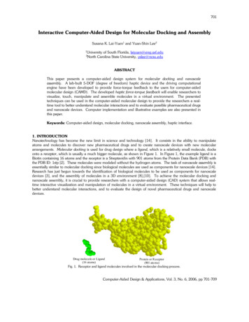
1.INTRODUCTION1.1ObesityOverweight and obesity result from a chronic imbalance betweenenergy intake and energy expenditure, leading to an excessive accumulationof fat in adipose tissue. The current classification of overweight and obesity inadults is based on the body mass index (BMI). BMI, indeed, is an indirectmeasure of body fat, described as the person's weight in kilograms, divided bythe square of height in meters (kg/m2) (World Health Organization 2014).Thus, according to World Health Organization (WHO) classification (WorldHealth Organization 2014), overweight is defined as a BMI of 25 or more andincludes pre-obesity (25-29.9) and obesity that is divided into threecategories: Class I (30–34.9), Class II (35–39.9) and Class III ( 40) (James etal. 2004) (Table 1).ClassificationUnderweightNormal rangeOverweightPre-obeseObese IObese IIObese IIIBMI (Kg/m2 ) 18.518.5-24.9 2525-29.930-34.935-39.9 40Table 1. Classification of adult underweight, overweight and obesity according to BMI.According to WHO, overweight is defined as a BMI equal to or more than 25 whereas obesity asa BMI equal to or more than 30.The incidence of obesity and overweight is increasing dramatically in westernsociety; it was estimated that the worldwide prevalence of obesity has doubledbetween 1980 and 2014. WHO (World Health Organization 2014) estimatesthat the 38% of males and 40% of women were overweight while the 11% ofadult males and the 15% of adult females were obese in 2014 (Figure 1).10
Figure 1. Prevalence of obesity in adulthood. The maps indicate the adult prevalence ofobesity respectively in males and females (World Health Organization 2014).Obesity is not a single disorder but a heterogeneous disease with multiplecauses. Body weight, indeed, is regulated by a complex interaction betweengenetic, societal, environmental, and political influences (Frood et al. 2013).On the genetic level, many evidence has shown that predisposing genes cancontribute significantly to the development of obesity (Silventoinen et al. 2009;Silventoinen et al. 2010) by interacting with environmental factors such asphysical inactivity and unhealthy diet (Choquet and Meyre 2011).Twin studies suggest a heritability of fat mass of between 40% and 70% with aconcordance rate of 0.7-0.9 between monozygotic twins compared to 0.35-0.45among dizygotic twins (Stunkard et al. 1986). Furthermore, it has been alsoseen that if one’s parent is obese, the own risk to develop obesity significantlyincreased compared with age and sex-matched individuals without familiarityof obesity, and if obesity is present during the early childhood, the individualchances to be obese throughout his/her own life are high compared with theirlean counterparts.11
In addition to genetics, also societal changes that modify individual behaviours,leading to a more sedentary life (Bauman et al. 2011), physical inactivity(Hansen et al. 2012), and poor dieting (Caraher et al. 2010; Perez-Escamilla etal. 2012), may also contribute to the increase of obesity incidence (Swinburn etal. 2011).Many evidence suggests a strong correlation between BMI and the incidence ofseveral chronic diseases, such as cancer, hypertension, coronary heart disease(CHD), respiratory complications and diabetes mellitus (Figure 2).This relationship is approximately linear for a range of BMI less than 30, butthe risk to develop the obesity-related comorbidities greatly increase forsubjects with a BMI above 29, independently of gender (Willett et al. 1995;Willett et al. 1999). The development of these complications is strictlycorrelated with the increase of body fat that impairs the physiologicalfunctions. Indeed, an increased amount of fat results in alterations of totalblood volume and cardiac function, whereas the distribution of fat aroundthoracic cage and abdomen impairs respiratory function by the reduction ofrespiratory compliance. Moreover, intra-abdominal visceral deposition ofadipose tissue plays an important role in the development of hypertension,insulin resistance, hyperlipidemia and diabetes mellitus (Kopelman 2000).Figure 2. Representation of the most important comorbidities associated with obesity.Obesity has a multitude of adverse metabolic health consequences such as diabetes mellitus,hypertension, dyslipidemia, and metabolic syndrome (Kopelman 2000).12
1.2DiabetesDiabetes mellitus is a group of chronic metabolic disorderscharacterized by elevated blood glucose levels (hyperglycemia) caused bydefects in insulin secretion, insulin action or both (American DiabetesAssociation 2010).The vast bulk of cases of diabetes falls into two main etiopathogeneticcategories (American Diabetes Association 2010).Type 1 diabetes occurs predominantly in young people and is characterized byan absolute deficiency of insulin, resulting from an autoimmune mediateddestruction of β cells. Its frequency is low relative to type 2 diabetes, whichaccounts for over 90% of cases globally, and requires a daily injection ofinsulin.Type 2 diabetes is the most common form of diabetes mellitus and ischaracterized by increased levels of blood glucose to which individuals arepredisposed by an interaction between genetic, social and environmental riskfactors (Khodabandehloo et al. 2016).In type 2 diabetes, the hyperglycemia is initially due to a decreased insulinsensitivity, as a result of insulin resistance, which leads to hyperinsulinemia.Later on, when the pancreatic β cells are not capable of producing the amountof insulin needed to maintain normal glycemic status, chronic hyperglycemiaand type 2 diabetes occur (Kahn S et al. 2006).Primarily strategy for type 2 diabetes treatment consists of an individualizedapproach, consisting initially of dietary control and lifestyle modifications,followed by oral type 2 diabetes medications and insulin therapy (AmericanDiabetes Association 2015).The worldwide rapid increasing prevalence of type 2 diabetes is one of themajor and serious public health burden (Tabish 2007). Type 2 diabetes reachedan epidemic stage in line with the increasing obesity prevalence and has beenparticularly accelerated in the developing countries.According to the International Diabetes Federation Atlas (InternationalDiabetes Federation Atlas 2013), there are 382 million people in the worldliving with diabetes, defined as a fasting plasma glucose value 7.0 mmol/L(126 mg/dl) or 2-h plasma glucose 11.1 mmol/l (200 mg/dl) after a load ofglucose (World Health Organization 2006), and it is expected to afflict around592 million people by 2035 (International Diabetes Federation Atlas 2013)(Figure 3). This rise is largely driven by modifiable risk factors, such asphysical inactivity, overweight and diet high in fat and salt (World HealthOrganization 2010).13
Figure 3. Prevalence of diabetes in the world. The IDF Atlas (International DiabetesFederation Atlas 2013) figure provides a worrying indication of the future impact of diabetes onthe global development.Diabetes and its complications are the major cause of morbidity and mortalityaround the world and result in higher health care costs.Diabetes is the well-recognised cause of premature death and disability and ispowerfully linked with both microvascular and macrovascular complications.Microvascular complications affect small blood vessel and include damage tothe nervous system (neuropathy), renal system (nephropathy) and eye(retinopathy) while macrovascular complications comprise coronary arterydisease (CAD), stroke and myocardial infarction (World Health Organization2010). Thus, looking at the rapid growth in the number of type 2 diabetics, theevolution and clinical implementation of effective psychosocial interventionsare needed (Debono and Cachia 2007).1.3The relationship between obesity and type 2 diabetesAs previously mentioned, the worldwide increase in the prevalence ofobesity largely explains the epidemic of type 2 diabetes over the past twentyyears (Eckel et al. 2011). According to Ford and colleagues (Ford et al. 1997),the risk to develop diabetes increases between 4.5 and 9% for every kilogramof weight increase.In 1995, the worldwide obese population was calculated to be around 200million, while the subjects considered obese, in 2014, are 600 million.14
Strikingly similar trends have been observed in type 2 diabetes statistics, andfuture predictions are alarming (King et al. 1998).For this reason, Sims and colleagues (Sims et al. 1973), coined the term‘diabesity’ to explain the close relationship existing between these twodiseases. Obesity and type 2 diabetes often occur together and the risk todevelop type 2 diabetes for a BMI over 35, compared to a BMI less than 23,increases by 50-80 fold (Figure 4) (Chan et al. 1994).Figure 4. BMI and type 2 diabetes risk. The relationship between BMI and risk to developtype 2 diabetes, derived from data extracted from Chang and colleagues (Chan et al. 1994).Therefore, excess weight is the primary risk factor for type 2 diabetes.Nevertheless, obesity is not a homogeneous condition. Indeed, approximately10-30% of obese individuals do not show the associated metaboliccomplications and, for this reason, are defined as metabolically healthyindividuals (Kloting et al. 2010). Nevertheless, a similar number of non-obeseindividuals show a dysmetabolic state and reduced insulin sensitivity (Scott etal. 2014). Thus, BMI per se is not a sufficiently sensitive marker of individualrisk for obesity-related metabolic complications.Conversely, increasing evidence suggests that adipose tissue distribution mightbe a more relevant marker for obesity-related metabolic complications. Indeed,abdominal distribution markedly increases both cardiovascular and diabetesrisk (Fox et al. 2009; Despres and Lemieux 2006).This finding has raised much interest in the possible role of regionaldifferences in adipose tissue metabolism and, in particular, in the role of intraabdominal and visceral fat in metabolic complications (Després and Lemieux2006; Neeland et al. 2012).15
1.4Adipose tissueAdipose tissue is a complex organ with heavy effects on physiology andpathophysiology. For many years, adipose tissue has been ignored because wasconsidered just as a form of connective tissue, containing lipid droplets butwithout any link with the metabolism of the organisms (Rosen and Spiegelman2014; Rosen and Spiegelman 2006). Nevertheless, this notion gradually beganto shift with the realization that adipose tissue takes on a critical role in energyhomeostasis and in the regulation of metabolic functions.Adipose tissue is indeed a metabolically active tissue, consisting of lipid-filledcells, called adipocytes, that make up the 90% of the tissue volume (Lee et al.2010). In addition, to mature adipocytes, fat tissue contains severalmetabolically active and inflammatory cells, including stromal vascular cellssuch as fibroblasts, leukocytes, macrophages, pericytes, endothelial cells, andpreadipocytes, which take on a significant part in the integrity of tissue (Katz2002).The mammalian adipose tissue is divided into two functionally distinct formsof fat: white and brown. White adipose tissue is the primary site of energystorage (Ronti et al. 2006; Rosen and Spiegelman 2006) but can act also as athermal insulator and protect other organs from mechanical damage (Trayhurn2007). Brown adipose tissue, on the other hand, is highly specialized in thedissipation of stored chemical energy by the generation of heat by the actionsof mitochondrial uncoupling protein-1 (UCP-1), a brown adipose tissuespecific protein located within the mitochondria (Rosen and Spiegelman 2014;Rosen and Spiegelman 2006). Nevertheless, the concept of two different formsof adipose tissue has been altered after the recognition that brown fat cellsmight be interspersed in the white adipose tissue to form “beige cells”(Boström et al. 2012). A recent study has shown that the fully stimulatedbrown and beige adipocytes contain comparable amounts of UCP-1, suggestingthat they have a similar thermogenic capacity (Wu et al. 2012).1.4.1 White adipose tissue as link between obesity and type 2 diabetesAs above mentioned, the main functions of white adipose tissue are: i)to store calories in the form of lipid droplets after food intake; ii) to release fatduring the fasting state; iii) to cushion the body parts and to protect delicateorgans from mechanical stress.Furthermore, white adipose tissue plays a critical role in the energy balanceand nutritional homeostasis (Rosen and Spiegelman 2014).In humans, white adipose tissue constitutes the largest endocrine organ and iscertainly so in overweight and obese individuals. Therefore, even minormetabolic changes in such a large secretory organ can impact on the wholebody metabolism (Trayhurn 2007).16
White adipocytes in addition to store fatty acids, produce and release also awide range of lipid, hormones and cytokines (adipokines) such as leptin,interleukin 6, adiponectin, monocyte chemoattractant protein 1 and tumournecrosis factor alpha, that are required for many biological functions as glucoseand lipid metabolism, insulin sensitivity, inflammation, coagulation, bloodpressure and feeding behaviour (Van Gaal et al. 2006; Müller et al. 2012;Wildman et al. 2008; Klein et al. 2004) (Figure 5).Figure 5. Adipose Tissue as an endocrine organ. Adipose tissue is an endocrine organ thatsecretes various hormones and adipokines involved in the many physiological and metabolicprocesses (Trayhurn 2013).The white adipose organ includes multiple discrete locations (Lee et al. 2014;Shen et al. 2003). The most common classification distinguishes betweensubcutaneous and visceral fat (Figure 6) that are characterized by functionaldifferences. Subcutaneous adipose tissues store more than 80% of total bodyfat in the body and is located beneath the skin; while visceral fat is aroundinternal organs and comprises less that 10% of the total fat mass.Being involved with its expansion in the development of obesity, white adiposetissue is now seen as the epicenter of the disorder and its associatedcomorbidities (Greenberg and Obin 2006).Many studies have, indeed, shown that total adiposity is strongly associatedwith metabolism and cardiovascular risk. More, it has been also determinedthat the various fat depots contribute differently to this risk (Hassan et al.2012). Indeed, the accumulation of visceral fat seems to correlate with anincreased prevalence of insulin resistance, metabolic syndrome, andcardiovascular complications (Lemieux et al. 1996) whereas the fataccumulation in the peripheral subcutaneous adipose tissue does not seem to beassociated with many of these obesity-related comorbidities (Porter et al.2009).17
A key role in the regional variation in fat tissue function is also played both byregional differences in preadipocytes characteristics (replication,differentiation, subtype abundance, susceptibility to apoptosis or senescenceand gene expression) and by some aspects of adipocyte behavior (adipokinesecretion, rates of lipolysis and triglyceride synthesis) (Tchkonia et al. 2010;Tchkonia et al. 2013).Figure 6. Representation of the anatomical localization of the main abdominal adiposetissue depots. There are two major types of fatty tissue in the body: visceral and subcutaneousadipose tissue (Lee et al. 2014; Shen et al. 2003).1.4.2 White adipose tissue remodeling in obesityWhite adipose tissue is characterized by an incredible ability to changeits dimension (Rosen and MacDougald 2006; Rosen and Spiegelman 2014;Rosen and Spiegelman 2006), in response to nutrient excess or insufficiency(Martinez-Santibañez and Nien-Kai Lumeng 2014); this is essential for themaintenance of whole-body energy homeostasis.White adipose tissue expansion is primarily achieved i) by increasing the sizeof individual adipocytes (hypertrophy), or ii) by recruiting new adipocytesfrom the resident pool of progenitors (hyperplasia) (Rosen and Spiegelman2014).In response to overfeeding, adipose depots expand first by hypertrophy, until acritical threshold 0.7-0.8 µg/cell) is reached (Krotkiewski et al. 1983).As shown by several studies, the balance between these two mechanismsdepends on the location of the fat pad. However, in humans, the mechanisms ofadipose tissue expansion during obesity are not fully delineated yet.Anyway, many evidence suggests that overfeeding for several months inducesan increase in cell size (Salans et al. 1971). A late version of this studysuggests that acute excess of calories induces hypertrophy in the upper bodysubcutaneous fat and hyperplasia in the fat depots located below the waist(Tchoukalova et al. 2010).18
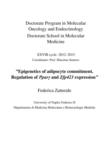
More recently, Spalding and colleagues (Spalding et al. 2008), shown that“new adipocytes are generated constantly to replace lost adipocytes” andestimated that the half-life of the average adipocyte is in the order of 8.3 years(Sun et al. 2011). Moreover, it has been demonstrated that the number ofadipocytes becomes already fixed in childhood and early adulthood (Sun et al.2011; Spalding et al. 2008) and that significant weight loss is associated onlywith reduction of adipocyte volume and not of their number.The last suggests that hypertrophy of adipocytes is the most significantmechanism by which white adipose tissue mainly undergoes alterations duringadulthood (Spalding et al. 2008; Sun et al. 2011).Nevertheless, as stated previously, the mechanisms by which adipose tissueexpands in humans in obesity are not fully defined, and the patterns of fatdeposition and expansion vary between people. This variance in adipose tissuedistribution appears to determine the differences found in metabolic diseaserisk seen among individuals with a similar level of adiposity.Many evidence suggests that in obese individuals, the metabolic dysfunction isrelated to large adipocytes, and is associated with the impaired ability todevelop new cells (Hammarstedt et al. 2012; Isakson et al. 2009).On the contrary, others indicate that an overabundance of small adipocytes isassociated with a more unhealthy metabolic phenotype (McLaughlin et al.2010; McLaughlin et al. 2007). In both instances, the final result is the same.Indeed, the ability of the white adipose tissue to act as an efficient storagedepot is impaired, inducing other peripheral tissues to compensate byaccumulating inappropriately lipids.Thus, the impairment in the ability of white adipose tissue to generate newcells through differentiation of preadipocytes is associated with thedevelopment of insulin resistance and diabetes mellitus (Karastergiou andMohamed-Ali 2010) and the enlargement of adipocytes (hypertrophy) iscorrelated with death of adipocytes by necrosis, leading to local inflammationand recruitment of monocytes as a response (Strissel et al. 2007; Cinti et al.2005).1.4.2.1 Hypertrophic obesity: restricted adipogenesis and inflammatoryresponseAs above mentioned, white adipocyte hypertrophy is characterized bythe presence of large, lipid-laden adipocytes and may result from anintrinsically reduced capacity of de novo adipogenesis (Klöting and Blüher2014).In addition to the last observations, Smith and colleagues, analysingadipogenesis in stromal vascular cells from human abdominal subcutaneousdepots (Gustafson and Smith 2012), have found poor differentiation inindividuals with large subcutaneous adipose cells (hypertrophic obesity),whereas small adipose cells were associated with good adipogenesis,19
suggesting thus a causal relationship between restricted adipogenesis andadipose cell volume (Figure 7).Furthermore, these differences were not due to lack of precursor cells inhypertrophic obesity, but rather to the inappropriate signaling of pathwayswhich promotes precursor cell differentiation and/or which enhances inhibitorysignals promoting dedifferentiation (Talchai et al. 2012).Unlike to hyperplastic obesity, many human studies have demonstrated thatadipocyte hypertrophy is negatively correlated with the impairment of glucoseand lipid metabolism and with the loss of insulin sensitivity, independently bythe grade of obesity measured as BMI (Smith and Hammarstedt 2010; Blüher2013; Klöting and Blüher 2014) and, plays a key function as an independentpredictor of future type 2 diabetes onset (Lönn et al. 2010).Figure 7. The distinction of human preadipocytes from the abdominal subcutaneousadipose tissue is concerned to the adipose cell size of the donors. Purified stromalvascular cells were differentiated for 21 days in vitro and then stained with Oil Red O to showthe lipid droplets (top).The negative correlation between the amount of lipids accumulatedduring differentiation and the adipose cell size of the donors is shown (bottom) (Gustafson andSmith 2012).Bluher and colleagues (Klöting and Blüher 2014), have furthermoredemonstrated that adipocyte volume is associated
Luca Parrillo, Valerio Costa, Gregory Raciti, Michele Longo, Rosa Spinelli, . physical inactivity and unhealthy diet (Choquet and Meyre 2011). Twin studies suggest a heritability of fat mas