Transcription
Epigenetic Silencing of Ankrd26 Gene Contributes toAdipose Tissue Inflammation in Obesity
TABLE OF CONTENTSPageLIST OF PUBBLICATIONS1ABSTRACT314BACKGROUND1.1 Obesity41.2 Biology and pathophysiology of obesity71.2.1 Adipose Tissue71.2.2 Genetic of obesiy121.2.3 Epigenetics: gene-environment interactions in obesity151.3 Epigenetics191.3.1 Epigenetic modifications201.3.2 DNA Methylation221.3.3 DNA Methylation and obesity241.4 ANKRD26 gene1.4.1 ANKRD26 as a candidate (epi)gene for obesity27292 AIMS OF THE STUDY323 MATERIALS AND METHODS333.1 Animal study, diet protocols and metabolic tests333.2 Western blotting analysis333.3 Real Time PCR (qPCR)343.4 Methylated DNA Immunoprecipitation (MeDIP)343.5 Bisulfite sequencing353.6 Cloning strategy, site-direct mutagenesis and in vitro methylation353.7 Luciferase assay353.8 Chromatin Immunoprecipitation and Micrococcal Nucleaseassays363.9 Electrophoretic mobility shift assay (EMSA)363.10 Primer sequences37
3.11 Cell culture and transfection383.12 Fatty Acid/BSA complex solution preparation393.13 Patient enrollment and tests393.14 Statistical analysis404 RESULTS414.1 Animal study design414.2 Metabolic characteristics of standard and high fat diet mice414.3 Effect of HFD on Ankrd26 gene expression in visceral adiposetissue424.4 Effect of HFD on DNA methylation at Ankrd26 promoter454.5 Role of free fatty acids on epigenetic regulation of Ankrd26 gene474.6 Methylation of specific CpG sites controls Ankrd26 geneexpression4.7 DNA methylation suppresses Ankrd26 promoter activity byimpairing the binding of co-activator/acetyltransferase p3004.8 HFD-induced differential recruitment of DNMTs and MBD2initiates the repression and silencing of Ankrd26 gene4.9 HFD increases nucleosome occupancy at the Ankrd26 promoter4.10 HFD changes histone acetylation and RNA Pol II binding at theAnkrd26 promoter4.11 Ankrd26 silencing induces a pro-inflammatory cytokine profilein cultured adipocytes4.12 ANKRD26 expression negatively correlates with BMI andinflammatory markers in obese subjects484952535455575 DISCUSSION596 CONCLUSION627 REFERENCES63ACKNOWLEDGEMENTS71
LIST OF PUBLICATIONS(Years 2014-2017)The Thesis is based on the following publications:1.Raciti GA*, Spinelli R*, Desiderio A, Longo M, Parrillo L, Nigro C,D'Esposito V, Mirra P, Fiory F, Pilone V, Forestieri P, Formisano P, Pastan I,Miele C, Beguinot F. Specific CpG hyper-methylation leads to Ankrd26 genedown-regulation in white adipose tissue of a mouse model of diet-induced obesity.Sci Rep. 2017 Mar 7;7:43526. doi: 10.1038/srep43526. *The authors Theseauthors contributed equally to this work.2.Desiderio A, Spinelli R, Ciccarelli M, Nigro C, Miele C, Beguinot F,Raciti GA. Epigenetics: spotlight on type 2 diabetes and obesity. J EndocrinolInvest. 2016 Oct;39(10):1095-103. doi: 10.1007/s40618-016-0473-1.3.Parrillo L, Costa V, Raciti GA, Longo M, Spinelli R, Esposito R, NigroC, Vastolo V, Desiderio A, Zatterale F, Ciccodicola A, Formisano P, Miele C,Beguinot F. Hoxa5 undergoes dynamic DNA methylation and transcriptionalrepression in the adipose tissue of mice exposed to high-fat diet. Int J Obes (Lond).2016 Jun;40(6):929-37. doi: 10.1038/ijo.2016.36.Other publications:4.Longo M, Raciti GA, Zatterale F, Parrillo L, Desiderio A, Spinelli R,Hammarstedt A, Hedjazifar S, Hoffmann JM, Nigro C, Mirra P, Fiory F,Formisano P, Miele C, Smith U, Beguinot F. Epigenetic modifications of theZNF423 gene control adipogenic commitment and are dysregulated in murineadipocytes and human hypertrophic obesity.5.Fiory F, Spinelli R, Raciti GA, Parrillo L, D'esposito V, Formisano P,Miele C, Beguinot F. Targetting PED/PEA-15 for diabetes treatment. Expert OpinTher Targets. 2017 Jun;21(6):571-581. doi: 10.1080/14728222.2017.1317749.Epub 2017 Apr 21. Review. PubMed PMID: 28395542. Accepted for publicationin Diabetologia.6.Longo M*, Spinelli R*, D'Esposito V, Zatterale F, Fiory F, Nigro C,Raciti GA, Miele C, Formisano P, Beguinot F, Di Jeso B. Pathologic endoplasmicreticulum stress induced by glucotoxic insults inhibits adipocyte differentiationand induces an inflammatory phenotype. Biochim Biophys Acta. 2016 Jun;1863(6PtA):1146-56. doi: 10.1016/j.bbamcr.2016.02.019. *The authors These authorscontributed equally to this work.1
7.D'Esposito V, Liguoro D, Ambrosio MR, Collina F, Cantile M, SpinelliR, Raciti GA, Miele C, Valentino R, Campiglia P, De Laurentiis M, Di Bonito M,Botti G, Franco R, Beguinot F, Formisano P. Adipose microenvironment promotestriple negative breast cancer cell invasiveness and dissemination by producingCCL5. Oncotarget. 2016 Apr 26;7(17):24495-509. doi: 10.18632/oncotarget.8336.8.Borriello F, Longo M, Spinelli R, Pecoraro A, Granata F, Staiano RI,Loffredo S, Spadaro G, Beguinot F, Schroeder J, Marone G. IL-3 synergises withbasophil-derived IL-4 and IL-13 to promote the alternative activation of humanmonocytes. Eur J Immunol. 2015 Jul;45(7):2042-51. doi: 10.1002/eji.201445303.2
ABSTRACTEnvironmental factors interact with the genome to influence gene expression,tissue function and also disease risk. External stimuli may affect the phenotypethrough epigenetic mechanisms that provide an interface with the genome. Indeed,epigenetic modifications represent a mechanism through which both genetic andenvironmental cues, including dietary factors, are integrated at specific genomicloci and affect individual phenotypes, contributing to susceptibility to obesity andtype 2 diabetes. Recently, alterations of Ankyrin repeat domain 26 (Ankrd26) geneexpression and function have been associated with the onset of these metabolicdisorders. However, whether external cues can affect its expression remainsunclear. Therefore, the aim of this study is to evaluate whether the administrationof a high fat diet (HFD) in mice could affect Ankrd26 expression and function, andwhether chromatin remodeling and epigenetic modifications take part in thisregulation. Particularly, I examined the correlation between obesity andinflammation with defective regulation of Ankrd26 gene in visceral adipose tissue(VAT) depots, aiming at identifying epigenetic mechanism that might underpin thedevelopment of VAT dysfunctions induced by overnutrition. Using the HFDinduced obesity mouse model, I reported evidence of detailed epigenetic changesof Ankrd26 promoter. Indeed, I demonstrated that the HFD-induced Ankrd26down-regulation in VAT was directly caused by specific hyper-methylation of 436 and -431 cytosine residues at its promoter region, that was followed bychromatin reorganization. Indeed, DNA methylation of these specific CpG sitesimpaired binding of the histone acetyltransferase/transcriptional coactivator p300to this region, both in vitro and in vivo, causing hypo-acetylation of histone H4 atthe Ankrd26 promoter and loss of binding of RNA Pol II at the Ankrd26Transcription Start Site in obese mice. Furthermore, the HFD treatment increasedbinding of DNA methyl-transferases 3a and 3b and methyl-CpG-binding domainprotein 2 to the Ankrd26 promoter. To evaluate the functional consequences ofthese changes, the HFD-induced Ankrd26 down-regulation was mimicked bysilencing Ankrd26 gene expression in vitro in 3T3-L1 adipocytes. This silencingcaused enhanced secretion of the pro-inflammatory chemokines KC/IL-8, Eotaxin,MCP1 and Rantes. The relevance of these observations to humans is supported byother findings in obese individuals, revealing that the reduction of ANKRD26expression in VAT negatively correlates with serum concentrations ofinflammatory markers. Taken together, my data provide evidence forenvironmentally induced DNA changes at Ankrd26 promoter and, for the first time,highlight a role for Ankrd26 epigenetic silencing in raising and/or sustaining VATinflammation following unhealthy dieting and in development of obesity-relatedinsulin resistance. Since direct evidence linking specific environmental cues andmetabolic disorders are still limited, addressing this issue will provide new insightinto the molecular basis of obesity as well as create novel translational perspective.3
1. BACKGROUND1.1ObesityObesity, considered by the World Health Organization (WHO) as a 21st centuryglobal epidemic [1], is defined as excessive adiposity in relation to lean massthroughout the body, that is usually accompanied by mild, chronic, systemicinflammation [2]. The current classification of overweight and obesity in adults isbased on the body mass index (BMI) [3,4]. BMI is an indirect measure of body fat,equals an individual's weight in kilograms, divided by the square of height inmeters (kg/m2). According to WHO classification [3], overweight is defined as aBMI of 25 or more, and includes pre-obesity (25-29.9), and obesity that is dividedinto three categories: Class I (30–34.9), Class II (35–39.9) and Class III ( 40) [5].High BMI is an important risk factor for cardiovascular and kidney diseases,diabetes, some cancers, and musculoskeletal disorders. Concerns about the healthand economic burden of increasing BMI have led to adiposity being includedamong the global non-communicable diseases, with a target of halting, by 2025,the rise in the prevalence of obesity at its 2010 level [1].Increased fat deposition, according to a simplistic view based on the first law ofthermodynamics, results from an imbalance between the caloric intake and energyexpenditure, driven by the consumption of high-energy-yielding foods above theneeds of the individual and a sedentary lifestyle [6]. However, the etiology ofobesity is more complex. Indeed, circumstances such as socioeconomic status,environment and personal behaviors, and genotype–phenotype interactions haveto be taken into account to understand obesity, as all these factors affect foodintake, nutrient turnover, thermogenesis, lipid utilization, and also differential fatstorage in regional adipose depots versus non-adipose tissues [2]. Multiple andcomplex mechanisms have evolved to regulate energy intake and expenditure andfat deposition to maintain body weight [7]. Several cues affect these regulatoryprocesses: environmental factors, diet quality, level of physical activity, the gutmicrobiota, endocrine disruptors, drugs, reproductive factors and assortativemating, intrauterine and epigenetic intergenerational effects (fig. 1) [2]. Anotherproblem concerning the obesity study and management is the markedheterogeneity of individuals with obesity. Examples of obesity types aresubcutaneous obesity, in which excess subcutaneous fat is found around the hipand thigh areas, and visceral obesity, in which fat is mainly accumulated in theabdominal region. Visceral obesity is more common in men and tends to be morepernicious in health terms [2]. However, different strategies and treatments at theindividual level have been developed and prescribed: dietary education andcontrol, physical activity programs, pharmacological treatments and bariatricsurgery.4
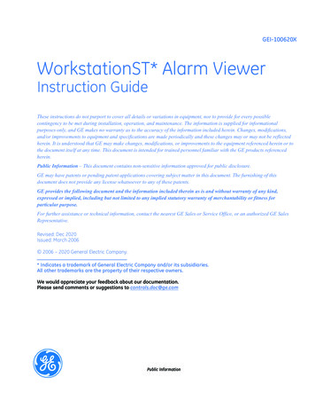
Figure 1. Key factors involved in the regulation of energy balance. The energy balance isinfluenced by several biological factors. The pyramid holding the balance emphasizes the notionthat we need to go beyond individual factors to ultimately have an optimal effect on the energybalance equation. Nat Rev Dis Primers. 2017; 3:17034.Actually, 39% of the world population is obese or overweight, despite decadesof efforts to slow the progress of the epidemy. According to the WHO, 2.1 billionadults were overweight or obese globally in 2014, of which 1.5 billion wereoverweight and 640 million were obese [3]. In the same year, the age-standardizedprevalence of obesity in 2014 was estimated to 10.8% among adult men and 14.9%among adult women. These data suggest that female sex is associated with higherrisk of obesity [1]. Furthermore, several studies conducted in the United States,indicate that African Americans exhibited a higher prevalence of extreme obesitythan other ethnicities [8]. Asian populations have lower BMI than whiteindividuals, but they have been shown to be prone to visceral fat deposition,making this populations more susceptible to developing T2D at lower BMI thanwhite individuals [9]. Although the prevalence of obesity is likely to increase asthe population ages dramatically in coming years [10], in 2013–2014, theworldwide number of children and adolescents with obesity has doubled since1980 and was estimated to be 110 million [2]. Childhood and adolescence obesityhas been associated with metabolic complications and chronic disease inadulthood.Critical windows in the onset of obesity have been identified in the prenatalperiod, infancy, childhood and adolescence. The programming of metabolismduring the prenatal and neonatal periods at both the genomic and epigenomic level(metabolic imprinting) might permanently influence future disease risk and health,according to the developmental origins of health and disease hypothesis [11,12].Indeed, it has been shown in literature that the type and amount of energy-yieldingnutrients consumed by the mother during pregnancy and lactation, and even byboth parents before pregnancy, is associated with the development of metabolic5
complications in adulthood [13,14]. Also, an earlier adiposity during childhoodhas been associated with a high BMI, a high amount of subcutaneous adipose tissueand a high waist circumference in adulthood [15].State‑of‑the-art epidemiological approaches, such as integrated bioinformaticssystem analyses, need to be used to better understand the causes of obesity,including the complex interplay between behavioral, environmental,physiological, genetic, social and economic factors.Several biological and environmental factors have an effect on energy intake andexpenditure (fig. 1). For example, eating behaviors are determined not only bygenetic factors and other biological factors but also by the opportunities to eat andfood availability. The food choices among energy-yielding food is enormous.Physical activity, another behavioral component, is also conditioned bysocioeconomic and cultural factors. An increasing trend toward sedentarylifestyles coupled with increases in energy intake is able to explain the observedenhancement in mean BMI in high-income countries, but also probably in mostlow- and middle-income countries [16]. In addition to the amount of calories, thetype and quality calorie, such as saturated versus non-saturated lipids, as well asthe source of fats, carbohydrates and proteins also affects the energy balance andbody weight [17]. However, this area is still controversial; indeed, whether thecaloric overload with the diet or the impact of a particular nutrient by a directmetabolic effect, is important in obesity development, is unclear [18]. A highquality dietary intake. defined by a balanced consumption of macronutrients, isinversely associated with weight gain and the risk of developing obesity. Severallines of evidence support the importance and benefits of consuming health mealsand high-quality, sustainable dietary regimens in preventing obesity [19,20].Obesity prevention should focus on maintaining weight loss or controllingexcessive weight gain. In general, the preventive strategies are health promotionprograms or marketing, addressing lifestyle behaviors and policies that target theenvironment. At the population level, public policies and economical strategies areneeded to improve food and physical environments, the food system and the healthsystem to control the global obesity epidemic. The complexity of obesity requiresmultilevel approaches by acting on multiple frameworks: in practice, going fromthe individual, often children or adolescents, schools or occupational environmentsand the communities. Drugs, such as amphetamines, methamphetamine andphenmetrazine, are approved by the US FDA for short-term treatment (usually 12weeks) of the patient with obesity for the treatment of obese patients, as adjunctsto diet and exercise [21]. Use of bariatric surgery has become rapidly adopted forthe treatment of severe obesity, and this has increased with the reduction oflaparoscopic procedure risk [21]. Several studies have reported improvements inbiochemical and metabolic outcomes following bariatric surgery. Mortality wasreduced by 24%, mainly by reducing the risk of myocardial infarction and, inwomen, cancer, and many other comorbidities, such as T2D are also ameliorated[22].To improve prevention and treatment strategies, a better knowledge of factors6
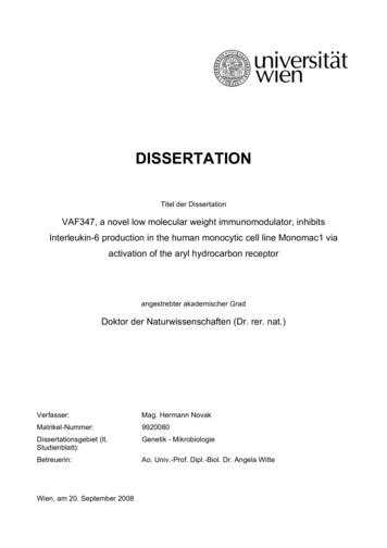
contributing to the development of obesity is essential. A deeper understanding ofappetite and energy expenditure, as well as the physiopathological role of adiposetissue in the regulation of metabolic function is needed. In this regard, the role ofgenetics, epigenetics, nutrigenomics or personalized nutrition is crucial to be betterunderstood to improve obesity management.1.2Biology and pathophysiology of obesityBeing a complex and heritable disorder, obesity results from the interplaybetween genetic susceptibility, epigenetics, metagenomics and the environment.These factors act through physiological mediators of food intake and energyexpenditure, affecting fat deposition [2].1.2.1 Adipose TissueAdipose tissue is a complex organ with profound effects on physiology andpathophysiology. This tissue has a central role in the regulation of energy balanceand nutritional homeostasis, serving as site of calorie storage after feeding and assource of circulating mediators, by which critical metabolic processes arecoordinated locally and systemically [23]. The mammalian adipose tissues havebeen divided into two major types: the white adipose tissue (WAT) and brownadipose tissue (BAT), that are functionally involved in sensing and responding tochanges in systemic energy balance [2,23]. Brown adipocytes contribute to energyexpenditure via thermogenesis to maintain body temperature. BAT is the site ofadaptative thermogenesis, providing extra heat in hibernating animals, coldexposed mammals and human newborns (cold-induced thermogenesis), as well asmaintaining energy balance in response to diet (diet-induced thermogenesis). Thebrown fat depots are abundant in newborn babies and are located in theinterscapular and supraclavicular regions, as well as around the several organs;these depots decrease with age, but can still be found in adults [24,25]. AlthoughBAT has been negatively correlated with the BMI [25], its role in the etiology ofobesity is still unclear. White adipocytes are the most abundant adipocytes inhumans. The main functions of WAT are: i. to store calories as lipid droplets afterfeeding; ii. to release free fatty acids during fasting; and, iii. to cushion body partsexposed to high levels of mechanical stress [23]. However, recent research hasrevealed that white adipocytes synthetize and secrete various bioactive mediators,hormones and cytokines (adipocytokines) such as leptin, interleukin 6 (IL6),adiponectin, monocyte chemoattractant protein 1 (MCP1) and tumor necrosisfactor alpha (TNFα), required for the regulation of metabolic processes, such asglucose and lipid metabolism, insulin sensitivity, feeding behavior, inflammation,immunity, angiogenesis and blood pressure (fig. 2) [23,26].7
Figure 2. Physiological and metabolic processes regulated by adipocyte-secreted mediators.The interactions may be autocrine, paracrine, or endocrine. Physiol Rev. 2013; 93(1):1-21.Intracellular WAT lipid homeostasis is a key determinant of body weight andinsulin sensitivity both in mice and humans. Excessive lipid load causes adipocytestress, which in turn accounts for many adverse effects of obesity, particularlyalterations in adipocytokine release and a low-grade inflammatory response,ultimately leading to the development of metabolic dysfunction such as insulinresistance and glucose intolerance. Due to their critical role in the maintenance ofproper adipocyte function, both storage and release of lipids in WAT arephysiologically under tight hormonal control [27].WAT develops in multiple discrete and specific depots [23]. Based on theiranatomic locations, the most common classification distinguishes betweensubcutaneous adipose tissue (SAT) and visceral adipose tissue (VAT) [2,23]. SATaccumulates more than 80% of total body fat in the body and is located beneaththe skin. VAT include intra-abdominal (mainly mesenteric adipose tissue),perirenal and pericardial adipose tissue and comprises less than 10% of the totalfat mass. However, the visceral versus subcutaneous scheme is oversimplified. Inaddition, many depots in human have no precise correlation in mice, an vice versa.A large percentage of VAT in humans is contained in the omentum, which isscarcely present in rodents; conversely, the large epidydimal adipose tissue (eAT),representative of VAT in mice, does not exist in humans [23].Subcutaneous and visceral adipose depots are very different in terms of theireffects on metabolism [28]. In mice, transplantation of subcutaneous fat into thevisceral depot of recipient mice caused improvements in glucose homeostasis aswell as decreased body weight and fat mass; conversely, placing visceral fat into asubcutaneous depot of recipient mice, had very little effect [29]. In human,peripheral obesity is characterized by an accumulation of SAT and is more frequentin women. This type of obesity is not associated with an increased risk of relatedpathologies. However, central or abdominal obesity is more common in men andconsists of an accumulation of VAT [28]. This type of obesity has been associated,through epidemiological studies, with a higher risk of diseases such as insulin8
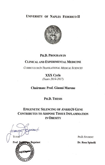
resistance, T2D and hypertension [28], and is closely correlates with thedevelopment of the various metabolic abnormalities, commonly referred to as themetabolic syndrome [2].Adipose tissue has the unique attribute of remodeling, by changing itsdimensions in response to nutritional demands. Adipose tissue remodeling can beaccomplished by increasing the size of cells (hypertrophy) or by recruiting newadipocytes from the resident pool of precursor cells (hyperplasia). In response tocaloric overload, adipose depots expand first by hypertrophy until a criticalthreshold is reached. The hypertrophy is followed by cell death and by secretionof several mediators, that increase the proliferation and/or differentiation ofpreadipocytes [23]. This cycling between hypertrophy and hyperplasia as obesityprogresses is supported by rodent studies indicating that eAT, along with othermurine VAT depots, responds to high-fat diet (HFD) feeding through differenttime-dependent changes, contributing to the inflammatory and metaboliccomplications in obesity [30,31]. It has been demonstrated that upon short-termHFD exposure (8-12 weeks), the eAT expansion is accompanied by hypertrophy.However, after a more prolonged exposure (20 weeks), the eAT is characterizedby an increased rate of adipocyte dying (up to 80%), paralleled by enhancedadipogenesis, so that the fat mass continues to expand as obesity progresses[23,30,31]. Emerging evidence in literature indicates a role for themicroenvironment and the transient local inflammation in triggering the adiposetissue remodeling in the context of hyperplastic expansion [30]. Remarkably, theobservation that obesity can be associated with adipocyte hyperplasia has beensupported by findings showing that manipulation of several genes causes obesityin vivo and increases adipogenesis in vitro. The increased adipogenesis is not theprimer driver of obesity in these in vivo models: the overnutrition is the culprit andthe adipogenesis is induced by the need to store excess calories (fig.3) [23].9
Figure 3. Obesity in light of the adipose tissue expandability hypothesis. Obesity is caused byadipose tissue dysfunction due to insufficient storage capacity, and this results in dyslipidemia,systemic inflammation and altered adipokine profiles. Combination of these factors promotes thedevelopment of insulin resistance and the metabolic syndrome, which further impairs adipose tissuefunction, creating a futile cycle. HIF, hypoxia-inducible factor; IL-6, interleukin-6; TNF-a, tumornecrosis factor-alpha; CRP, C-reactive protein. Adapted from EMBO J. 2017; 36(14):1999-2017.Although it is difficult to translate findings from rodents when examiningvisceral (epidydimal) and subcutaneous (inguinal) fat, consistent evidence inhumans shows that the number and severity of health complications, associatedwith obesity, seem to be dependent on the additional presence of excess fat storedin the VAT depots and in and around lean tissues, such as the heart, the liver andthe kidneys (ectopic fat deposition) [23,32]. Also in humans, the number ofadipocytes depends on the balance between cell death and adipogenesis. Adipocyteprogenitors have been recently identified in vivo as immature mesenchymalstromal cells associated with the vasculature. The turnover rate in adult humans10
has been estimated at 10% per year and, in any case, the size of fat depots correlateswith the number of adipocytes [33]. Importantly, obese individuals have highertotal adipocyte numbers, and this becomes evident already in childhood obesity[34]. Human studies focusing on adipose tissue cellularity have demonstrated amarked fat depot-specificity of the hyperplastic response to the diet. Indeed,increased adipocyte number has been detected in the femoral subcutaneous fatdepot in humans but not in the upper abdominal subcutaneous depot [35]. As abovementioned, in male mice, higher rate of proliferation/adipogenesis has been shownin the intraabdominal gonadal fat depot but not in the posterior subcutaneous depot[30,31,33]. These differences can have substantial impact on the systemicadaptation of metabolism given the pathophysiological relevance of the individualdepots. Accumulation of fat in the intra-abdominal/visceral depots is a centralfeature of the metabolic syndrome and is associated with insulin resistance andincreased risk for diabetes and cardiovascular disease. Conversely, highersubcutaneous fat mass is associated with improved metabolic parameters includinginsulin sensitivity and blood lipid profiles. Taken together, location specifichyperplastic expansion of adipose tissue can affect body fat distribution, contributeto systemic metabolic homeostasis and influence pathogenesis [33]. Furthermore,Lotta et al. [36] have identified a genetic association of 53 loci with traits relatedto insulin resistance, T2D and coronary heart disease. Remarkably, geneticpredisposition for insulin resistance predicted by these loci was associated withreduced fat deposition in peripheral “metabolically safe” depots. Moreover, thesame study revealed common links to insulin resistance associated withlipodystrophy, highlighting the importance of adipose tissue function formaintenance of systemic homeostasis [36].Taken together, these observations emphasize the role of adipose tissue depotsas key checkpoints in systemic energy homeostasis. The way by adipose tissuedepots manages the excess calories and the oxygen tension generated by thepathological growth of the adipocytes impacts on individual’ s cardiometabolicrisk, insulin sensitivity and dyslipidemia [32,37]. When the SAT responds toenergy excess by fat cell hyperplasia, acting as a ‘metabolic sink’ , lean tissues areprotected against ectopic fat deposition. Conversely, if SAT cannot properlyexpand through hyperplasia, the stored triglycerides drive adipocyte hypertrophyuntil these enlarged fat cells become saturated, leading to either death andmacrophage invasion and/or increased release of pro-inflammatory adipokines.These phenomena determine a pro-inflammatory and insulin-resistant milieu ininner adipose depots [35]. Furthermore, the excess triglyceride molecules, thathave no place in the SAT, will be ectopically stored contributing to an atherogenic,diabetogenic and inflammatory environment in the cell [23,37]. By comparison,increased risk of metabolic dysfunction, as obesity, is associated with expansionof the omentum and increased deposition of fat in the liver, heart and kidneys(fig.4) [38]. However, more complex scenarios are also plausible, because geneticand environmental factors may influence the individual’ s regional adipose tissueaccumulation capacity.11
Figure 4. Pathological changes in adipose tissue. The value of visceral fat area correlated withthe risk of obesity-associated complications. Adipose tissues in subcutaneous fat obesity mightfunction normally with the expected release of anti-inflammatory adipokines, whereas adiposetissues in visceral fat obesity is increased the secretion of pro-inflammatory adipokines andsuppressed the secretion of anti-inflammatory adipocytokines, raising and sustaining the low-gradeinflammation, which contributes to obesity-related disorders. Pathological changes in visceraladipose tissue show higher levels of adipocyte necrosis and the recruitment of macrophages withan inflammatory phenotype (M1 macrophages). IL-6, interleukin-6; TNF, tumour necrosis factor;PAI1, prothrombin activator inhibitor 1. Adapted from Nat Rev Dis Primers. 2017; 3:17034.1.2.2 Genetic of obesityGenetic factors contribute to obesity-susceptibility, with heritability estimatesranging between 40 and 70% across populations [2,39,40]. The evidence forgenetic component of BMI/overall obesity is substantial and come from a varietyof population, family and twin-based studies [41]. In 1894, for the first time, SirFrancis Galton reported the familial aggregation of body size [42]. Since then,family history of obesity has become a well-established risk factor for childhoodobesity [43]. The ethnic dependent pattern of obesity prevalence further supportsan important contribution of genes in obesity heritability [43]. Furthermore, twinstudies have shown that monozygotic twins display more similar body fatacquisition in comparison with dizygotic twins, indicating that this trait is highlyin
1. Raciti GA*, Spinelli R*, Desiderio A, Longo M, Parrillo L, Nigro C, D'Esposito V, Mirra P, Fiory F, Pilone V, Forestieri P, Formisano P, Pastan I, Miele C, Beguinot F. Specific CpG hyper-methylation leads to Ankrd26 gene down-regulation in white adipose tissue of