Transcription
A Patient’s Guide toProton RadiosurgeryNortheast ProtonTherapy CenterMassachusettsGeneral HospitalBoston,MassachusettsH EALTHC ARE
NORTHEAST PROTON THERAPY CENTERThe Northeast Proton Therapy Center (NPTC) has beendesigned specifically to provide proton treatments in ahospital setting with state-of-the-art technology and a fullrange of patient and research support services.The centeris used for both cancer therapy research and for treatmentsalready proven to be effective.The NPTC opened inNovember 2001 and when fully operational will have thecapacity to treat 1,000 patients per year.The program builds on the MGH experience gained usingthe 160 MeV proton beam at the Harvard CyclotronLaboratory (HCL), Harvard University. There, MGH physiciansand physics personnel, along with a clinical support team,collaborated to treat both benign and malignant diseasefrom 1961 to the closing of HCL in April 2002. During thatperiod 9,116 patients were treated.Therapy Center (NPTC) hasbeen designed specifically toprovide proton treatmentsin a hospital setting withstate-of-the-art technologyand a full range of patientand research supportservices.Northeast Proton Therapy CenterThe Northeast Proton1
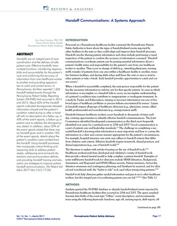
HISTORYIn 1961, MGH physicians from the Department ofNeurosurgery began treating patients with the protonbeam at HCL using a method known as stereotacticradiosurgery (treatment delivered in one or two high-precisionsessions). By 1993, about 3,000 patients with a variety ofbrain tumors and vascular malformations had been treatedby proton radiosurgery.At that time the Departments ofNeurosurgery and Radiation Oncology collaborated toinaugurate an improved system of radiosurgical treatmentknown as STAR (StereotacticAlignment for Radiosurgery). Bythe time HCL closed its doors in2002, almost 800 patients had beentreated using the STAR system.With the opening of NPTC, the STARprogram is being transferred to thenew facility.In 1974, MGH physicians from theDepartment of Radiation Oncologytreated the first patient usingfractionated therapy (treatmentdelivered in 10 to 46 precision sessions). In 1975,physicians from the MGH and the Massachusetts Eyeand Ear Infirmary, treated the first ocular melanoma patient.From 1971-75, the National Science Foundation fundedthe development of techniques required to treat large targetvolumes and the treatment of the first few patients withfractionated proton therapy. The National Cancer Institutefunded the development of fractionated proton therapyfrom 1971-75 and has funded clinical research from 1974to the present time.At HCL the relatively low proton energy and the fixedhorizontal beams, which required rotating patients to aimthe proton beam from various directions limited the typesof patient treatments that could be delivered. However,the success of the HCL program led to the building of adedicated proton therapy facility, the NPTC, on the maincampus of MGH.
NORTHEAST PROTON THERAPY CENTERThe construction costs of the NPTC were jointly fundedby the National Cancer Institute and the MGH eachcontributing 20 million. MGH, through its DevelopmentOffice, has been encouraged by efforts to recover its 20 million contribution and is seeking funds from loyalfriends of the hospital for the remaining amount.The NPTC has many advantages over HCL. The maximumproton energy at the NPTC is 230 MeV, resulting in a maximumtissue penetration of 32 cm. This allows tumors in most partsof the body to be treated. The proton beam at NPTC can bedelivered by a rotating gantry, which allows the radiation to bedirected toward the patient from any angle, a technique thatgreatly improves the ease and versatility of treatments. Thereare two such gantry treatment rooms, and a total of four treatmentlocations. This makes it more possible to meet the large andincreasing medical demand for proton treatments. Becausethe NPTC is located on the MGH campus, patients and theirfamilies have easy access to all hospital support services. TheNPTC will become an integral part of the Yawkey Center forOutpatient Care, now under construction.RADIOSURGERYRadiosurgery may be done as an alternative to conventionalneurosurgery for certain types of medical conditions. In othercircumstances it may be used in conjunction with fractionatedradiotherapy and/or surgery. The determining factors are thenature of the disease, its location, and its extent. Brain tumorscommonly treated by radiosurgery include meningiomas,acoustic neuromas, and pituitary adenomas, as well as avariety of malignant tumors including gliomas and metastases.Vascular abnormalities of the brain, especially arteriovenousmalformations, are also frequently treated.NORTHEAST PROTON THERAPY CENTERRadiosurgery is a procedure that uses a neurosurgicaltechnology known as stereotaxis to precisely aim anintense dose of radiation into a targeted abnormality, suchas a brain tumor or vascular malformation. This is done sothat the radiation dose to normal tissues surrounding thetarget is minimized. Radiosurgical treatments are typicallyperformed in one or two sessions.3
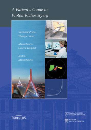
Radiosurgery can be performed with any type of beam ofionizing radiation. This includes photon beams such as gammarays and x-rays, as well as particle beams, which includeprotons. When gamma or x-rays are directed at tissue, theradiation dose received by that tissue is most intense nearthe point of penetration. Progressively less radiation isdelivered to the tissue as the beam passes more deeplyinto it. When protons are directed at tissue, the radiationdose gradually increases as the beam passes more deeply,then drops to near zero beyond the target depth.In orderto deliver a high dose of radiation to a target deep withinthe brain or other organ while sparing thesurrounding tissues from the same radiation dose,it may be necessary to aim the beam at the targetfrom multiple directions, thus focusing an intensespot of radiation on the target. This also allows theradiation dose to conform more closely to the margins ofthe target.PROTON RADIOSURGERYSmall targets that tend to be spherical can be effectivelytreated using gamma or x-ray radiosurgery. However, withlarger and more irregularly shaped targets, it becomesincreasingly difficult to deliver a uniform dose of radiationwithin the target and spare surrounding normal tissues. In
NORTHEAST PROTON THERAPY CENTERthis circumstance, the unique characteristics of protonradiation are a significant advantage for performing radiosurgery.Protons have a physical advantage over x-rays and gammarays when it comes to sparing normal tissue. Protonsdeposit most of their radiation energy in what is knownas the Bragg peak, which occurs at the point of greatestpenetration of the protons in tissue. The exact depth towhich protons penetrate and at which the Bragg peakoccurs is dependent on the energy of the beam. Thisenergy can be very precisely controlled to cause theBragg peak to fall within the tumor or other tissue that istargeted to receive the radiation dose. Because the protonsare absorbed at this point, normal tissues beyond the targetreceive no radiation.In order to further reduce the amount of radiation receivedby normal tissues in the path of the proton beam, beamsare aimed at the target from multiple directions. For eachof these directions special devices called apertures arefabricated to shape the radiation to the target profile.Other devices called compensators control the radiationdepth within tissue. These devices are custom designed tofurther help each beam conform to the unique shape of thetarget volume.MGH / ers.orgNeurosurgeryOffice of Dr. Paul Chapman (617) 726-3887Secretary: Sylvia Weldhttp://neurosurgery.mgh.harvard.edu/Radiation OncologyOffice of Dr. Jay Loeffler(617) 724-1548Nurses: Patricia McManusEna Chang(617) 726-0922(617) 726-0923http://cancer.mgh.harvard.edu/cancer home.htmNortheast Proton Therapy CenterGENERAL INFORMATION5
REGISTRATIONAll MGH outpatients must have a current blue hospital cardfor hospital services. You may get one at Patient Registrationin the Cox Building Lobby or on the first floor of the WangBuilding next to the outpatient pharmacy, Monday throughFriday from 8:00 am to 4:30 pm. Please arrive 15-30 minutesprior to your scheduled appointment if you need to registerfor a blue card. Remember to bring all your insurance cardswith you. You may also register in advance by phone bycalling Patient Registration at (617)726-9090 or toll free at(877)726-9090. Please remember to bring your blue hospitalcard for all visits.When you arrive, please check in with the receptionist.You will find it helpful to bring a date book or calendar tokeep track of your appointments and tests. We also encourageyou to bring someone with you.
NORTHEAST PROTON THERAPY CENTERRESTRICTIONSUnless otherwise advised, none of the procedures leadingto your treatment will require you to restrict your diet.INITIAL CONSULT/REVIEWA senior neurosurgeon and radiation oncologist reviewseach case. These physicians will assess your medical andtreatment history, medications and prior imaging studies todetermine the best treatment course for your particularcondition. Further diagnostic imaging such as MRI or angiographymay be required. You will have the opportunity to meet withthese physicians to discuss their recommendations.SKULL MARKERS (FIDUCIALS)Depending on the location of the treatment site within theskull, three or four tiny (1mm diameter) stainless steel beadsmay be implanted in the surface of your skull using a smallneedle and local anesthesia. The procedure is done on anoutpatient basis and takes about 20 minutes. These markersallow us to aim the proton beam very precisely at thetarget when you are treated. The alignment process usessophisticated computer technology similar to that used byglobal positioning satellites. As part of the pre-treatmentwork-up, a complete blood test will also be done.ANDCT SCANAfter the skull markers have been placed, a CT scan willbe performed, usually on the same day. The purpose of theCT scan is to carefully identify the area to be treated andto create a precise three-dimensional picture, which includesthe skull with the implanted markers, the brain, and thelesion to be treated. This becomes the framework for makingthe necessary radiation dose calculations and customdesigning the treatment to be given. At the time of the CTscan, you will also have a dental mold and head cup fabricated.These immobilization devices are used to hold your headstill during the CT scan and later at the time of your treatment.Please let us know about any concerns or discomforts,since the immobilization devices being made will be usedfor your CT imaging and treatment.Northeast Proton Therapy CenterHEAD IMMOBILIZATION7
TREATMENT PLANNINGThe physicians will use the CT scan, in addition to otherstudies you have had, to outline the target and to noteimportant brain structures. The size of the target, as wellas its relationship to these structures, is critical in calculatingthe prescribed radiation dose. It also determines the directionsfrom which the proton beam will be aimed through thebrain to the target. The medical physicist will now design thetreatment plan, which is reviewed once again by the physicians.Once the treatment plan has been finalized, customizedequipment is fabricated which shapes the proton beam sothat the radiation dose matches as closely as possible theshape of the target. This equipment is designed for eachdirection from which the beam will be aimed.CONSENTPrior to the treatment a physician will discuss the detailsof your treatmentwith you. The physician will remind youabout the potential risks and benefits of the treatment. Youwill need to sign a consent form, which says that thesedetails have been reviewed with you. Please feel free toask any questions and express any concerns you may haveat this time.TREATMENTTreatment does not involve any invasive procedures. Thehead frame with its dental mold, which was fitted at thetime of your CT scan, will again be strapped to your headin a manner which is as comfortable as possible. You will thenbe positioned for treatment with the head frame secured,which will help you keep your head as still as possible duringthe treatment. X-rays will be taken to locate the skull markersand the bony anatomy. Based on your position, adjustmentswill be made to bring the lesion we are treating into theproper alignment in relation to the proton beam. The beamwill then be turned on to deliver the precise radiation dose.This process is repeated two to four times in order to aimthe beam at the target from a number of different directions.
NORTHEAST PROTON THERAPY CENTEREach direction from which the beam is aimed is called a field.The time from the initial x-ray to treatment of the first fieldis usually 15 minutes. Setting up and treating each fieldthereafter takes about 10 minutes per field. The total timerequired for a typical treatment is about one hour.During the treatment you will not feel the radiation. Althoughthe head frame is fixed to the bed and you are required toremain as quiet as possible, medical personnel will be inthe room with you much of the time. They will be very alertto your needs throughout the treatment. We ask that youdo not speak once the procedure has started, since yourpositioning relies on the dental mold and any motion willprolong the procedure. You will be given a hand-held buzzerto signal any problems.FOLLOW-UPAfter the treatment, your physician will discuss your furthercare with you, including any immediate precautions and follow-uprecommendations. You will be provided with the necessaryinstructions in the event of further symptoms related to yourillness and/or treatment.Northeast Proton Therapy Center9
Northeast Proton Therapy CenterMassachusetts General HospitalFruit StreetBoston, MA 02114Brochure prepared by Marc Bussiére, MSc, DABR
as the Bragg peak, which occurs at the point of gr eatest penetration of the protons in tissue. The exact depth to which protons penetrate and at which the Bragg peak occurs is dependent on the energy of the beam. This energy can be very precisely controlled to cause the Bragg