Transcription
llReviewMechanics of DevelopmentKatharine Goodwin1 and Celeste M. Nelson2,3,*1Lewis-SiglerInstitute for Integrative Genomics, Princeton University, Princeton, NJ 08544, USAof Chemical and Biological Engineering, Princeton University, Princeton, NJ 08544, USA3Department of Molecular Biology, Princeton University, Princeton, NJ 08544, USA*Correspondence: vcel.2020.11.0252DepartmentSUMMARYMechanical forces are integral to development—from the earliest stages of embryogenesis to the construction and differentiation of complex organs. Advances in imaging and biophysical tools have allowed us todelve into the developmental mechanobiology of increasingly complex organs and organisms. Here, wefocus on recent work that highlights the diversity and importance of mechanical influences during morphogenesis. Developing tissues experience intrinsic mechanical signals from active forces and changes to tissuemechanical properties as well as extrinsic mechanical signals, including constraint and compression,pressure, and shear forces. Finally, we suggest promising avenues for future work in this rapidly expandingfield.INTRODUCTIONMuch of developmental biology has sought to uncover the rolesof morphogens and the molecular programs that drive development (Briscoe and Small, 2015; Freeman and Gurdon, 2002; Kicheva and Briscoe, 2015). More recently, attention has turned tostudying how mechanical forces influence developing tissuesand understanding the physical mechanisms of morphogenesis(Anlaş and Nelson, 2018; Mammoto and Ingber, 2010; StookeVaughan and Campàs, 2018). These two schools of thoughtare not necessarily separate; indeed, local mechanical properties and their effects on tissue topology can shape morphogenfields (Manning et al., 2015; Shyer et al., 2015), and cell displacements can change how a tissue experiences a static moleculargradient (Nerurkar et al., 2019).Mechanical forces influence all stages of development, fromthe early embryo, to gastrulation and establishment of thebody plan, to organogenesis. Pioneering reviews laid the foundation for our current appreciation of the role of mechanicalforces in development (Beloussov et al., 1994; Keller et al.,2003; Wolpert, 1971), and over the past 10 years, advances inanimal models, live-imaging approaches, and biophysical measurements have shed light on the mechanics of morphogenesis(Ayad et al., 2019). In particular, these advances have allowed usto extend principles of developmental mechanobiology thatwere first established primarily in invertebrate embryos (Heisenberg and Bellaı̈che, 2013; Lecuit and Lenne, 2007; Mammotoand Ingber, 2010) to the morphogenesis of larger, more complex,and less accessible organs and organisms.Here, we provide an overview of different kinds of mechanicalforces that drive morphogenesis and highlight recent progress inunderstanding how they regulate developmental processes—from the earliest stages of embryogenesis to the finishingtouches of organogenesis. We include examples of intrinsic mechanics driving morphogenesis, including the generation of240 Developmental Cell 56, January 25, 2021 ª 2020 Elsevier Inc.active forces and tuning of tissue fluidity, as well as examplesof extrinsic mechanical signals, such as physical constraintsand microenvironment mechanics, static and dynamic pressures, and shear forces from fluid flow. In this review, we focuson how mechanical forces directly influence tissue form. However, it is important to note that mechanical forces also influenceanother key aspect of development—cellular differentiation, inwhich physical forces activate intracellular signaling cascadesor change nuclear envelope mechanics to regulate the activationor nuclear localization of mechanosensitive transcription factors,leading to downstream changes in gene expression and alteringcell behavior and fate (Hampoelz and Lecuit, 2011; Kumar et al.,2017; Vining and Mooney, 2017). These changes at the nuclearlevel could indirectly influence the material properties of the tissue and thus changes in tissue form.Tension and Active ForcesActomyosin networks coupled to cell-cell and cell-extracellularmatrix (ECM) adhesions are responsible for generating the forcesrequired for cells to pull on their neighbors or on their underlyingsubstratum (Hoffman et al., 2011). Actomyosin contractions canlocally constrict parts of the cell, as in apical constriction, in orderto change cell shape or allow it to exert traction in order to moveover a surface, as in cell migration. These active forces at the levelof single cells can be translated into dramatic changes in shape atthe tissue level. Actomyosin networks often contract with pulsatiledynamics. Indeed, pulsed actomyosin contractions are an evolutionarily conserved mechanism for driving tissue morphogenesis.These contractions promote ventral-furrow ingression inDrosophila (Martin et al., 2009), compaction in the mouse blastocyst (Maı̂tre et al., 2015), and have even been observed in collectives of unicellular organisms which form sheets that can fold inresponse to light (Brunet et al., 2019).Active forces generated by actomyosin networks orchestratecell-shape changes required for transforming the early mouse
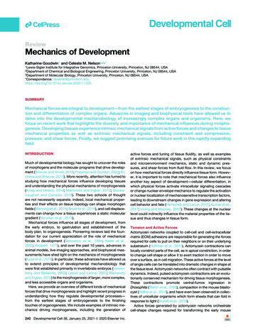
llReviewθcmcell-mediuminterfacial tensionθccθcccell-cell interfacial tensionadherens andtight & pullingAMLPmyosin contractility& actin polymerizationDmigration* * ** * *blastodermdeep EVLcellstensioncell area*P hindgut*Vθcccell-mediumcell-cellactive sealingmyosinpullingtime ( F-actinexpandθcmtensionCtensionintensitytime ( 102 s)passive expansionBcompactionangleAposition along AP axisyolk cellcell divisions & loss ofcell-cell junction integrityfluidizationEVL expandsdeepcellscompressedextracellularspaceFigure 1. Intrinsic Mechanical Forces Driving Morphogenesis(A) During compaction of the 8-cell embryo, tension at the cell-medium interface increases, changing the angles between cells. The increase in tension isaccompanied by pulsatile actin dynamics at the cell cortex.(B) Apical actin rings in the 16-cell mouse embryo expand toward intercellular junctions to seal the blastocyst.(C) Hindgut morphogenesis in the chick embryo requires the gut endoderm to fold over onto itself to form the gut tube. This movement is generated by collectivecell migration resulting from a gradient of cell contractility that interacts with a stable morphogen gradient to drive tissue flow.(D) Deep cells fluidize at the onset of zebrafish epiboly as they undergo divisions and lose integrity of their cell-cell contacts. As a result, the expanding EVLcompresses the deep-cell layer to drive blastoderm thinning.embryo from an agglomeration of roughly spherical cells into acompacted embryo and then into a hollow blastocyst. As the embryo compacts, the angle between adjacent cells at the peripheryof the embryo increases and the surface tension at the cell-medium interface increases (Figure 1A; Table 1) (Maı̂tre et al., 2015).Consistently, actomyosin activity is enriched at the cell-mediuminterface compared with cell-cell interfaces, and the increase intension at the cell-medium interface requires actin polymerizationand myosin activity. Strikingly, this increase in tension is cell autonomous (occurs even in dissociated cells) and is accompanied byoscillatory cell deformations and actin accumulation; whether theoscillations themselves are required for compaction has yet tobe shown. Cells of the 8-cell embryo also extend cadherin-containing filopodia over their neighbors that deform the apical partof the cell to drive compaction (Fierro-González et al., 2013).From the 8-cell stage to the 16-cell stage, a subset of cellsends up in the center of the embryo. These inner cells give riseto the embryonic tissues and the primitive endoderm, whereasthe outer cells (the trophectoderm) give rise to extraembryonictissues that encapsulate the embryo. Inner cells are positionedby apical constriction (Maı̂tre et al., 2016; Samarage et al.,2015). Myosin is enriched at constricting intercellular junctions,and laser ablation revealed more recoil (a proxy for cortical tension) along junctions between constricting cells and their neighbors than between non-constricting cells. Furthermore, ablations oriented perpendicular to junctions between constrictingcells caused more recoil of the cortical actin network than ablations parallel to junctions, demonstrating that the apical cortex ofconstricting cells is under greater tension. Specification andpositioning of trophectoderm cells may also be mechanicallyregulated—keratin-rich intermediate filaments stabilize F-actinwithin the apical domain and additionally serve as an asymmetrically inherited structural component that predicts trophectoderm fate (Lim et al., 2020).During the next major phase of mouse embryogenesis (16- to32-cell stages), the embryo transforms from a solid mass of cells(called the morula) to a fluid-filled blastocyst with an inner cellmass (ICM) and an outer layer of cells called the trophoblast.During this transition, the embryo undergoes additional actomyosin-dependent remodeling in order to seal junctions betweenthe outer cells and to facilitate the formation of a fluid-filled cavity— called the ‘‘blastocoel’’—within the center of the embryo(Zenker et al., 2018). In the apical (outward-facing) domains ofthe cells of the 16-cell embryo, actin rings assemble andpassively expand until they reach cell-cell junctions(Figure 1B). Actin ring expansion appears to be driven indirectlyby constriction of the basal (inward-facing) portion of the cells,which causes the apical portion of the cell to expand and pushthe actin ring outward. Active processes are then initiated—myosin becomes enriched at the contact between the actinring and the membrane and helps to capture rings in neighboringcells and pull them to the same contact point. The two actin ringsare then zippered together in a myosin- and tension-dependentprocess that helps to seal the cell-cell junction by recruiting andstabilizing adherens and tight junction components.Active forces drive the formation of increasingly complex tissue architectures throughout embryogenesis. Actomyosin activity is required for several aspects of neural tube closure,including apical constriction, apical-basal cell lengthening, andcell migration (Nikolopoulou et al., 2017), and for formation ofthe gut tube (Nerurkar et al., 2019). After gastrulation, the endoderm, which gives rise to the digestive tract and lungs, is transformed from a sheet of cells into a tube. Morphogenesis of thehindgut from the endoderm during embryonic development ofthe chick involves a gradient in contractile forces that drives coordinated migration of the gut endoderm sheet, allowing it to foldover on itself to generate the gut tube (Nerurkar et al., 2019)(Figure 1C). Cells of the endoderm are exposed to a gradientDevelopmental Cell 56, January 25, 2021 241
llReviewTable 1. Physical Concepts and Equations Related to the Mechanics of DevelopmentExamples of Related BiologicalProcessesPhysical ConceptsRelevant QuantitiesEquationsViscoelasticityStress (s)Elastic modulus (E)Viscosity (m)Displacement (ε)Time (t)Elastic elements EεViscous elementdss mdtCortical tension and recoil after laserablation (Samarage et al., 2015;Zenker et al., 2018)Shape index and thejamming transitionShape index (p)Cell area (A)Cell perimeter (P)Critical shape index (p0)Pp pffiffiffiffiAif p p0, the tissue is fluid-likeDrosophila germband extension(Wang et al., 2020), zebrafish tailbudelongation (Mongera et al., 2018),avian gastrulation (Firmino et al.,2016; Saadaoui et al., 2020)Differential stiffnessdriving buckling of atube attached to a sheetBuckling wavelength (l)Elastic modulus of the tube (Et)Elastic modulus of the sheet (Es)Moment of inertia of the tube (It)Thickness of the sheet (h)Differential growthdriving buckling of asheet attached to aviscoelastic foundationBuckling wavelength (l)Elastic modulus of the sheet (E)Critical in-plane load (N, scaleswith growth rate)Pressure in a sphericalvesselStress (s)Pressure (P)Radius of the vessel (R)Thickness of the walls (t)Young-Laplace equationas applied to micropipetteaspirationSurface tension (g)Applied pressure (P)Radius of curvature of theaspirated portion of the cell (Rc)Micropipette radius (Rp)Fluid shear stressShear stress (t)Viscosity (m)Fluid velocity (u)Height above the boundary (y)if p p0, the tissue is solid-like lf 1 3rffiffiffiffiENs PR2tP g 112 Rp Rct mof fibroblast growth factor 8 (FGF8) that is highest at the posteriorend of the tissue. Posterior-most cells contract, pulling their trailing neighbors into the region of high FGF8 concentration,causing them to contract as well. This positive feedback generates a gradient of contractile forces that drives tissue elongation.Actomyosin-driven apical constriction is responsible forgenerating tissue topology in many different organs. Pulsedactomyosin contractions promote apical constriction duringinvagination of the Drosophila salivary gland (Chung et al.,2017). Surprisingly, apical constrictions are not strictly requiredfor invagination in this context (tissue-wide compressive forcescan compensate to induce buckling—a concept discussedbelow) but are necessary to achieve the correct tissuemorphology. In the avian lung, lateral branches form by apicalconstriction of epithelial cells in the primary bronchus (Kimet al., 2013). This active tissue folding requires growth factorsignaling and myosin contractility, but how growth factorsignaling elicits localized apical constriction remains unclear.During postnatal development of the murine intestine, cryptsform through apical constriction of epithelial cells and ‘‘hinge’’cells basally constrict to generate a boundary between crypts242 Developmental Cell 56, January 25, 2021Et ItEs hlfdudyLooping of the avian gut(Savin et al., 2011)Branching of the mesenchyme-freemurine airway epithelium(Varner et al., 2015)Pressurization of the blastocyst(Chan et al., 2019; Dumortier et al.,2019; Leonavicius et al., 2018),zebrafish inner ear morphogenesis(Hoijman et al., 2015; Mosaligantiet al., 2019)Surface tension at the cell-mediuminterface of early mouse embryo(Maı̂tre et al., 2015)Zebrafish heart valvemorphogenesis (Boselli et al., 2017)and villi (Sumigray et al., 2018). The initial step requires myosinactivity, whereas the subsequent step depends upon remodeling of cell-cell adhesion; both are necessary to achieve correcttissue architecture and spacing between villi.Aside from apical constriction, actomyosin-based contractilitycan drive other cellular behaviors that are important for tissuemorphogenesis. For example, contractility drives condensationof dermal progenitors in the avian skin, inducing a mechanosensitive response in the neighboring epidermal cells to initiatespecification of feather follicles (Shyer et al., 2017). In the murinehair follicle, actomyosin-mediated rearrangements of epidermalfollicle progenitors lead to rotational cell flows that relocate anterior cells to the periphery of the follicle and posterior cells to thecenter; these rearrangements are required to establish an asymmetric follicle morphology and the constituent cell fates (Ceteraet al., 2018). In this case, active forces drive morphogenesis bypromoting cell migration and, furthermore, promote patterneddifferentiation of the murine hair follicle.Cellular mechanosensing of physical signals such as substratum stiffness and tension can inform fate decisions, even in cellsthat may not be actively contributing to morphogenetic
llReviewmovements. For example, airway epithelial cells cultured intubes of developmentally relevant geometries (wider to mimicproximal airways, narrower to mimic distal airways) experiencecurvature-dependent tension that is interpreted to dictate theexpression of either proximal or distal fate markers (Soleaset al., 2020). Future work will elucidate whether and how theactive forces that generate complex tissue topologies are alsodirectly sensed and interpreted by differentiating cells.Fluid-to-Solid TransitionsFluidity and solidity have recently emerged as useful descriptorsof cell migration that can predict whether cells migrate independently or collectively (or somewhere in between). In fluid-like tissues, cells can rearrange and move past their neighbors,whereas in solid-like tissues, neighbors are maintained and rearrangements are minimal (Bi et al., 2015; Schötz et al., 2013). Thetransition between these two states is called the ‘‘jamming transition’’ and can be predicted in diverse biological systems bychanges in cell geometry (Atia et al., 2018; Bi et al., 2015;Wang et al., 2020). This framework has been applied to studiesof cancer (Haeger et al., 2014) and asthma (Park et al., 2015)and is increasingly being applied to studies of development.In simple two-dimensional cell sheets, the jamming transitionis predicted to occur when the shape index—a simple parameterthat depends on the cell perimeter and area—surpasses a specific value (Table 1). Tissues composed of hexagonal cells have ashape index below this threshold and exhibit solid-like behavior,whereas those with irregularly shaped or elongated cells have ashape index above the threshold and exhibit fluid-like behavior(Bi et al., 2015). However, recent work in Drosophila germbandextension has shown that the parameters regulating jammingmay depend on whether the tissue is experiencing isotropic oranisotropic forces (Wang et al., 2020). Convergent extension isan anisotropic process, and cell shape is insufficient to predictthe jamming transition that marks the onset of tissue flow inthe germband. An additional parameter describing cell-shapealignment is required to predict these changes and altered cellbehavior in mutants—snail mutants, which fail to undergo mesoderm invagination, have a lesser degree of cell-shape alignmentand a delayed onset of tissue flow.During epiboly, the first stage of zebrafish embryogenesis, theblastoderm (composed of an epithelial enveloping layer [EVL]covering deep cells) spreads over the massive yolk cell by expanding the EVL and radially contracting via intercalation ofdeep cells (Bruce, 2016; Morita et al., 2017). The active forcesthat drive this process are well appreciated; additionally, recentwork showed that tuning the mechanical properties of deep cellsallows for their passive deformation at the onset of epibolybefore radial intercalation has begun (Petridou et al., 2019).Deep cells lose cell-cell junctional integrity as they divide, leading to lower tissue viscosity and fluidization and allowing the expanding EVL to compress the deep-cell layer and drive blastoderm thinning (Figure 1D).Axis elongation in zebrafish embryos involves extensive collective cell migration and depends upon tissue fluidity (Lawtonet al., 2013; Mongera et al., 2018). Cells of the tailbud migrateposteriorly in a coherent fashion until they reach the tip wheretheir movement becomes less coherent, a hallmark of fluidization (Lawton et al., 2013). The observed patterns of cell migrationpoint to a role for graded tissue mechanics in regulating axiselongation. Recently, new biophysical tools have been appliedto uncover the mechanical properties of the tailbud and have revealed that axis elongation involves a jamming transition alongthe anterior-posterior axis (Mongera et al., 2018). A deformable,magnetic oil droplet injected into the tailbud was used to measure local stresses (from passive droplet deformations at rest)and mechanical properties (from active droplet deformationupon application of a magnetic field). These experiments revealed that the fluid-like cells at the tip of the tailbud exhibit lowermechanical integrity than the more solid-like anterior cells thatcomprise the presomitic mesoderm (PSM). Furthermore, recentwork has shown that these tissue mechanical properties are critical components of signal transmission in the tailbud. The cells atthe tip of the tailbud form the tailbud organizer and have longbeen appreciated as an important source of morphogensrequired for axis elongation. Mechanical information can becommunicated between the cells of the tailbud, thus extendingthe influence of the tail organizer cells beyond the range ofmorphogen diffusion (Das et al., 2019). The transmission of thismechanical information is likely dependent on the fluidity of thetissue.The zebrafish tailbud has proven useful for uncovering thephysical parameters that regulate fluidity in embryonic tissues.Similar to the deep-cell layer during early epiboly (Petridouet al., 2019), there are greater spaces between cells at the tipof the tailbud that allow for fluid-like behavior (Mongera et al.,2018). Additionally, cell-cell contact lengths are more variableand fluctuate to a greater extent at the tip of the tailbud, and inhibiting myosin activity abrogates this behavior and solidifies thetissue. Finally, cell-cell adhesion supports tissue coherence andsolidity in the tailbud, likely by reducing the space betweenneighboring cells (Lawton et al., 2013; Mongera et al., 2018).Jamming transitions in embryogenesis are also regulated bycell division and changes in cell phenotype. During gastrulationin chick embryos, cells of the epiblast exhibit dramatic,counter-rotational flows called ‘‘polonaise movements’’ thatspecify the embryonic midline and allow mesendoderm initiation. These rotational movements require cell divisions, withoutwhich the epithelial layer is too stable (or solid) for flows to occur(Firmino et al., 2016). Furthermore, recent work combining quantitative live imaging, mechanical modeling, and laser ablations toinfer tissue strains in quail embryos showed that the epiblast issurrounded by a supracellular tensile ring that contracts to driverotational tissue flows and gastrulation (Saadaoui et al., 2020).The onset of neural crest cell migration requires an epithelialto-mesenchymal transition, which involves a reduction in cellcell adhesion. In Xenopus embryos, internalization of N-cadherinallows neural crest cells to fluidize and thus migrate collectivelythrough narrow spaces (Kuriyama et al., 2014).Thus far, there are few examples of jamming transitions androles for tissue solidity and fluidity in organogenesis. In the avianlung, fluidity of the pulmonary mesenchyme helps to distributethe large ECM protein, tenascin, around extending branches(Spurlin et al., 2019). During murine eyelid closure, collectivemigration of vast epithelial sheets is driven by intercalations ofcells at the eyelid front, which may involve localized changes intissue fluidity (Heller et al., 2014). Jamming transitions havebeen shown to occur in airway epithelial remodeling in asthmaDevelopmental Cell 56, January 25, 2021 243
llReview(Park et al., 2015), raising the possibility that airway epithelialfluidity may also be developmentally regulated as the lungbranches and differentiates during embryogenesis.Physical Constraints and MicroenvironmentalMechanicsDeveloping tissues are subject to a variety of physical constraints, including wrapping by ECM, attachment to a neighboring tissue, and sculpting by surrounding contractile tissues.Physical signals from the tissue microenvironment can elicitcellular behaviors via mechanotransduction (DuFort et al.,2011), and physical constraints can prevent or direct tissuegrowth or lead to mechanical instabilities and subsequent buckling of tissues (Nelson, 2016).Changes to basement membrane composition, stiffness, andmechanical integrity provide physical signals that guide tissuemorphogenesis (Dzamba and DeSimone, 2018; Walma and Yamada, 2020). The basement membrane around the post-implantation mouse embryo is remodeled to shape the embryo prior togastrulation and then locally weakened at the posterior pole ofthe embryo to allow for extension of the primitive streak (Kyprianou et al., 2020). Initially, perforations in the basement membrane around the epiblast allow growth of the embryo along itslong axis. As the anterior visceral endoderm migrates from its position at the base of the embryo to the anterior pole, it locally inhibits matrix metalloproteinases, limiting the domain of perforations to the posterior pole. This local reduction in the mechanicalintegrity of the basement membrane is critical for gastrulationand extension of the primitive streak. Similar basement membrane perforations have been observed at branch tips in thedeveloping salivary gland and are thought to be important forpermitting branch growth (Harunaga et al., 2014).Alterations to tissue mechanical properties can also serve asphysical signals that influence the behavior of neighboring tissues. Neural crest cells in the Xenopus embryo sense stiffeningof the underlying head mesoderm tissue via cell-matrix adhesions (Barriga et al., 2018). This stiffening is required for neuralcrest development and is itself promoted by morphogeneticmovements—convergent extension of the head mesodermbeneath the neural crest cells increases cell density, thus leadingto increased stiffness (Barriga et al., 2018). Similar to theexample above, stiffening of the underlying endoderm can drivemesenchymal-to-epithelial transition in heart progenitor cellsduring Xenopus embryogenesis (Jackson et al., 2017). Finally,proliferation generates a stiffness gradient in the developingbrains of Xenopus embryos that is sensed by the retinal ganglioncell axons that migrate along the brain surface (Thompson et al.,2019). Formation of the gradient leads to axon turning, directingthem to their destination at the optic tectum.Growing tissues that are subject to compressive forces fromthe ECM or from neighboring tissues can fold to relieve the resultant strains. In the developing eye of the chick embryo, the ECMconstrains the growing optic vesicle and surface ectoderm, forcing them to invaginate and form the concave optic cup (Olteanet al., 2016). During intestinal morphogenesis in the chick embryo, the small intestine grows faster than the attached dorsalmesentery, resulting in compressive forces along the length ofthe intestinal tube that lead to looping of the gut into stereotypedpatterns (Savin et al., 2011). The wavelength of gut looping de244 Developmental Cell 56, January 25, 2021pends on differential growth rates between the intestine andthe dorsal mesentery, which are regulated by bone morphogenetic protein (BMP) signaling (Nerurkar et al., 2017) and canalso be explained by the differences in stiffness between thesetwo connected tissues (Table 1) (Savin et al., 2011). When it isisolated from the surrounding mesenchyme, the murine airwayepithelium also undergoes predictable buckling behavior with awavelength that depends on in-plane compressive forces thatare proportional to its growth rate (Table 1) (Varner et al., 2015).Compressive forces that lead to buckling are often driven bytissue growth and can also be influenced by differences in tissuestiffness or contractility. Constrained growth and mechanicalforces imposed by surrounding smooth muscle drive epithelialmorphogenesis in the avian intestine (Shyer et al., 2013) and inthe murine airway epithelium (Goodwin et al., 2019; Kim et al.,2015). In each of these cases, a uniformly growing epithelialtube achieves complex topologies (folds and zigzags in the intestine, branches and bifurcations in the lung) due to spatiotemporally patterned differentiation of stiff smooth muscle layersfrom the surrounding mesenchyme.Forces from the growth of neighboring tissues direct morphogenesis and can also influence differentiation. For example,smooth muscle differentiation in the chick intestine drivesepithelial buckling and is itself regulated by strain that resultsfrom growth of the epithelial tube (Huycke et al., 2019). Circumferential and longitudinal smooth muscle layers differentiatesequentially during intestinal morphogenesis. Continuous strainfrom growth of the epithelium aligns the first layer of smoothmuscle cells circumferentially and, subsequently, periodic contractions of this first layer longitudinally align the second, outerlayer of smooth muscle (Figure 2A).Static and Dynamic PressureFluid-filled lumens play diverse mechanical and biochemicalroles during development, and several experimental techniqueshave been used to quantify pressure within them (Chan and Hiiragi, 2020). Lumens form when cells release ions and solutes intothe extracellular space, generating an osmotic gradient that directs fluid flow. The fluid within the lumen is contained by tightjunctions between cells of the tissue surrounding them, and itsexpansion generates hydrostatic pressure that exerts stress onthe cells surrounding the lumen (Table 1). The mechanisms bywhich cells sense pressure have yet to be fully elucidated andlikely vary depending on the context. Pressure may deform thecell cortex, leading to mechanotransduction via actomyosin networks or mechanosensitive ion channels. Osmotic gradientsgenerated by fluid-filled lumens can also affect cytoplasmic fluidcontent and cell volume, thus changing the pH, salt content, andconcentration of proteins within the cytoplasm (Chan and Hiiragi,2020; Guo et al., 2017).Recently, several studies have beautifully delineated roles forfluid pressure in lumen coalescence, junctional maturation, andcell allocation during blastocyst development (Chan et al.,2019; Dumortier et al., 2019; Leonavicius et al., 2018; Ryanet al., 2019). Pressure buildup in the blastocyst also helps theembryo to hatch from the zona pellucida, a thick membranethat surrounds the embryo before implantation (Leonaviciuset al., 2018). Prior to blastocyst formation, cells of the mouse embryo produce actin-coated vesicles to secrete fluid into the
llReviewAcontinuousstraincircumferentialsmooth musclealignmentouter chymesmooth mu
Review Mechanics of Development Katharine Goodwin1 and Celeste M. Nelson2 ,3 * 1Lewis-Sigler Institute for Integrative Genomics, Princeton University, Princeton, NJ 08544, USA 2Department of Chemical and Biological Engineering, Princeton University, Princeton, NJ 08544, USA 3Department of Molecular Biology, Princeton