Transcription
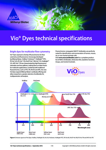
Vio Dyes technical specificationsBright dyes for multicolor flow cytometryFluorochrome-conjugated MACS Antibodies are perfectlysuited for identification and enumeration of human, mouse,rat, or non-human primate cells.Visit www.macsantibodies.com for a complete productlist of MACS Antibodies, Detection Kits, Cytokine SecretionAssays, and Control Cocktails.Vio Dyes represent a family of fluorochromes for flowcytometry and fluorescence microscopy developedby Miltenyi Biotec. VioBlue , VioGreen , VioBright FITC,PE-Vio 615, PE-Vio 770, PerCP-Vio 700, Vio 515, VioBright 515, APC-Vio 770 are characterized by high fluorescenceintensities and low spillover, making them an ideal choicefor multicolor applications. Combined with traditionalfluorochromes, such as FITC, PE, PerCP, and APC, the newVio Dyes expand Miltenyi Biotec’s antibody offering andallow researchers a greater selection of antibodies formultiparameter cell analysis.Figure 1: Emission spectra of Vio Dyes: VioBlue, VioBright 515, Vio 515, VioGreen, VioBright FITC, PE-Vio 615, PerCP-Vio 700, PE-Vio 770 and APC-Vio 770Vio Dyes technical specifications September 20161/18Copyright 2016 Miltenyi Biotec GmbH. All rights reserved.
Overview of flurochromes available from Miltenyi BiotecFluorochromeExcitationlaser (nm)Exmax (nm)Emmax (nm)MACSQuant AnalyzerChannelFilter (nm)MACSQuant VYBChannelFilter 520V2525/50V2525/50VioBright 515488488514B1525/50B1525/50Vio 515488488514B1525/50B1525/50VioBright B1525/50PE488 or 561565578B2585/40Y1586/15PE-Vio 615488 or 730N/AN/APerCP-Vio 700488482704B3655–730N/AN/APE-Vio 770488 or 561565775B4750 LPY4750 LPAPC561 or 635652660R1655–730Y3661/20APC-Vio 770561 or 635652775R2750 LPY4750 LPTable 1: Fluorochrome specifications.Miltenyi Biotec dyesOther dyesVioBlue Alexa Fluor 405, BD Horizon , V450, BV 421 , Calcein Violet 450 AM, Cascade Blue , DAPI, Vybrant ,DyeCycle Violet, eBFP, eFluor 450, Hoechst Dyes, Pacific Blue , Zombie Violet VioGreen Alexa Fluor 430, AmCyan, BD Horizon V500, BV510 , Cascade Yellow , Krome Orange ,Pacific Orange , Qdot 525 Zombie Aqua FITC, VioBright FITC, Vio 515,VioBright 515Alexa Fluor 488, Calcein AM, DyLight 488, CFSE, GFP, SYTOX Green, Vybrant DyeCycle Green, YFP,Zombie Green , BD Horizon Brilliant Blue 515PECy 3, Vybrant DyeCycle OrangePerCP, PerCP-Vio 700PE-Cy 5, PE-Cy 5.5, Per-Cy 5.5, Per-CP-eFluor 710, Propidium iodide, 7-AADPE-Vio 615ECD, PE-Texas Red , PE-CF594, PE/Dazzle 594, PE-eFluor 610PE-Vio 770PE-Alexa Fluor 750, PE-Cy 7APCAlexa Fluor 647, Alexa Fluor 700, APC-Alexa Fluor 700, Cy 5, DRAQ5 , eFluor 660APC-Vio 770APC-Alexa Fluor 750, APC-Cy 7, APC-eFluor 780, APC-H7, Zombie NIR Table 2: Available Vio Dye fluorochromes.Support Material Fluorescence spectra overview poster Fluorochromes and instruments poster Fluorochrome brightness indexVio Dyes technical specifications September 2016 Lot-to-lot consistency data Staining enhancement products Viability dyes2/18Copyright 2016 Miltenyi Biotec GmbH. All rights reserved.
Violet laser dyesAIntensityVioBlue and VioGreen Fluorochromes are a novelrange of violet laser excited dyes developed exclusivelyby Miltenyi Biotec. Designed to maximize the potentialof a flow cytometer’s violet laser, they not only showsuperior optical performance compared with manyother 405 nm excited fluorochromes, but also significantadvances in brightness, signal-to-noise ratios, and intralaser compensation requirements. In addition, neitherfluorochrome exhibits significant levels of photoinduceddegradation, and can consequently be used for manydifferent applications, such as fluorescent microscopy.330VioBlue DyePacific Blue Coumarin-based dye with excitation and emissionwavelengths of 405 nm and 455 nm, respectively Superior alternative to Pacific Blue, Alexa Fluor 405,or BD Horizon V450 Multiplexing of VioBlue with other fluorochromesis easily possible, adding to the variety of markercombinations for multiparameter cell analysis Combined use with the VioGreen DyeAbsorptionmax. (nm)Emissionmax (nm)VioBlue400455Pacific Blue401452Cascade Blue401419Alexa Fluor 405401421eFluor 450405450BD Horizon V450404448BD Horizon Brilliant Violet 421407421Alexa Fluor 405VioBlueB380430480530Wavelength (nm)Pacific BlueTable 3: Absorption and emission maximums of fluorochromes relatedto VioBlue.Cascade BlueAlexa Fluor 405VioBlueFigure 2: Absorption (A) and emission (B) spectra of VioBluecompared to Pacific Blue, Cascade Blue, and Alexa Fluor 405.The blue box represents the 450/50 nm filter.Laser and filter compatibilityWith a maximum absorption and emission at 400 nm and 455nm respectively, VioBlue Fluorochromes are fully compatiblewith standard filter sets from all major flow cytometryhardware providers, giving researchers the flexibility to usethe VioBlue Dye with existing laboratory platforms (figure 2).Vio Dyes technical specifications September 2016Cascade BlueIntensityFluorochrome430380Wavelength (nm)Enhanced brightnessHuman peripheral blood mononuclear cells (PBMC) and mousesplenocyte (MS) cells stained with VioBlue exhibit excellentdiscrimination between positively and negatively stainedpopulations over a wide range of antibody specificities(figure 3). Furthermore, VioBlue provides a superior alternativeto many spectrally similar conjugates for the V1 channel,further increasing the options of multicolor analysis (figure 4).3/18Copyright 2016 Miltenyi Biotec GmbH. All rights reserved.
750750Side scatterB 1000Side scatterA 10005002500-1 0 110¹10²High stability during fixationIt is of crucial importance for a conjugate to retain itsfluorescent properties after fixation, in order to allowresearchers to maximize the use of biological samples.The VioBlue Dye has a very high stability after fixation withparaformaldehyde (figure 5), equal to or exceeding manyother spectrally similar fluorochromes.5002500-1 0 110³CD20-VioBlue10¹10²10³CD123-VioBlueAC 1000D 1000750750B10³10³25010²2500-1 0 110³CD8-VioBlue10¹10²10³10¹10²10³-1 0 110¹10²10³CD123-VioBlueCD90.2-VioBlueFigure : CD123-VioBlue staining before (A) and after (B) fixation with3.7% paraformaldehyde, indicating a minimal decrease in fluorescenceafter fixation.Lot-to-lot variationReproducible lot-to-lot antibody performance ensures theintegrity of long-term studies, and is crucial in comparingexperimental results over a period of time. All MACS Antibodies are vigorously tested to ensure consistentquality, thus eliminating the need for repeat testingarising from reagent degradation or failure (figure 6).10¹10²A10³BRelative cell numberRelativeRelative cell numberVioBluePacific BlueVioBlue-1 0 110-1CD123-VioBlueFigure 3: Human peripheral blood mononuclear cells (PBMC) or mousesplenocyte (MS) cells were stained with CD20-VioBlue (A; PBMCs), therare cell marker CD123-VioBlue (B; PBMCs), CD -VioBlue (C; PBMCs),or CD90.2-VioBlue (D; MS) and analyzed by flow cytometry using theMACSQuant Analyzer.-1 0 110-110¹Pacific BlueFigure : Fluorescence intensity of cells labeled with CD14 antibodiesconjugated to either VioBlue or Pacific Blue.Relative cell number10¹10¹-1 0 110¹10²-1 0 110³10¹10³CD4-VioBlueCD3-VioBlueDecreased spilloverVioBlue Dyes exhibit minimal spillover into the V2 channel,making them perfect candidates for multicolor panels,which utilize both violet channels. Furthermore, VioBlueis negligibly excited by the 488 nm laser, and thus requiresno compensation between the V1 and B1 channels.10²DRelative cell numberCRelative cell number0-1 0 150010²CD303-PE500CD303-PESide scatterSide scatter10²-1 0 110¹10²CD20-VioBlue10³-1 0 110¹10²10³CD45-VioBlueFigure 6: Lot-to-lot variation for different lots of CD3 (A), CD4 (B), CD20(C), and CD45 (D) antibodies conjugated to VioBlue. Black and blue linesrepresent different antibody lots.Vio Dyes technical specifications September 20164/18Copyright 2016 Miltenyi Biotec GmbH. All rights reserved.
VioGreen Dye Absorptionmax. (nm)Emissionmax. (nm)VioGreen388520Pacific Orange400551Krome Orange398528AmCyan457491Horizon V500415500BD Horizon Brilliant 510405510A 1000B 1000750750Side scatterFluorochromeSide scatterLarge Stokes shift fluorochrome, emitting strongfluorescence at 520 nm upon excitation at 405 nmSignificantly increased mean fluorescence intensitiesand higher stain indices over Pacific Orange,Krome Orange, and BD Horizon V500Non-protein fluorochromeSuitable for ethanol fixationCombined use with the VioBlue DyePerfectly suited for multiparameter cell analysisusing all current flow cytometers5002500-1 0 110¹10²5002500-1 0 110³CD14-VioGreenC 1000D 1000750750Side scatterTable : Absorption and emission maximums of fluorochromescomparable to comparable to VioGreen .Laser and filter compatibilityWith a standard 525/50 filter set, the VioGreen Dye exhibitsa better spectral profile than Pacific Orange or AmCyan(figure 7).5002500-1 0 110¹10²10¹10²10³CD20-VioGreenSide scatter Enhanced brightnessLike most dyes designed for the violet laser, VioGreenshows a lower fluorescence intensity compared to otherwell-established fluorochomes, such as PE. However,specific human and mouse cellular populations are easilydistinguishable between positively and negatively labeledcells (figure 8). In addition, many VioGreen Dyes exhibitbrighter fluorescence compared to spectrally similarconjugates, such as Pacific Orange (figure 9 and table 5),AmCyan, and Horizon V500, when measured by meanfluorescence intensity (MFI) or stain index (normalizedsignal-to-noise ratio).50025010³CD15-VioGreen0-1 0 110¹10²10³CD8a-VioGreenA300350400450Relative cell numberIntensityFigure 8: Human peripheral blood mononuclear cells (PBMC) ormouse splenocyte (MS) cells were stained with CD14-VioGreen (A;PBMCs), CD20-VioGreen (B; PBMCs), CD15-VioGreen (C; PBMCs), orCD a-VioGreen (D; MS) and analyzed by flow cytometry using theMACSQuant Analyzer.500Wavelength (nm)BIntensityVioGreenHorizon V500Krome OrangeFigure 9: Analysis of human PBMCs using CD3 antibodies conjugatedto either VioGreen, Horizon V500, or Krome Orange.419519619694Wavelength (nm)VioGreenAmCyanPacific OrangeFigure 7: Absorption and emission spectra of VioGreen.Vio Dyes technical specifications September 20165/18Copyright 2016 Miltenyi Biotec GmbH. All rights reserved.
ConjugateCloneMFIStainindexBlue laser 50.5CD3-Horizon V500UCHT11198.01.5CD3-Krome D8-Pacific OrangeBW135/8010113.00.5VioBright FITC, VioBright 515 andVio 515 DyesVioBright FITC, VioBright 515 and Vio 515 are revolutionaryblue laser (488 nm) excitable dyes, developed exclusively byMiltenyi Biotec. The innovative VioBright technology allowsfor an increased number of fluorochrome molecules perantibody, as compared to conventional conjugation. Thisresults in dyes that emit a signal detected in the standardFITC channel, but with brightness levels similar to PE. Theseblue laser dyes expand the dimensions of multicolor flowanalysis and provide a bright alternative for confidentdetection of rare cells, dim, and uncharacterized markers.Table : MFI, stain indices, and % spillover of CD3-VioGreen,CD3-Horizon V500, CD3-Krome Orange, CD -VioGreen, andCD -Pacific Orange.High stability during paraformaldehydeand ethanol fixationVioGreen Dyes exhibit strong photostability duringfixation, with only very minimal photodegradation, thushighlighting VioGreen’s suitability as a fluorochrome foruse in studies that require fixation (figure 10). It is alsosuitable for ethanol fixation.750750Side scatterB 1000Side scatterA 10005002500-1 0 110¹10²CD14-VioGreen10³Dye5002500-1 0 110¹10²FeaturesApplicationVioBright FITC(1st generationVioBright dye) High brightnessDetection ofextracellularmarkersVioBright 515(2nd generationVioBright dye) Very highbrightness Low spill over High fixation- &photostabilityDetection ofextracellularmarkersVio 515(Organic dye) Moderatebrightness, Low spill over High fixation- &photostabilityDetection ofintracellularmarkers10³Table 6: Features and application specifications for VioBright FITC,VioBright 515 and Vio 515 in comparison.CD14-VioGreenFigure 10: CD14-VioGreen staining before (A) and after (B)paraformaldehyde fixation, indicating a minimal decrease influorescence after fixation.Laser and filter compatibilityExcitable by the blue laser (488 nm), VioBright 515 and Vio 515display peak excitation and emission at 488 nm and 514 nm,respectively (figure 11 and table 7). VioBright FITC is excitedat 496 nm with a peak emission at 522 nm. The high intensityfluorescent signals can be detected with a standard FITC filtersuch as 525/50 on MACSQuant Instruments. Thus, no change inthe filter or cytometer is required. Along with PE, the VioBrightFITC, VioBright 515 and Vio 515 dyes enhance the potential ofthe blue laser, one of the most common laser lines available forsingle laser to multi-laser instruments.FluorochromeAbsorptionmax. (nm)Emissionmax. (nm)VioBright 515488514Vio 515488514VioBright FITC496522FITC495520Alexa Fluor 488495519BD Horizon Brilliant Blue 515490515Table 7: Absorption and emission maximums of fluorochromescomparable to VioBright FITC, VioBright 515 and Vio 515Vio Dyes technical specifications September 20166/18Copyright 2016 Miltenyi Biotec GmbH. All rights reserved.
AAbsorption 4-APC10²0.80010¹10-1-1 0 10.40010¹10²10¹10-1-1 0 110³CD25-PE10¹10²10³CD25-VioBright FITC0.200Figure 13: Human PBMCs were stained with CD25 antibodiesconjugated to PE or VioBright FITC, as well as with CD4-APC, andanalyzed by flow cytometry using the MACSQuant Analyzer.Cell debris and dead cells were excluded from the analysis basedon scatter signals and propidium iodide velength (nm)Emission 20530540550560570Wavelength (nm)VioBright FITCVioBright 515Figure 1 : Human whole blood was stained with CD3-VioBlue andCD56-VioBright 515 or CD56-BB515, respectively.Vio 515FITCReliable analysis of rare cells requires specific identificationof low frequency cellular subset and clear target populationresolution. Thus, highly specific antibodies together withbright conjugates are a prerequisite for detection of suchlow frequency populations. As demonstrated in figures11–14, VioBright FITC and VioBright 515 represent bright dyealternatives for optimal detection of your subpopulation ofinterest and detailed phenotypic analysis.Figure 11: Absorption (A) and emission (B) spectra of VioBright FITC,VioBright 515 and Vio 515 compared to FITC.Enhanced brightnessVioBright FITC, VioBright 515 and Vio 515 are superiordyes for detection of low-expressed markers, as shownin figure 12 and 13. With stain indices (SI) and meanfluorescent intensities similar to PE, staining with thesenovel dyes allows for an excellent resolution of positiveand negative populations.ABCD34-VioBlue10³10²10¹10-1-1 0 D5B-V551C6D5-BCD56-VioBright 1-1 0 110¹10²10³CD133/1-VioBright FITCFigure 1 : CD34 enriched human PBMCs were stained with CD133/1antibodies conjugated to PE or VioBright FITC as well as with CD34VioBlue and analyzed by flow cytometry using the MACSQuantAnalyzer. Cell debris and dead cells were excluded from the analysisbased on scatter signals and propidium iodide fluorescence.15B5Figure 12: Human PBMCs were stained using CD56 antibodiesconjugated to FITC, VioBright FITC, VioBright 515, or BB515 andanalyzed by flow cytometry using the MACSQuant 10 Analyzer.Vio Dyes technical specifications September 20167/18Copyright 2016 Miltenyi Biotec GmbH. All rights reserved.
MFIVioBright 74.74.7ConjugateFixation stabilityBright fluorochrome conjugates, such as PE and APC, areoften sensitive to fixatives like Methanol. For a successfulflow cytomeric analysis, which often involves stainingof intracellular markers, stability of the fluorochromeconjugates is critical. VioBright 515 conjugates show excellentstability to methanol- and paraformaldehyde-based fixativeswith only little compromise in brightness (figure 16).Stain indexVioBright FITCPETable 8: MFI and stain indices of CD25 (clone 4E3), CD335 (clone 9E2),and CD133/1 (clone AC133) antibodies conjugated to either VioBrightFITC or PE.Spill overCompared to standard FITC, VioBright 515 and Vio 515 dyesdisplay reduced spill over into the PE channel (table 8). Incontrast, VioBright FITC has a slightly higher spill over intothe PE channel. Thus, with nominal change in compensationsettings, these innovative dyes offer the benefit of abrighter staining.35Stain Index302520151050Sample alyzer 10Compensation inchannelB2 (%)MACSQuantVYBCompensation inchannelY1 ––CD56(REA196)-VioBright 515MFI10.6StainindexMACS QuantCompensationin ChannelB2 [%]CD56-FITCREA19618.3CD56-VioBright FITCAF1215.822.68.6CD-VioBright A196)-PEPhoto stabilityThe stability of VioBright 515 upon exposure to confocallaser light was analyzed at different time points on a ZeissLSM710 confocal microscope. Significantly higher meanfluorescence intensity (MFI) compared to FITC conjugate,were demonstrated for these time points (figure 17).VioBright 515 shows comparable stability to Alexa Fluor 488conjugated antibodies indicating the excellent suitabilityfor immunofluorescence microscopy.% 1006.6Normalized IntensityCloneMethanolFixationFigure 16: Human PBMCs were stained using CD56 antibodies (cloneREA196) conjugated to VioBright 515 and PE. In addition, cells wereanalyzed before and after fixation using paraformaldehyde and 90%methanol. Stained cells were analyzed by flow cytometry using theMACSQuant 10 AnalyzerTable 9: Compensation requirements in PE channel (B2) of CD4 (cloneVIT4) antibodies conjugated to VioBright FITC, FITC, or PE.ConjugatePFA FixationNo FixativeTable 10: Mean fluorescent intensities (MFI) and stain indices ofanti-CD56 antibodies conjugated to FITC, VioBright 515, and BB515.80604020002:00VioBright 51507:00FITC12:00Min17:00Alexa Fluor 488Figure 17: Human PBMCs were stained with fluorochrome-conjugatedCD4 antibodies. Stained cell samples were fixed with p-formaldehydefor 20 min and transferred to a 96-well plate for analysis. The plottedcurve is normalized over multiple time points.Vio Dyes technical specifications September 20168/18Copyright 2016 Miltenyi Biotec GmbH. All rights reserved.
Blue laser tandem conjugatesAPE-Vio 615 Tandem Conjugate1.0PE-Vio 615 is a tandem dye with PE as the donor dye andVio 615 as the acceptor dye. This tandem dye is optimizedfor efficient donor-to-acceptor dye energy transfer, highfluorescent intensity, and low spillover into the donor dyedetection channel. Designed to be a superior alternativeto ECD, PE-Texas Red , PE-efluor 610, PE-CF594 and PE/Dazzle 594, this Vio Dye expands the options for flexiblemulticolor panel design and provides a bright dye forconfident detection of dim and rare 0600650700B Optimal excitation with blue (488 nm),green (532 nm), and yellow green (561 nm)laser lines for maximum flexibility.Excellent brightness for confident detection of dim,rare, and uncharacterized markers.Extensive portfolio of PE-Vio 615 dye conjugated torecombinantly engineered REAfinity Antibodies forhigher reproducibility.FluorochromeAbsorptionmax (nm)Emissionmax (nm)PE-Vio 615565619ECD, PE-Texas Red565613PE-efluor 610565606PE-CF594565614PE/Dazzle 5945666121.00.80.60.40.20400— PE-CF594— PE-Vio 615— ECDFigure 18: Absorption (A) and emission (B) spectra of PE-Vio 615 andcomparable fluorochromes.ATable 11: Absorption and emission maximums of fluorochromescomparable to PE-Vio 615.B10³10³Laser and filter compatibilityThe PE molecule shows peak absorption at twowavelengths, 496 nm and 565 nm, respectively. This allowsfor optimal excitation of PE-containing tandem dyes suchas PE-Vio 615 using blue, green, and yellow green laser lines(488-561 nm). This enables the utilization of the maximumpotential of your instrument with flexible choice of laserlines (figure 19). As depicted in figure 15, the extent ofabsorption at 565 nm is greater than 496 nm nm and thus,the maximum excitation of PE-Vio 615 can be achieved withinstruments such the MACSQuant VYB, which uses yellowlaser lines for excitation of PE and all PE containing tandemdyes. The emission signal can be detected using typicalfilters design to detect PE-Texas Red, such as 615/20 nm.10²CD3-FITCCD3-FITC10²Vio Dyes technical specifications September 2016— PE-Dazzle 59410¹10-1-1 0 110¹10²CD4-PE-Vio 61510³10¹10-1-1 0 110¹10²10³CD4-PE-Vio 615Figure 19: Human PBMCs were stained using CD4 antibodies (cloneVIT4) conjugated to PE-Vio615 followed by analysis using MACSQuantVYB (A) and MACSQuant Analyzer (B).9/18Copyright 2016 Miltenyi Biotec GmbH. All rights reserved.
Enhanced brightnessPE-Vio 615 is designed to offer a bright alternative tocomparable dyes such as PE-efluor 610, PE-CF594, ECD,and PE/Dazzle 594 (figure 20). PE-Vio 615 not only allows forhigh fluorescent intensities but also excellent separation ofpositive population for higher stain indices (figure 21). Thisproperty of PE-Vio 615 makes it an excellent dye for analysisof dim and difficult to characterize markers (figure 22).10³CD335-VioBright FITCCD335-VioBright FITC10³10²10¹10-1-1 0 110¹10²10³10²10¹10-1-1 0 1CD56-PE-Vio 73.5Q30.31Figure 20: Human PBMCs were stained using CD4 antibodiesconjugated to PE-eFluor610, PE-CF594, PE-Vio615, ECD, PE-Dazzle594and analyzed by flow cytometry using the MACSQuant VYB.StainindexMACS Quant VYBCompensation inchannel Y1 [%]CD4-PEVio 15Figure 22: PBMCs were stimulated for 6 hours. After 2 hours, 1 µg/mLBrefeldinA was added for the last 4 hours. Cells were surface stainedwith CD4-FITC, fixed and permeabilized with the Inside Stain Kit(Miltenyi Biotec) and stained intracellularly with CD154-VioBlue andAnti-IL-4-PE or Anti-IL-4-PE-Vio615 (clone: 7A3-3). Cells were acquired ona BD Fortessa. Cells are gated on CD4 lymphocytes, numbers indicatefrequency among CD4 cells.Data courtesy: Petra Bacher, Clinic for Rheumatology and ClinicalImmunology, Charité – University Medicine Berlin, Berlin, GermanyCD4-PE-Vio 615MFI10³Q125.2Anti-IL-4-PEClone10²Figure 21: Human PBMCs were stained using CD56 antibodies (cloneREA196) conjugated to either PE-Vio 615 or PE and CD335-VioBrightFITC. Stained cells were then analyzed by flow cytometry using theMACSQuant VYB.PE-eFluor610PE-CF594PE-Vio 615ECDPE-Dazzle 594Conjugate10¹CD56-PETable 12: MFI and stain indices of CD4 antibodies conjugated toPE-Vio 615, ECD, PE-CF594, PE-eFluor610, PE/Dazzle594.Vio Dyes technical specifications September 201610/18Copyright 2016 Miltenyi Biotec GmbH. All rights reserved.
Fixation stabilityTandem conjugates are often sensitive to fixatives. Thus,for a successful flow cytomeric analysis, stability of tandemconjugates is critical. PE-Vio 615 conjugate shows excellentstability to paraformaldehyde-based fixative without anyincrease in background signal (figure 23).Without fixationWith fixation10³10¹10-1-1 0 110¹10²10³10²10¹10-1-1 0 1CD56-PE10¹10²10³10¹10-1-1 0 110¹10²10³10¹10²10³CD56-PE-Vio 61510²10¹10²10³10¹10-1-1 0 110¹10²10³10²10¹t 24 h10-1-1 0 1CD56-PE-Vio 61510¹10²10³CD56-PE-Dazzle59410¹10-1-1 0 110¹10²10³10²10¹10-1-1 0 1CD56-PE-Dazzle59410¹10²10³CD56-PE-Vio 615CD56-PE-Dazzle594Compensation (%)10²Decrease in MFI (%)10³CD335-VioBright FITC10³t010-1-1 0 110³CD335-VioBright FITC10²10¹10¹CD56-PE-Dazzle59410³10-1-1 0 110²CD56-PE-Vio 615CD335-VioBright FITCCD335-VioBright FITC10²CD56-PE10³CD335-VioBright FITC10³CD335-VioBright FITC10²10³CD335-VioBright FITCCD335-VioBright FITC10³CD335-VioBright FITCPhoto stabilityThe stability of PE-Vio 615 upon exposure to ambientlight ( 850 Lux) was analyzed at different time points.Significantly higher mean fluorescence intensities (MFI)and stain indices (SI) for these time points, compared tocommercially available alternatives, were also demonstrated(figure 23).CD56-PE-Vio 615CD56-PE-Dazzle594CD56-PE-Dazzle594Light exposing time (h)without fixationFigure 2 : Human PBMCs were stained, using CD56 antibodiesconjugated to PE-Vio615 (clone REA196) or PE-Dazzle594 (clone HCD56).Stained cells were then exposed to ambient light ( 50 Lux) followedby analysis at different time points by flow cytometry using theMACSQuant VYB. Spill over in the Y1 channel, which is optimized fordetection of PE signal was also analyzed after exposure to light.with 56-PEREA19612.033.211.932.7CD56-PEVio 42.231.554.0Light exposing time (h)Figure 23: Human PBMCs were stained using CD56 antibodies (cloneREA196) conjugated to PE, PE-Vio615 or PE-Dazzle594. In addition, cellswere stained with CD335-VioBright FITC and analyzed before and afterfixation using paraformaldehyde. Stained cells were analyzed by flowcytometry using the MACSQuant VYB.Vio Dyes technical specifications September 201611/18Copyright 2016 Miltenyi Biotec GmbH. All rights reserved.
PerCP-Vio 700 Tandem Conjugate Absorptionmax. (nm)Emissionmax. (nm)PerCP482675PerCP-Vio 700482704PerCP-Cy5.5490695A 1000B 1000750750Side scatterFluorochromeTable 13: Absorption and emission maximums of fluorochromesrelated to PerCP-Vio700.Side scatterCombines the peridinin chlorophyll protein (PerCP)and the new Vio 700 dyeEmits strong fluorescence at 655–730 uponblue laser excitation at 488 nmSuitable for the B3 channel of the MACSQuant Analyzer5002500-1 0 110¹10²5002500-1 0 110³CD20-PerCP-Vio 700C 1000D 1000750750Side scatterLaser and filter compatibilityUsing a standard 655–730 bandpass filter, PerCP-Vio 700exhibits a very narrow fluorescence spectral profile, thusallowing the majority of light to be captured and retained(figure 25).5002500-1 0 de scatter Enhanced brightnessHuman peripheral blood mononuclear cells (PBMC)and mouse spenocyte (MS) cells were stained withPerCP-Vio 700, and consequently exhibited excellentseparation between positive and negative populationsover many different antibody specificities (figure 26).5002500-1 0 110³CD45-PerCP-Vio 70010¹10²10³CD90.2-PerCP-Vio 700Intensity7060Figure 26: Human peripheral blood mononuclear cells (PBMC) ormouse spenocyte (MS) cells were stained with CD20-PerCP-Vio 700(A; PBMCs), CD4-PerCP-Vio700 (B; PBMCs), CD45-PerCP-Vio 700(C; PBMCs) or CD90.2-PerCP-Vio 700 (D; MS) and analyzed by flowcytometry using the MACSQuant Analyzer.50403020High stability during fixationPerCP-Vio 700 shows excellent fixation stabilities, with onlyminimal decreases in fluorescence after fixation (figure 27).100350500650800Wavelength (nm)A 1000B 1000Figure 2 : Absorption and emission spectra of PerCP-Vio 700.The blue box represents the 655–730 filter.750750Side scatterEmissonSide scatterAbsorption5002500-1 0 110¹10²CD8-PerCP-VioPP-Vio70010³5002500-1 0 110¹10²10³CD8-PerCP-VioPP-Vio 700Figure 27: CD -PerCP-Vio 700 before (A) and after (B) fixation with3.7% paraformaldehyde, indicating a minimal decrease in fluorescenceafter fixation.Vio Dyes technical specifications September 201612/18Copyright 2016 Miltenyi Biotec GmbH. All rights reserved.
Photo-induced conjugate degradationAnalysis of the photo-induced degradation of CD14-PerCPVio 700 indicated no discernable changes after up to fourhours of continuous exposure to ambient light ( 850 Lux).Significantly higher mean fluorescence intensities (MFI)and stain indices (SI) for these time points, compare tocommercially available alternatives (BD Biosciences),were also demonstrated (figure 28).t 0 minCD14-PerCP-Cy5.5CD14-PerCPCD14-PerCP-Vio 700t 120 mint 240 PerCP-Vio 700TÜK4757575CD14-PerCP-Cy5.561D3363128t 0 minStain indext 120 min t 240 minFigure 28: Photo-induced conjugate degradation of PerCP, PerCP-Vio700, and PerCP-Cy5.5 with corresponding MFI and SI. Conjugateswere exposed to ambient light for up to 4 hours, with negligibledegradation rates.Vio Dyes technical specifications September 201613/18Copyright 2016 Miltenyi Biotec GmbH. All rights reserved.
PE-Vio 770 Tandem ConjugateEnhanced brightnessPE-Vio 770 provides the greatest fluorescence intensity ofthe Vio Dye family, with excellent separation of positivelyand negatively stained cells (figure 30). When comparedto other spectrally similar tandem conjugates, such asPE-Cy7 (figure 31) or PE-Alexa Fluor 750, PE-Vio 770 exhibitssignificantly higher mean fluorescence intensities (MFI)and stain indices, and requires less compensation (table 14).These properties make PE-Vio 770 a far superior tandemconjugate compared to PE-Cy7 or PE-Alexa Fluor 750.FluorochromeAbsorptionmax. (nm)B 1000750750Side scatterA 1000Side scatterThe PE-Vio 770 Dye is a tandem conjugate, like PE-Cy 7,that exploits the principle of fluorescence-resonanceenergy-transfer (FRET) allowing for large Stokes shifts betweenthe absorbed energy of a fluorescence donor (PE) and theemission wavelength of a suitable acceptor (Vio 770) dye. Excitation with the blue (488 nm) or yellow (561 nm)laser, emission in the near-infrared region at 775 nm The combination of Vio 770 as the acceptor dye and anoptimized chemistry have furnished a tandem dye thatis characterized
of a flow cytometer's violet laser, they not only show superior optical performance compared with many other 405 nm excited fluorochromes, but also significant advances in brightness, signal-to-noise ratios, and intra-laser compensation requirements. In addition, neither fluorochrome exhibits significant levels of photoinduced
