Transcription
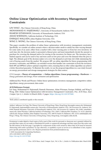
19.3 SPECTROSCOPY OF ALDEHYDES AND KETONES8953O3S%C %d–d bond dipoleof the C O bondEPM of acetoneBecause of their polarities, aldehydes and ketones have higher boiling points than alkenes oralkanes with similar molecular masses and shapes. But because aldehydes and ketones are nothydrogen-bond donors, their boiling points are considerably lower than those of the corresponding alcohols.CH3CH A CH2boiling pointdipole momentboiling pointdipole moment-47.4 C0.4 DCH2S%C %H3CCH3-6.9 C0.5 DCH3CH A O20.8 C2.7 DOS%C %H3CCH356.5 C2.7 DCH3CH2OH78.3 C1.7 DH3COH"MCH %82.3 C1.7 DCH3Aldehydes and ketones with four or fewer carbons have considerable solubilities in waterbecause they can accept hydrogen bonds from water at the carbonyl oxygen.HO L HH L OH3O3SH3C L C L CH3Acetaldehyde and acetone are miscible with water (that is, soluble in all proportions). Thewater solubility of aldehydes and ketones along a series diminishes rapidly with increasingmolecular mass.Acetone and 2-butanone are especially valued as solvents because they dissolve not onlywater but also a wide variety of organic compounds. These solvents have sufficiently low boiling points that they can be easily separated from other less volatile compounds. Acetone, witha dielectric constant of 21, is a polar aprotic solvent and is often used as a solvent or co-solvent for nucleophilic substitution reactions.19.3SPECTROSCOPY OF ALDEHYDES AND KETONESA. IR SpectroscopyThe principal infrared absorption of aldehydes and ketones is the CA O stretching absorption,a strong absorption that occurs in the vicinity of 1700 cm 1. In fact, this is one of the most important of all infrared absorptions. Because the C A O bond is stronger than the C A C bond,the stretching frequency of the CA O bond is greater.
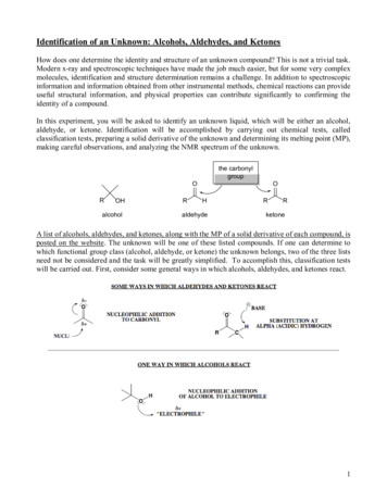
896CHAPTER 19 THE CHEMISTRY OF ALDEHYDES AND KETONES. CARBONYL-ADDITION REACTIONS2.6 2.8 33.5wavelength, micrometers5 5.5 6784 4.59 101112 13 14 1516percent transmittance1008060O40H Cstretch20C OstretchCH3CH2CH2CHO03800 3400 3000 2600 2200 2000 1800 1600 1400 1200 1000wavenumber, cm 1800600Figure 19.3 The infrared spectrum of butyraldehyde with key absorptions indicated in red.The position of the CA O stretching absorption varies predictably for different types ofcarbonyl compounds. It generally occurs at 1710–1715 cm 1 for simple ketones and at1720–1725 cm 1 for simple aldehydes. The carbonyl absorption is clearly evident, forexample, in the IR spectrum of butyraldehyde (Fig. 19.3). The stretching absorption of the carbonyl–hydrogen bond of aldehydes near 2710 cm 1 is another characteristic absorption; however, NMR spectroscopy provides a more reliable way to diagnose the presence of this type ofhydrogen (Sec. 19.3B).Compounds in which the carbonyl group is conjugated with aromatic rings, double bonds,or triple bonds have lower carbonyl stretching frequencies than unconjugated carbonyl compounds.OScL C L CH3OSH2C A CH L C L CH3compare:H2C A CHCH2CH3OSH3C L C L CH2CH33-buten-2-one1-butene1685 cm 11670 cm 1—1715 cm 1 1 C A C 1600 cm1613 cm 11642 cm 1— C A O2-butanoneacetophenone(19.2)The carbon–carbon double-bond stretching frequencies are also lower in the conjugated molecules. These effects can be explained by the resonance structures for these compounds. Because the CA O and CA C bonds have some single-bond character, as indicated by the following resonance structures, they are somewhat weaker than ordinary double bonds, andtherefore absorb in the IR at lower frequency.
19.3 SPECTROSCOPY OF ALDEHYDES AND KETONESsingle-bondcharacter3O3SH2C A CH L C L CH33O2 3" H2C L CH A C L CH3897(19.3)In cyclic ketones with rings containing fewer than six carbons, the carbonyl absorption frequency increases significantly as the ring size decreases. (See Further Exploration 19.1.)Further Exploration 19.1Ocyclohexanone1715 cm 1(normal)PROBLEMVSOH2C A C A Ocyclopropanone1850 cm 1ketene2150 cm 1SSSOIR Absorptions ofCyclic KetonesOcyclopentanone1745 cm 1cyclobutanone1780 cm 1(19.4)19.3 Explain how IR spectra could be used to differentiate the isomers within each of thefollowing pairs.(a) cyclohexanone and hexanal (b) 3-cyclohexenone and 2-cyclohexenone(c) 2-butanone and 3-buten-2-olB. Proton NMR SpectroscopyThe characteristic NMR absorption common to both aldehydes and ketones is that of the protons on the carbons adjacent to the carbonyl group: the a-protons. This absorption is in thed 2.0–2.5 region of the spectrum (see also Fig. 13.4 on p. 580). This absorption is slightly farther downfield than the absorptions of allylic protons because the C A O group is more electronegative than the CA C group. In addition, the absorption of the aldehydic proton is quitedistinctive, occurring in the d 9–10 region of the NMR spectrum, at lower field than most otherNMR absorptions.a-protonsOSH3C L C L CH3d 2.17d 2.17a-protonsOSH3C L C L CH2 L CH3d 2.15d 2.40 d 0.99a-protonsaldehydicprotonOSH3C L C L Hd 2.2d 9.8In general, aldehydic protons have very large chemical shifts. The explanation is the same asthat for the large chemical shifts of protons on a carbon–carbon double bond (Sec. 13.7A).However, the carbonyl group has a greater effect on chemical shift than a carbon–carbon double bond because of the electronegativity of the carbonyl oxygen.
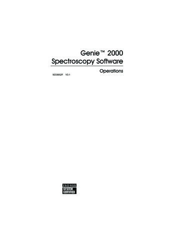
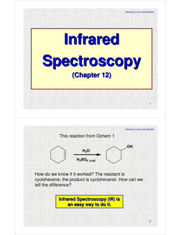
898CHAPTER 19 THE CHEMISTRY OF ALDEHYDES AND KETONES. CARBONYL-ADDITION REACTIONS2400210018001500chemical shift, Hz12009006003006HJ 7.1 HzJ 1.2 Hz0 HzJ 7.1 HzJ 1.2 Hzd 9.65(off scale)1H1H876543chemical shift, ppm (d)210Figure 19.4 The NMR spectrum for Problem 19.4(a). The relative integrals are indicated in red over theirrespective resonances.PROBLEM19.4 Deduce the structures of the following compounds.(a) C4H8O: IR 1720, 2710 cm 1NMR in Fig. 19.4(b) C4H8O: IR 1717 cm 1NMR d 0.95 (3H, t, J 8 Hz); d 2.03 (3H, s); d 2.38 (2H, q, J 8 Hz)(c) A compound with molecular mass 70.1, IR absorption at 1780 cm 1, and thefollowing NMR spectrum: d 2.01 (quintuplet, J 7 Hz); d 3.09 (t, J 7 Hz). The integral of the d 3.09 resonance is twice as large as that of the d 2.01 resonance.(d) C10H12O2: IR 1690 cm 1, 1612 cm 1NMR d 1.4 (3H, t, J 8 Hz); d 2.5 (3H, s); d 4.1 (2H, q, J 8 Hz); d 6.9 (2H, d,J 9 Hz); d 7.9 (2H, d, J 9 Hz)C. Carbon NMR SpectroscopyThe most characteristic absorption of aldehydes and ketones in 13C NMR spectroscopy is thatof the carbonyl carbon, which occurs typically in the d 190–220 range (see Fig. 13.20, p. 624).This large downfield shift is due to the induced electron circulation in the p bond, as in alkenes(Fig. 13.14, p. 613), and to the additional chemical-shift effect of the electronegative carbonyloxygen. Because the carbonyl carbon of a ketone bears no hydrogens, its 13C NMR absorption, like that of other quaternary carbons, is characteristically rather weak (Sec. 13.9). Thiseffect is evident in the 13C NMR spectrum of propiophenone (Fig. 19.5).The a-carbon absorptions of aldehydes and ketones show modest downfield shifts,typically in the d 30–50 range, with, as usual, greater shifts for more branched carbons. Thea-carbon shift of propiophenone, 31.7 ppm (Fig. 19.5, carbon b) is typical. Because shifts inthis range are also observed for other functional groups, these absorptions are less useful thanthe carbonyl carbon resonances for identifying aldehydes and ketones.
89919.3 SPECTROSCOPY OF ALDEHYDES AND KETONESc,ddfe(offset)d 200.1CgdeOcbaCH2CH3cbafg200 190 180 170 160 150 140 130 120 110 100 90 80 70 60 50 40 30 20 10chemical shift, ppm (d)0Figure 19.5 The 13C NMR spectrum of propiophenone.Two particularly important features of the spectrum arethe large downfield shift of the carbonyl carbon g, and the small resonances for the two carbons (f and g) that bearno protons. Recall from Sec. 13.9 that absorption intensities in 13C NMR spectra generally do not accurately correspond to numbers of carbons, and quaternary carbons generally have considerably weaker absorptions than proton-bearing carbons.PROBLEMS19.519.6Propose a structure for a compound C6H12O that has IR absorption at 1705 cm 1, no protonNMR absorption at a chemical shift greater than d 3, and the following 13C NMR spectrum:d 24.4, d 26.5, d 44.2, and d 212.6. The resonances at d 44.2 and d 212.6 have very low intensity.The 13C NMR spectrum of 2-ethylbutanal consists of the following absorptions: d 11.5,d 21.7, d 55.2, and d 204.7. Draw the structure of this aldehyde, label each chemically nonequivalent set of carbons, and assign each absorption to the appropriate carbon(s).D. UV SpectroscopyThe p T p* absorptions (Sec. 15.2B) of unconjugated aldehydes and ketones occur at about150 nm, a wavelength well below the operating range of common UV spectrometers. Simplealdehydes and ketones also have another, much weaker, absorption at higher wavelength, inthe 260–290 nm region. This absorption is caused by excitation of the unshared electrons onoxygen (sometimes called the n electrons). This high-wavelength absorption is usually referred to as an n T p* absorption.(CH3)2C A O22n electronsnp*271 nm (e 16) (in ethanol)This absorption is easily distinguished from a p T p* absorption because it is only 10 2 to10 3 times as strong. However, it is strong enough that aldehydes and ketones cannot be usedas solvents for UV spectroscopy.
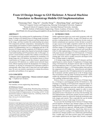
CHAPTER 19 THE CHEMISTRY OF ALDEHYDES AND KETONES. CARBONYL-ADDITION REACTIONS1.00.9O0.8CCH3pp*0.7absorbance9000.6803 320340350wavelength (l), nmFigure 19.6 The ultraviolet spectrum of 1-acetylcyclohexene [1-(cyclohexen-1-yl)ethanone] in methanol. Thespectrum of a more concentrated solution (red) reveals the “forbidden” n T p* absorption, which is so weak thatit is not apparent in the spectrum taken on a more dilute solution (black).Like conjugated dienes, the p electrons of compounds in which carbonyl groups are conjugated with double or triple bonds have strong absorption in the UV spectrum. The spectrumof 1-acetylcyclohexene (Fig. 19.6) is typical. The 232-nm peak is due to light absorption bythe conjugated p-electron system and is thus a p T p* absorption. It has a very large extinction coefficient, much like that of a conjugated diene. The weak 308-nm absorption is an n Tp* absorption.conjugatedp-electron system3O3SL C L CH3lmax 232 nm (e 13,200)lmax 308 nm (e 150)(in hanone]The lmax of a conjugated aldehyde or ketone is governed by the same variables that affectthe lmax values of conjugated dienes: the number of conjugated double bonds, substitution onthe double bond, and so on. When an aromatic ring is conjugated with a carbonyl group, thetypical aromatic absorptions are more intense and shifted to higher wavelengths than those ofbenzene.clmax 204 (e 7900)254 (e 212)OScL C L CH3lmax 240 (e 13,000)278 (e 1100)319 (e 50) (np*)
19.3 SPECTROSCOPY OF ALDEHYDES AND KETONES901The p T p* absorptions of conjugated carbonyl compounds, like those of conjugatedalkenes, arise from the promotion of a p electron from a bonding to an antibonding (p*) molecular orbital (Sec. 15.2B). An n T p* absorption arises from promotion of one of the n (unshared) electrons on a carbonyl oxygen to a p* molecular orbital. As stated previously in thissection, n T p* absorptions are weak. Spectroscopists say that these absorptions are forbidden. This term refers to certain physical reasons for the very low intensity of these absorptions. The 254-nm absorption of benzene, which has a very low extinction coefficient of 212,is another example of a “forbidden” absorption.PROBLEMS19.7 Explain how the compounds within each set can be distinguished using only UVspectroscopy.(a) 2-cyclohexenone and 3-cyclohexenone(b)(O(Oand(c) 1-phenyl-2-propanone and p-methylacetophenone19.8 In neutral alcohol solution, the UV spectra of p-hydroxyacetophenone and p-methoxyacetophenone are virtually identical. When NaOH is added to the solution, the lmax of p-hydroxyacetophenone increases by about 50 nm, but that of p-methoxyacetophenone is unaffected. Explain these observations.19.9 Which one of the following compounds should have a p T p* UV absorption at the greaterlmax when the compound is dissolved in NaOH solution? Explain.CH3OHOHOCHOCH3OvanillinACHOisovanillinBE. Mass SpectrometryImportant fragmentations of aldehydes and ketones are illustrated by the electron-impact (EI)mass spectrum of 5-methyl-2-hexanone (Fig. 19.7, p. 902). The three most important peaksoccur at mÜz 71, 58, and 43. The peaks at mÜz 71 and mÜz 43 arise from cleavage ofthe molecular ion at the bond between the carbonyl group and an adjacent carbon atom by twomechanisms that were discussed in Sec. 12.6C: inductive cleavage and a-cleavage. Inductivecleavage accounts for the mÜz 71 peak. In this cleavage, the alkyl fragment carries thecharge and the carbonyl fragment carries the unpaired electron. 3O8S(CH3)2CHCH2CH2 L C L CH3 3O3"(CH3)2CHCH2CH2 L C L CH38molecular ion from loss of unshared electroninductivecleavage3O3S(CH3)2CHCH2CH2 8 C L CH3m!z 71 (19.5)
902CHAPTER 19 THE CHEMISTRY OF ALDEHYDES AND KETONES. CARBONYL-ADDITION REACTIONS10043relative 5060708090 100mass-to-charge ratio m/z110120130140Figure 19.7 The EI mass spectrum of 5-methyl-2-hexanone. The odd-electron ion at mÜz 58 results from aMcLafferty rearrangement of the molecular ion.a-Cleavage accounts for the mÜz 43 peak. In this case the same molecular ion undergoesfragmentation in such a way that the carbonyl fragment carries the charge and the alkyl fragment carries the unpaired electron: a-cleavage3O8S 8(CH3)2CHCH2CH2 L C L CH3(CH3)2CHCH2CH2 3 O ' C L CH3m!z 43(19.6)An analogous cleavage at the carbon–hydrogen bond accounts for the fact that many aldehydes show a strong M - 1 peak.What accounts for the mÜz 58 peak? A common mechanism for the formation of oddelectron ions is hydrogen transfer followed by loss of a stable neutral molecule (p. 564
12O that has IR absorption at 1705 cm _1, no proton NMR absorption at a chemical shift greater than d 3, and the following 13C NMR spectrum: d 24.4, d 26.5, d 44.2, and d 212.6. The resonances at d 44.2 and d 212.6 have very low in-tensity. 19.6 The 13C NMR spectrum of 2-ethylbutanal consists of the following absorptions: d 11.5,