Transcription
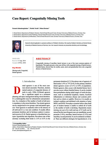
February 2015, Volume 12, Number 1Case Report: Congenitally Missing TeethFatemeh Jahanimoghadam 1*, Moolok Torabi 2, Shima Rostami 31. Dental School, Department of Pediatric Dentistry, Oral & Dental Research Center, Kerman University of Medical Sciences, Kerman, Iran.2. Dental School, Department of Oral Pathology, Oral & Dental Research Center, Kerman University of Medical Sciences, Kerman, Iran.3. Dental School, Department of Orthodontics, Oral & Dental Research Center, Kerman University of Medical Sciences, Kerman, Iran.Fatemeh Jahanimoghadam is assistant professor of Pediatric Dentistry. She teaches Pediatric Dentistry at Dental School,University of Medical Sciences of Kerman, Iran. Her research interests are preventive dentistry and microbiology.Article info:Received: 11 Agu. 2014Accepted: 29 Nov. 2014Key Words:Missing teeth, Congenital,Dental agenesisABSTRACTCongenitally missing of maxillary lateral incisors is one of the most common patterns ofhypodontia. This paper presents a nine year old boy with congenital missing of lateral incisors.Familial history showed that, his mother, aunts, uncle and grandmother have also congenitalabsence of lateral incisors.1. IntroductionDental agenesis is one of the most common dental anomalies Therefore, dentistsstarted analysis of congenital absence ofteeth in early 1990s [1].Dental agenesishas a significant impact on a treatmentplanning and space management during mixed dentition.It’s a challengeable issue for orthodontists and pedodontists. So, evaluation of the number of teeth in both jawsis mandatory in the mixed dentition. True dental agenesiscan be categorized in two groups; total and partial agenesis: Total agenesis or anadontia refers to the absence ofall teeth and it is a very rare condition. It usually affectspermanent dentition and its heritance pattern is autosomal recessive [2-4]. Partial agenesis is classified into twotypes: hypodontia and oligodotia. Prevalence rate of thisanomaly excluding third molar is 3.5% to 6.5% in thepermanent dentition [5-7]. Prevalence rate of agenesis ofthird molar is 9% to 37% [8-9]. In the primary dentitiondental agenesis occurs in 0.1% to 0.9% of population.Partial agenesis often occurs with familial history but itcan also occur without familial history. It can be resultedfrom perturbation during initial stages of tooth development, such as ectodermal dysplasia, trauma, localizedinflammation or infectious disease and systemic problem such as rickets or syphilis. However, it is usually anisolated condition and attributed with mutation of geneMSX1 and PAX9. Nowadays scientists believe that smalljaw of modern human is unable to embed all of teethso the number of teeth is declining. Hypodontia meansmissing one up to five teeth and any of 32 teeth can bemissed but the most frequent ones are mandibular secondpremolar (3.4%), maxillary lateral incisors (2.12%) andmaxillary second premolar. It can be unilateral or bilateral. There are different etiologies for hypodontia [2, 4, 6].* Corresponding Author:Fatemeh Jahanimoghadam, PhDAddress: Dental School, Department of Pediatric Dentistry, Oral & Dental Research Center, Kerman University of Medical Sciences, Kerman, Iran.Tel: 98 (34) 32119021Tel: 98 (913) 3431507E-mail: fatemehjahani4@gmail.com45
Fatemeh Jahanimoghadam et al. Congenitally Missing Teethpernumerary was 3% in 3-5 year old children. A majorityof these anomalies were seen in boys. Missing and supernumerary were more frequent in maxilla [13].A research done by Partovi and others in Babol evaluated missing teeth in 12-17 year old individuals. Theystated the prevalence rate of missing teeth as 8.59%(8.4% in males and 8.8% in females). The most commonmissing teeth were mandibular second premolar (45.5%),maxillary lateral incisor (34%) and maxillary second premolar (10.6%) [14].Akhlaghi and et al studied the prevalence of missingteeth except third molar in girl students of high schoolsin Rasht. The prevalence of missing teeth was 8%. In thisstudy the most incidence of missing teeth was seen in age16. The most common missing teeth were second premolars and the least ones were second molars [15].Figure 1. Frontal view of the patient.Physical disruption of dental lamina may causes obliteration of tooth buds and agenesis of tooth. This condition isseen in orofaciodigital syndrome, Ellisvan syndrome andcleft lip & palate. It can also be the result of metabolicmisbalance that causes dental agenesis. Finally, hereditary defect of the underlying mesenchyme may lead tohypodontia.Hypodontia is more frequent in women so it is important to manage these teeth aesthetically. Prevalence rateof hypodontia is variable in different sexes and races.Some of the studies have investigated its prevalence insome races and sexes [1, 8, 10].2. Literature reviewHedayati and colleagues have done a research in Shiraz. They evaluated the prevalence of missing teeth inorthodontic patients .The prevalence rate of missing was7.66%. Patients with malocclusion Class III have theleast prevalence rate of missing (1%) and patients withmalocclusion Class II have the most prevalence rate ofmissing (4.68%). Majority of them had bilateral missingteeth. The most common missing teeth were lateral incisors [16].Many studies specify that women show more partialanadontia than men. Egermark and Erikson found the 3:2ratios of women to men in their studies but there wasno difference in prevalence rate of hypodontia in womenand men [17].Sofaer et al concluded that the missing of teeth on oneside of the dental segment induces a compensatory increase in the size of the teeth on the other side [18].Amini et al evaluated the prevalence of hypodontia inthe permanent dentition in Iranian dentition (3374 patients) in 2011.They concluded that the prevalence rate ofhypodontia in Iranian population was 5% and it is morefrequent in maxilla [11].Sheikhi et al reported prevalence of congenitally missing permanent teeth in Iran (Tehran) in 2013.They noteda 10.9% rate of congenital absence teeth. In this studythe most common missing teeth were mandibular secondpremolar and maxillary second premolar [12].Following a study performed by Afshar et.al in Tehran,it was found that the prevalence rate of missing and su-46Figure 2. Intraoral view of the patient.
February 2015, Volume 12, Number 1Figure 3. Panoramic radiograph of the patient.3. Case reportA 9-year-old boy was referred to the Department ofPediatric Dentistry, Faculty of Dentistry, Universityof Medical Sciences, Kerman, Iran complaining of anabscess and pain in right and left mandibular first andsecond molars (Figure 1). The patient’s medical historywas non-contributory for pertinent findings. Intra oral examination showed that his lateral incisors hadn’t eruptedyet (Figure 2). Due to his age it was a little suspicious.Therefore, panoramic radiograph was ordered (Figure 3).Panoramic radiograph showed that lateral incisors weremissing. Familial history revealed that his mother, aunts,grandmother, cousin and his uncle had congenital absence of lateral incisors (Figure 4, 5, 6, 7).On intra oral examination there was class III relationship on the left side and class I on the right side. Therewas 1 mm of over jet and 1 mm of overbite .The maxillary and mandibular arch were symmetrical with spacing in the anterior region. The oral hygiene was fair withmild gingivitis. There was no history of extraction ofpermanent teeth. The primary maxillary right and leftlateral incisors were still retained. In treatment planning,the first and second primary molars were extracted andlingual arch was placed as a space maintainer.Figure 4. Intraoral view of the patient’s mother.Figure 5. Panoramic radiograph of the patient’s mother.4. DiscussionHypodontia is the most common dental anomalies occurring in human dentition [19]. Hypodontia may occurin association with some other conditions, such as ectodermal dysplasia, down syndrome and cleft of lip & palate, although it usually occurs alone [20].Familial hypodontia is an isolated trait. This conditionmay occur in autosomal dominant, autosomal recessiveor sex-linked patterns of inheritance [21]. After thirdmolar, the most commonly missing permanent teeth aremandibular second premolars and maxillary lateral incisors or maxillary second premolars [22].Congenitally missing of maxillary lateral incisors is oneof the patterns of hypodontia. This trait can have an autosomal dominant pattern of inheritance, showing reducedpenetrance and variable expressivity. Also this can be arecessive or polygenic trait [21].The treatment plan for congenitally missing of maxillary incisors is challenging and requiring multidisciplinary approach to achieve a successful outcome. Oftenpediatric dentist initiates interdisciplinary treatment bythe diagnosis of hypodontia and maintenance of the primary teeth. Definitive treatment plan is often determinedFigure 6. Intraoral view of the patient’s aunt.47
Fatemeh Jahanimoghadam et al. Congenitally Missing TeethAdolescence. 4th edition. Philadelphia, PA: Mosby; 2005,pp: 61-73.[4] McDonald RE, Avery DR, Hartsfield JK. Acquired and de-velopmental disturbances of the teeth and associated oralstructures. In: Dean JA, Avery DR, McDonald RE, editors.McDonald and Avery Dentistry for the Child and Adolescent. 9th ed. St. Luis: Mosby; 2011, pp: 85–125.[5] Hosur MB, Puranik R, Vanaki S. Oligodontia: A case reportand review of literature. World Journal of Dentistry. 2011;2(3):259-262.Figure 7. Panoramic radiograph of the patient’s aunt.[6] Gibson A. Concomitant hypo-hyperodontia. British Journalof Orthodontics. 1979; 6(2):101-105.after the eruption of all permanent teeth [23]. The treatment options include space closure with canine substitution and space opening with prosthetic replacement.Determination of correct treatment depends on severalfactors including occlusion, degree of crowding, skeletaland dental development, facial profile, color and shape ofthe canines [22].[7] Lavelle C, Ashton E, Flinn R. Cusp pattern, tooth size andThe treatment plan for this case is encouraging thepermanent canines to erupt into the lateral incisors position. Therefore, alveolar bone is formed in the area ofthe missing teeth and then moving the canines distally toopen space for the implants. The implants should not beplaced until vertical growth is complete. The treatment ofthis condition is complex and needs coordinated interdisciplinary approach.[10] Rushmah M. Hypodontia of the primary and permanentAcknowledgements[13] Afshar H, Shahrabi M, Salami K. [Prevalence of missingThe authors wish to thank the patient for her assistancein all periods of study.Conflict of InterestThe authors of this manuscript declare no conflict ofinterest in this paper.third molar agenesis in the human mandibular dentition.Archives of Oral Biology. 1970; 15(3):227-237.[8] De Coster P, Marks LA, Martens LC, Huysseune A. Dentalagenesis: genetic and clinical perspectives. Journal of OralPathology & Medicine. 2009; 38(1):1-17.[9] Dahlberg AA. The changing dentition of man. The Journalof the American Dental Association. 1945; 32(11):676-690.dentition. The Journal of Clinical Pediatric Dentistry. 1991;16(2):121-123.[11] Amini F, Rakhshan V, Babaei P. Prevalence and pattern ofhypodontia in the permanent dentition of 3374 Iranian orthodontic patients. Dental Research Journal. 2012; 9(3):245.[12] Sheikhi M, Sadeghi MA, Ghorbani-Zadeh S. Prevalence ofcongenitally missing permanent teeth in Iran. Dental Research Journal. 2012; 9(Suppl 1):105-111.and supernumerary teeth in 3-5 years-old, kindergartenchildren of Tehran (Persian)]. Journal of Islamic Dental Association of Iran. 2009; 20(4):273-277.[14] Partovi M, Mehranfar P. [Prevalence of congenital missing of anterior and premolar permanent teeth among 12-17year old people in Babol (Persian)]. Journal of Babol University of Medical Sciences. 2002; 4(2):30-33.[15] Akhlaghi F, Yavari SAS, Eshaghi SM. Clinical prevalence ofmissing teeth (except third molar) in girl students at Rashthigh schools (1999-2000). Journal of Dental School. 2008;24(2):155-162.[16] Hedayati Z, Dashlibrun YN. The prevalence and distribution pattern of hypodontia among orthodontic patients inSouthern Iran. European Journal of Dentistry. 2013; 7(Suppl1):S78–S82.References[1] Reddy N. Congenitally missing teeth: a case report. Annalsand Essences of Dentistry. 2010; 2(1):5-9.[2] Silverman N, Ackerman J. Oligodontia: a study of its preva-lence and variation in 4032 children. American Society ofDentistry for Children. 1978; 46(6):470-477.[3] Dummett C Jr. Anomalies of the developing dentition.Pinkham JR, Casamassimo P, Fields HW, McTigue D,Nowak A, editors. Pediatric Dentistry: Infancy through48[17] Egermark-Eriksson I, Lind V. Congenital numerical variation in the permanent dentition. D. Sex distribution ofhypodontia and hyperodontia. Odontologisk Revy. 1970;22(3):309-315.[18] Sofaer JA, Chung CS, Niswander JD, Runck DW. Developmental interaction, size and agenesis among permanentmaxillary incisors. Human Biology. 1971; 43(1):36-45.[19] Cobourne MT. Familial human hypodontia--is it all in thegenes? British Dental Journal. 2007; 203(4):203-8.
February 2015, Volume 12, Number 1[20] Hartsfield JK. Genetics and orthodontics. In: Grabber LW,Vanarsdall RL Jr, Vigk WL, editor. Orthodontics: CurrentPrinciples and Techniques. 5th edition. Philadelphia, PA:Mosby Co; 2012, pp: 144-8.[21] Woolf CM. Missing maxillary lateral incisors: a genet-ic study. American Journal of Human Genetics. 1971;23(3):289.[22] Proffit WR, Fields HW Jr, Sarver DM. Contemporary orthodontics. 5th edition. St. Louis: Mosby Co; 2013, pp: 453-5.[23] Nunn JH, Carter NE, Gillgrass TJ. The interdisciplinarymanagement of hypodontia: background and role of paediatric dentistry. British Dental Journal. 2003; 194(5):245-51.49
50
Nowak A, editors. Pediatric Dentistry: Infancy through Adolescence. 4th edition. Philadelphia, PA: Mosby; 2005, pp: 61-73. [4] McDonald RE, Avery DR, Hartsfield JK. Acquired and de-velopmental disturbances of the teeth and associated oral structures. In: Dean JA, Avery DR, McDonald RE, editors. McDonald and A