Transcription
Cancer Reports and ReviewsResearch ArticleISSN: 2513-9290Bone metastases from head & neck squamous cellcarcinoma (HNSCC) – Reviewing the patients’background and imaging features mainly of whole bodyMRI (WBMRI)Katsuyuki Nakanishi1, Mio Sakai1, Hiromitsu Sumikawa1, Naoyuki Kanayama2, Kazuya Oshima3, Takashi Fujii4, Toshinari Yagi5, ShinichiNakatsuka6, Nobuo Kashiwagi7 and Noriyuki Tomiyama8Department of Diagnostic and Interventional Radiology, Osaka International Cancer Institute, 3-1-69, Otemae Chuo-ku, Osaka city, Osaka 541-8567, JapanDepartment of Radiation Oncology, Osaka International Cancer Institute, 3-1-69, Otemae Chuo-ku, Osaka city, Osaka 541-8567, Japan3Department of Orthopaedic Surgery, Osaka International Cancer Institute, 3-1-69, Otemae Chuo-ku, Osaka city, Osaka 541-8567, Japan12Department of Otolaryngology Head and Neck Surgery, Osaka International Cancer Institute, 3-1-69, Otemae Chuo-ku, Osaka city, Osaka 541-8567, JapanDepartment of Clinical Oncology, Osaka International Cancer Institute, 3-1-69, Otemae Chuo-ku, Osaka city, Osaka 541-8567, Japan6Department of Pathology, Osaka International Cancer Institute, 3-1-69, Otemae Chuo-ku, Osaka city, Osaka 541-8567, Japan7Department of Radiology, Kindai University Faculty of Medicine, 337-2, Ohno-higashi, Osakasayama city, Osaka 589-0014, Japan8Department of Diagnostic Radiology, Osaka University Graduate School of Medicine, 2 Yamadaoka, Suita-city Osaka 565-0871, Japan45AbstractThe aim of this article is to review images of bone metastases from Head & Neck Squamous Cell Carcinoma (HNSCC) mainly of whole body MRI (WB-MRI). 14cases of HNSCC were reviewed (2, nasopharynx; 3, oropharynx; 2, hypopharynx; 4, tongue; 2, larynx; 1, nasal cavity). In four of these 14 cases, only bone metastaseshave been found more than 14 months. In all cases, the metastatic lesions were seen as high intensity on diffusion-weighted images (DWI) on MRI. In five of our14 cases, osteosclerotic patterns were recognized in CT. In these five, two were nasopharyngeal carcinoma (NPC) and the other three were oropharyngeal carcinoma(OPC). In two cases of NPC, Epstein-Barr virus (EBV) and in three cases of OPC, Human papilloma virus (HPV) 16 were proven. In the other four cases, neitherosteosclerotic nor lytic patterns were shown. Hence, they were classified as intertrabecular patterns. In the other five cases, osteolytic patterns were recognized alongwith the lesions. Bone metastases from HNSCC are rare but need to be detected. It is often found with lung or liver metastases, but it can appear alone. Although,on CT bone metastases could appear in various patterns, these lesions appear consistently as high intensity on DWI of MRI.IntroductionHead and neck squamous cell carcinomas (HNSCC) are usuallylocally invasive, with a proclivity to metastasize to regional lymphnodes rather than to spread hematogenously [1-6]. Distant metastasesusually occur late during the course of disease, most commonly in thelungs, followed by the bones and liver. Whole body MRI (WB-MRI) isnow developing as a new non-invasive method, mainly for detectingbone metastases [2,7-9].In this paper, we retrospectively reviewed the imaging features ofbone metastases from HNSCC on WB-MRI mainly by reference to18F-Fluorodeoxyglucose-positron emission tomography/CT (FDGPET/CT) and CT .Materials and methodsPatients’ backgroundWe retrospectively reviewed the cases of WB-MRI for detecting bonemetastases from 2013-2016. Fourteen cases, which were histologicallyproven HNSCCs of primary sites, were selected. Details of these 14cases were summarized in Table 1. All cases had a follow-up of at leastsix months after initial examination and possible metastatic sites notCancer Rep Rev, 2017doi: 10.15761/CRR.1000132only bone but also other metastatic site such as lung and liver wereconfirmed radiologically and clinically adding either or both FDG-PETand contrast enhanced whole body CT. As far as we researched, otherprimary sites have not been found. All patients gave informed consentfor undergoing WB-MRI. This retrospective study was approvedby our institutional review board (IRB). Ethics Committee of OsakaInternational Cancer Institute approved this study in 13th May 2016, asNo. 160135027. This research on human subjects is in compliance withHelsinki Declaration [10].In all patients, we reviewed the span of the detection of bonemetastases, follow-up term and the existence of other metastases suchas lung and liver.Correspondence to: Katsuyuki Nakanishi MD, PhD, Department of Diagnosticand Interventional Radiology, Osaka International Cancer Institute, 3-1-69,Otemae Chuo-ku, Osaka city, Osaka 541-8567, Japan, Tel: 81669451181; Fax:81669453329; E-mail: je2k-nkns@asahi-net.or.jpKey words: head and neck squamous cell carcinoma (HNSCC), bone metastasis,intertrabecular metastases, whole body MRI, whole body DWIReceived: July 22, 2017; Accepted: August 21, 2017; Published: August 24, 2017Volume 1(6): 1-5
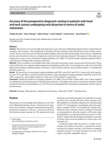
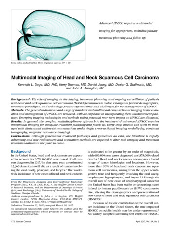
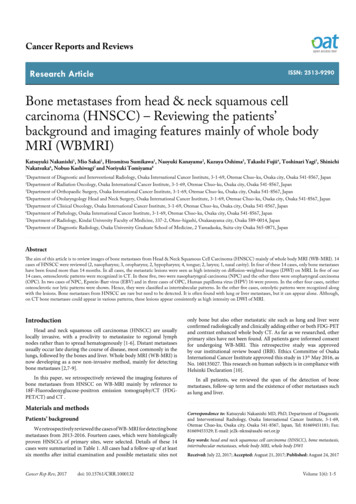
Nakanishi K (2017) Bone metastases from head & neck squamous cell carcinoma (HNSCC) – Reviewing the patients’ background and imaging features mainly ofwhole body MRI (WBMRI)Image parameter of WB-MRIDistribution of bone metastasesA 3 tesla MRI (Magnetom Trio 3T, Siemens, Erlangen, Germany)was utilized. The patients were covered with a 102 channel surfacecoil, positioned head first on an extended anatomical coverage table.Detailed imaging parameters were summarized in Table 2. Total spinesagittal images of STIR and T1WI (Turbo Spin Echo) (Table 2), coronalimages of in phase and out of phase T1WI (Table 3), and axial imagesof DWI from lower neck to proximal femur (Table 4) were captured.In 12 cases, multiple bone metastases were found on WB-MRI. Intwo cases, solitary metastasis was found (case 1 and 12).The time required by the patients to be immobile in the MRI borewas approximately 23 minutes. The total examination time, includingthe coil set up, patient positioning, image processing (MIP, MPR andFusion) and the image layout for reading, was less than 60 minutes.2) Intertrabecular pattern (N 4) (case 6,7,8 and 13): In four cases,CT showed neither osteosclerotic nor lytic changes which suggestedintertrabecular pattern. In these cases, the multiple lesions wereseen as high intensity on DWI and high accumulation on FDG-PETCT (Figure 3).Follow-up term and span of the detection of bone metastasesThe follow-up term from the initial detection ranged from six to97 months and eight patients died due to their primary diseases (cases4,6,7,8,9,11,12 and 13). The span from the initial staging to the detectionof bone metastases ranged from 0 to 42 months (average: 14.3 months).Onset of metastatic siteIn six of these 14 cases, bone metastases were independent newonset metastases (case 1,2,5,6,12 and 13). In four of these six (case2,5,6,12), only bone metastases have been found beyond 14 months.In two cases (case 3 and 4), bone and liver metastases were togetherthe new-onset metastases. Bone and lung metastases (case 8) and boneand adrenal metastases (case 7) were together the new-onset metastases.In three cases (case 9,10 and 11), bone metastases were foundsecondary to lymph node metastases and in one case, they were foundsecondary to lung metastases (case 14).ABEImage findings1) Osteosclerotic pattern (N 5) (case 1-5): In five cases, osteoscleroticpatterns are recognized diffusely in CT (Figures1 and 2). In thesefive cases, these lesions are shown as high intensity on DWI.3) Osteolytic pattern (N 5) (case 9-12 and 14): In the other fivecases, the lesions were shown as osteolytic pattern. In these cases,the multiple lesions were also shown as high intensity on DWI andhigh accumulation on FDG-PET CT (Figure 4).DiscussionIn the field of HNSCC, bone metastases have received less attentionthan pulmonary metastases in patients with HNSCC [1].In our study group, in four (case 2,5,6,12), metastases were foundin bone only. Kim, et al. found four patients of only bone metastases in17 of bone metastases [1].In our results, five cases of HNSCC were shown to be osteosclerotic(Case 1-5). In these five, two were nasopharyngeal carcinoma (NPC)and the other three were oropharyngeal carcinoma (OPC). Metastaticlesions of these five cases reveals as high intensity on DWI.CDFGFigure 1. 42-year-old female with NPC T1N2M0 (Case 2) [A: On initial CT, right Ruby ale lymph node swelling is shown (arrow) and SCC was proved by right nasopharyngeal wall biopsy;B: On PET-MIP image 10 months after primary site detection, accumulated areas are shown in the vertebral bodies (arrows); C: On DWI-MIP image 36 days after FDG-PET (Fig.1B),high intensity areas are also shown in sacrum, L3 and Th5 and are well correlated with PET-MIP image (arrows); D: On PET-CT at the L3 level, same examination of Fig.1B, accumulationis seen (arrow); E: On CT at the L3 level, same examination of Fig 1B and D, subtle osteosclerotic change is seen (white arrow); F: On axial DWI at the L3 level, same examination ofFig.1C, high intensity area is seen (arrow). These areas are judged comprehensively as osteosclerotic bone metastases; G: On DWI coronal reconstructed image 27 months after primarysite detection, new high intensity areas are shown in Th4 and Th10 (arrows). They are judged as new bone metastases.] This patient has been followed up 27 months and is confirmed to bealive. Only bone metastases are confirmed.Cancer Rep Rev, 2017doi: 10.15761/CRR.1000132Volume 1(6): 2-5
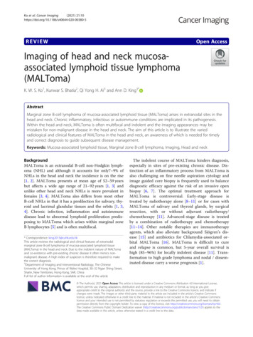
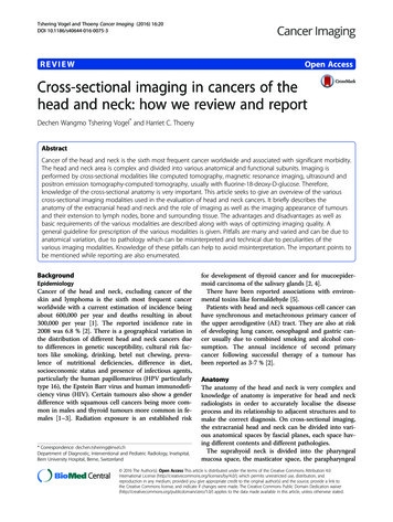
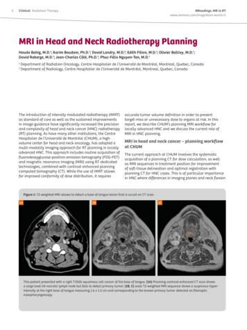
Nakanishi K (2017) Bone metastases from head & neck squamous cell carcinoma (HNSCC) – Reviewing the patients’ background and imaging features mainly ofwhole body MRI (WBMRI)ABCEDGFFigure 2. A 53-year-old male with OPC (Case 3). Its initial stage was T4aN2cM0 [A: On initial CT, mass is shown on the left oropharyngral wall (arrows) and SCC was proved by biopsy;B: On CT image 33 months after initial CT (Fig.2A), at the Th7 level, osteosclerotic pattern is shown (white arrow); C: On CT same examination of Fig.2B, at the right proximal femur level,osteosclerotic pattern is shown (white arrow). Also in the left ischium, osteosclerotic pattern is shown (white arrowhead); D: On WB-MRI examination 28 days after CT (Fig.2B andC), in coronal reconstructed fused T1WI and DWI, on the right pedicle of Th10, high intensity area is shown (white arrow) and diagnosed as bone metastasis; E: In same examination ofFig.2D, also on the right proximal femur, high intensity area is shown (white arrow) and diagnosed as bone metastasis. In both lesions, high accumulations are seen in PET-CT also andthey are judged comprehensively as osteosclerotic metastases; F: In same WB-MRI examination of Fig.2D and E, high intensity area is shown in the liver (white arrow); : G: On PET-CT,high accumulation is shown in the liver (white arrow) and it matches the MR Image. It is judged as liver metastasis.] This patient has been followed up 72 months and is confirmed to be alive.ADBECFFigure 3. A 60-year-old male with bone metastases from HPC (Case 7). The initial stage was T2N2bM0 [A: On initial CT, mass is occupied in the right piriform fossa (white arrow) andlymph node is also shown in the right jugular chain (arrow). SCC was proved by biopsy; B: On MIP image of FDG-PET 7 months after initial CT (Fig.3A), multiple accumulated areasare shown (arrows). These lesions were judged as multiple bone metastases, comprehensively; C: In WB-MRI examination 10 months after initial CT (Fig.3A), in MIP image of DWI,multiple high intensity areas are shown in ribs, bilateral femurs and ilium and so on (arrows). It correlated well with PET-MIP image; D: In original axial image of DWI at the ilium level,multiple high intensity areas of bone metastases are shown (arrows); E: In bone window CT, which is included in PET-CT examination of Fig.3C, on the almost same level of Fig. 3D,neither osteosclerotic nor lytic patterns were shown in the ilium. These lesions were classified as intertrabecular metastases; F: In PET-CT, on the level of adrenal gland, high accumulationis shown in the left adrenal gland and judged as adrenal metastasis (white arrow).] This patient died 20 months after primary site detection.Cancer Rep Rev, 2017doi: 10.15761/CRR.1000132Volume 1(6): 3-5
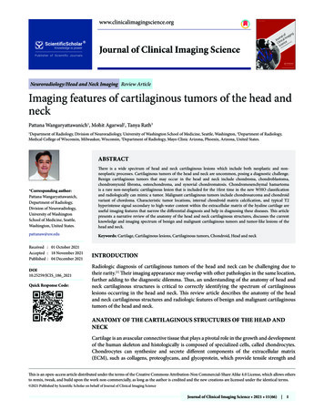
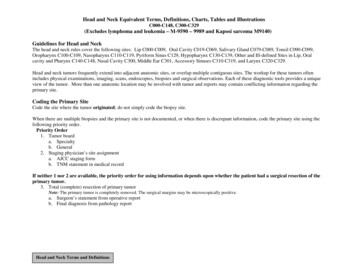
Nakanishi K (2017) Bone metastases from head & neck squamous cell carcinoma (HNSCC) – Reviewing the patients’ background and imaging features mainly ofwhole body MRI (WBMRI)ABCFigure 4. A 57-year-old female with bone metastases from tongue carcinoma (case 11). The initial stage was T2N0M0 [A: On initial PET-CT, high accumulation is shown in the middleapex of the tongue (arrow) and SCC was proved by biopsy; B: In CT, after 19 months after initial PET-CT, on the level of pelvis, osteolysis and discontinuity of cortex of the right ilium isshown (arrow) and also osteolysis is shown in the left sacrum (white arrow); C: In DWI of WB-MRI examination 15 days after CT (Figure 4B), high intensity areas are shown in the rightilium and left sacrum (arrows).] These areas were judged as osteolytic metastases comprehensively. In this case, lung and liver metastases appeared at the same time. This patient died 24months after primary site detection.Table 1. The list of the cases of bone metastases from rev. tr.Follow-up(mth.)Span(mth.)Onset of Metastaticsite ProgressionDWIsignalDistributionCT findingsRemarks154/MNPC/SCCT2bN2M0CRT38 (alive)0Bone 7 aN2cM0CRT72 (alive)25Bone &LiverMultiplehighScleroticHPV 16460/MOPC/SCCT3N2cM0CRT32 (died)11Bone & LiverMultiplehighScleroticHPV 16552/MOPC/SCCT2N2bM0RT30 (alive)12BoneMultiplehighScleroticHPV 16Intertrabecular669/MHPC/SCCT4N2bM0Op. RT97 (died)21BoneMultiplehigh760/ MHPC/SCCT2N2bM0CRT20 (died )5Bone. adr. gld. & CRT18 (died)14Bone & M0Op. RT14 (died)8LN Bone BrainSternum, C6, lthumeurshighLytic1036/FTongue/SCCT1N0M0Op RT34 (alive)24LNBone LiverMultiplehighLytic1157/FTongue/SCCT2N0M0Op.24 (died)16Bone, LN, Lung &LiverIlium, SacrumhighLytic126 6/MLarynx/ SCCT2N2bM0RT25 (died )11BoneL5highLytic1358/MLarynx/SCCT3N0M0CRT6 (died)4Bone LungMultiplehighIntertrabecular1445/MNasal cavity/SCCT2N0M0Op. RT67 (alive)42LungBoneC6, SacrumhighLyticNPC: Nasopahryngeal carcinoma; OPC: Oropha1yngcal carcinoma; HPC: Hvpopharyngeal carcinoma; Prev. tr.: Previous treatment; CRT: Chemotherapy and radiotherapy; RT:Radiotherapy; Op.: Operation; adr. gld.: Adrenal gland; LN: Lymph node; DWI: Diffusion-weighted images; EBV: Epstein-Barr virus; HPV16: Human pappillorna virus16Table 2. Protocol of WB-MRI: total spine sagittal sequences.Table 4. Protocol of WB-MRI: diffusion-weighted axial images.1.T1WI fast SE2. STIRTR/TE/Tl (msec.)14000/60/240TR/TE/Tl (msec.)600/9.46000/80/9.4ETL214ETL1Slice thickness(mm)44Slice thickness(mm)4 without interslice gapFOV of each station(mm)420420FOV of each station(mm)450Matrix336 448250 384No. of stations22Time of each station1 min. 14 sec.2 min. 2 sec.OtherFip angle 120 deg.Table 3. Protocol of WB-MRI: body Coronal sequences.TR/TE (msec.)120/1.2 (opposed phase), 2.5 (in phase)ETL1Slice thickness(mm)7FOV of each station(mm)480Matrix256 179No. of stations2 with 2 acquisitionsTime of each station (sec.)23 per an acquisitionOtherBreath hold scanThe numbers of these cases are too few. However, NPC is closelyrelated to Epstein-Barr virus (EBV), peculiar natural history and a goodprognosis [11] and OPC is also closely related with Human papillomavirus (HPV) 16 [12]. Indeed, in our two cases of NPC, EBV and alsoin our three cases of OPC, HPV 16 were proven. Hence, these findingsCancer Rep Rev, 2017doi: 10.15761/CRR.1000132Matrix79 140No. of stations4 or 5Time of each station2 min 39 sec.Otherb value 0 and 800 mmmight be the features of SCC of NPC and OPC, compared with theother HNSCC.We have to pay attention to the four cases of intertrabecular pattern.This pattern is clinically problematic in overall bone metastases, notexclusively in HNSCC [13,14]. In this pattern, WB-MRI includingDWI, as well as FDG-PET can detect this abnormality but conventionalCT or Technecium-99m bone scintigraphy cannot.SummaryBone metastases from HNSCC are rare but need to be detected. It isusually found with lung or liver metastases. However, bone metastasisalone is a possible condition. CT reveals as various patterns and DWIof MRI as high intensity, consistently. In the case of NPC and OPCwhich are closely related to viral infection, osteosclerotic metastasesVolume 1(6): 4-5
Nakanishi K (2017) Bone metastases from head & neck squamous cell carcinoma (HNSCC) – Reviewing the patients’ background and imaging features mainly ofwhole body MRI (WBMRI)might be characteristic. WB-MRI is a promising tool for detecting bonemetastases of HNSCC.AcknowledgmentAuthors acknowledge the MRI technical staff for cooperating withthe rapid and exact image processing of WB-MRI.References1. Kim MR, Roh JL, Kim JS, Choi SH, Nam SY, et al. (2008) 18F-fluorodeoxyglucosepositron emission tomography and bone scintigraphy for detecting bone metastases inpatients with malignancies of the upper aerodigestive tract. Oral Oncol 44: 148-152.[Crossref]2. Noij DP, Boerhout EJ, Pieters-van den Bos IC, Comans EF, Oprea-Lager D, et al.(2014) Whole-body-MR imaging including DWIBS in the work-up of patients withhead and neck squamous cell carcinoma: a feasibility study. EJR Eur J Radiol 83:1144-1151. [Crossref]3. Peters TT, Senft A, Hoekstra OS, Castelijns JA, Witte BI, et al. (2015) Pretreatmentscreening on distant metastases and head and neck cancer patients: Validation of riskfactors and influence on survival. Oral Oncol 51: 267-271. [Crossref]4. de Bree R, Haigentz M Jr, Silver CE, Paccagnella D, Hamoir M, et al. (2012) Distantmetastases from head and neck squamous cell carcinoma. Part II. Diagnosis. OralOncol 48: 780-786. [Crossref]5. Yi X, Fan M, Liu Y, Zhang H, Liu S (2013) 18 FDG PET and PET-CT for the detectionof bone metastases in patients with head and neck cancer. A meta-analysis. J MedImaging Radiat Oncol 57: 674-679. [Crossref]6. Buckley JG, Ferlito A, Shaha AR, Rinaldo A (2001) The treatment of distant metastasesin head and neck cancer--present and future. ORL J Otorhinolaryngol Relat Spec 63:259-264. [Crossref]7. Pearce T, Philip S, Brown J, Koh DM, Burn PR (2012) Bone metastases from prostate,breast and multiple myeloma: differences in lesion conspicuity at short-tau inversionrecovery and diffusion-weighted MRI. Br J Radiol 85: 1102-1106. [Crossref]8. Lecouvet FE, Mouedden J, Collette L, Coche E, Danse E, et al. (2012) Can whole-bodymagnetic resonance imaging with diffusion-weighted imaging replace Tc 99m bonescanning and computed tomography for single-step detection of metastases in patientswith high-risk prostate cancer? Eur Urol 62: 68-75. [Crossref]9. Nakanishi K, Kobayashi M, Nakaguchi K, Kyakuno M, Hashimoto N, et al. (2007)Whole-body MRI for detecting metastatic bone tumor: diagnostic value of diffusionweighted images. Magn Reson Med Sci 6: 147-155. [Crossref]10. Puri KS, Suresh KR, Gogtay NJ, Thatte UM (2009) Declaration of Helsinki, 2008:implications for stakeholders in research. J Postgrad Med 55: 131-134. [Crossref]11. Chiesa F, De Paoli F (2001) Distant metastases from nasopharyngeal cancer. ORL JOtorhinolaryngol Relat Spec 63: 214-216. [Crossref]12. Sanguineti G, Pai S, Agbahiwe H, Ricchetti F, Westra W, et al. (2014) HPV-relatedoropharyngeal carcinoma with Overt Level two? metastases at presentation: The riskof subclinical diseases in ipsilateral levels IB. Acta Oncol 53: 662-668. [Crossref]13. Yamaguchi T, Tamai K, Yamato M, Honma K, Ueda Y, et al. (1996) Intertrabecularpattern of tumors metastatic to bone. Cancer 78: 1388-1394. [Crossref]14. Yamaguchi T (2001) Intertrabecular vertebral metastases: metastases only detectableon MR imaging. Semin Musculoskelet Radiol 5: 171-175. [Crossref]Copyright: 2017 Nakanishi K. This is an open-access article distributed under the terms of the Creative Commons Attribution License, which permits unrestricteduse, distribution, and reproduction in any medium, provided the original author and source are credited.Cancer Rep Rev, 2017doi: 10.15761/CRR.1000132Volume 1(6): 5-5
8Department of Diagnostic Radiology, Osaka University Graduate School of Medicine, 2 Yamadaoka, Suita-city Osaka 565-0871, Japan Abstract The aim of this article is to review images of bone metastases from Head & Neck Squamous Cel