Transcription
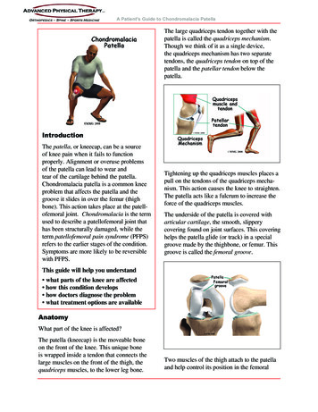
A Patient's Guide to Chondromalacia PatellaThe large quadriceps tendon together with thepatella is called the quadriceps mechanism.Though we think of it as a single device,the quadriceps mechanism has two separatetendons, the quadriceps tendon on top of thepatella and the patellar tendon below thepatella.IntroductionThe patella, or kneecap, can be a sourceof knee pain when it fails to functionproperly. Alignment or overuse problemsof the patella can lead to wear andtear of the cartilage behind the patella.Chondromalacia patella is a common kneeproblem that affects the patella and thegroove it slides in over the femur (thighbone). This action takes place at the patellofemoral joint. Chondromalacia is the termused to describe a patellofemoral joint thathas been structurally damaged, while theterm patellofemoral pain syndrome (PFPS)refers to the earlier stages of the condition.Symptoms are more likely to be reversiblewith PFPS.Tightening up the quadriceps muscles places apull on the tendons of the quadriceps mechanism. This action causes the knee to straighten.The patella acts like a fulcrum to increase theforce of the quadriceps muscles.The underside of the patella is covered witharticular cartilage, the smooth, slipperycovering found on joint surfaces. This coveringhelps the patella glide (or track) in a specialgroove made by the thighbone, or femur. Thisgroove is called the femoral groove.This guide will help you understand what parts of the knee are affected how this condition develops how doctors diagnose the problem what treatment options are availableAnatomyWhat part of the knee is affected?The patella (kneecap) is the moveable boneon the front of the knee. This unique boneis wrapped inside a tendon that connects thelarge muscles on the front of the thigh, thequadriceps muscles, to the lower leg bone.Two muscles of the thigh attach to the patellaand help control its position in the femoral
A Patient's Guide to Chondromalacia Patellagroove as the leg straightens. These musclesare the vastus medialis obliquus (VMO) andthe vastus lateralis (VL). The vastus medialisobliquus (VMO) runs along the inside of thethigh, and the vastus lateralis (VL) lies alongthe outside of the thigh. If the timing betweenthese two muscles is off, the patella may bepulled off track.CausesWhat can cause this problem?Problems commonly develop when the patellasuffers wear and tear. The underlying cartilage begins to degenerate, a condition mostcommon in young athletes. Soccer players,snowboarders, cyclists, rowers, tennis players,ballet dancers, and runners are affected mostoften. But anyone whose knees are under greatstress is at increased risk of developing chondromalacia patella.Wear and tear can develop for several reasons.Acute injury to the patella or chronic frictionbetween the patella and the femur can resultin the start of patellofemoral pain syndrome.Degeneration leading to chondromalacia mayalso develop as part of the aging process, likeputting a lot of miles on a car.The main cause of knee pain associated withpatellofemoral pain syndrome is a problem inthe way the patella tracks within the femoralgroove as the knee moves. Physical andbiomechanical changes alter the stress and loadon the patellofemoral joint.The quadriceps muscle helps control thepatella so it stays within this groove. If partof the quadriceps is weak for any reason,a muscle imbalance can occur. When thishappens, the pull of the quadriceps musclemay cause the patella to pull more to oneside than the other. This in turn causes morepressure on the articular cartilage on oneside than the other. In time, this pressure candamage the articular cartilage leading to chondromalacia patella.Weakness of the muscles around the hip canalso indirectly affect the patella and can leadto patellofemoral joint pain. Weakness of themuscles that pull the hip out and away fromthe other leg, the hip abductor muscles, canlead to imbalances to the alignment of theentire leg - including the knee joint and themuscle balance of the muscles around theknee. This causes abnormal tracking of thepatella within the femoral groove and eventually pain around the patella. Many patients areconfused when their physical therapist beginsexercises to strengthen and balance the hipmuscles, but there is a very good reason thatthe therapist is focusing on this area.A similar problem can happen when thetiming of the quadriceps muscles is off. There
A Patient's Guide to Chondromalacia Patellaare four muscles that form the quadricepsmuscle group. As mentioned earlier, the vastusmedialis obliquus (the muscle on the inside ofthe front of the thigh) and the vastus lateralis(the muscle that runs down the outside partof the thigh) are two of these four muscles.People with patellofemoral problems sometimes have problems in the timing betweenthe VMO and the VL. The VL contracts first,before the VMO. This tends to pull the patellatoward the outside edge of the knee. The resultis abnormal pressure on the articular surface ofthe patella.Another type of imbalance may exist due todifferences in how the bones of the knee areshaped. These differences, or anatomic variations, are something people are born with.Doctors refer to this the “Q angle”. Somepeople are born with a greater than normalangle where the femur and the tibia (shinbone)come together at the knee joint. Womentend to have a greater angle here than men.The patella normally sits at the center of thisangle within the femoral groove. When thequadriceps muscle contracts, the angle in theknee straightens, pushing the patella to theoutside of the knee. In cases where this angleis increased, the patella tends to shift outwardwith greater pressure. This leads to a similarproblem as that described above. As the patellaslides through the groove, it shifts to theoutside. This places more pressure on one sidethan the other, leading to damage to the underlying articular cartilage.Finally, anatomic variations in the bones ofthe knee can occur such that one side of thefemoral groove is smaller than normal. Thiscreates a situation where the groove is tooshallow, usually on the outside part of theknee. People who have a shallow groovesometimes have their patella slip sideways outof the groove, causing a patellar dislocation.This is not only painful when it occurs, but itcan damage the articular cartilage underneaththe patella. If this occurs repeatedly, degeneration of the patellofemoral joint occurs fairlyrapidly.SymptomsWhat does chondromalacia patella feel like?The most common symptom is pain underneath or around the edges of the patella.The pain is made worse by any activitiesthat load the patellofemoral joint, such asrunning, squatting, or going up and downstairs. Kneeling is often too painful to eventry. Keeping the knee bent for long periods, asin sitting in a car or movie theater, may causepain.There may be a sensation like the patella isslipping. This is thought to be a reflex responseto pain and not because there is any instability
A Patient's Guide to Chondromalacia Patellain the knee. Others experience vague pain inthe knee that isn't centered in any one spot.The knee may grind, or you may hear acrunching sound when you squat or go up anddown stairs. If there is a considerable amountof wear and tear, you may feel popping orclicking as you bend your knee. This canhappen when the uneven surface of the underside of the patella rubs against the femoralgroove. The knee may swell with heavy useand become stiff and tight. This is usuallybecause of fluid accumulating inside the kneejoint, sometimes called 'water on the knee'.This is not unique to problems of the patellabut sometimes occurs when the knee becomesinflamed.DiagnosisHow do doctors identify this problem?Diagnosis begins with a complete history ofyour knee problem followed by an examination of the knee, including the patella. X-raysmay be ordered on the initial visit to yourdoctor. An X-ray can help determine if thepatella is properly aligned in the femoralgroove. Several X-rays taken with the kneebent at several different angles can help determine if the patella seems to be moving throughthe femoral groove in the correct alignment.The X-ray may show arthritis between thepatella and thighbone, especially when theproblems have been there for awhile.Diagnosing problems with the patella canbe confusing. The symptoms can be easilyconfused with other knee problems, becausethe symptoms are often similar. In thesecases, other tests, such as magnetic resonanceimaging (MRI), may be suggested. The MRImachine uses magnetic waves rather thanX-rays to show the soft tissues of the body.This machine creates pictures that look likeslices of the knee. Usually, this test is done tolook for injuries, such as tears in the meniscior ligaments of the knee. Recent advances inthe quality of MRI scans have enabled doctorsto see the articular cartilage on the scan anddetermine if it is damaged. This test doesnot require any needles or special dye and ispainless.In some cases, arthroscopy may be used tomake the definitive diagnosis when there isstill a question about what is causing yourknee problem. Arthroscopy is an operation thatinvolves placing a small fiber-optic TV camerainto the knee joint, allowing the surgeon tolook at the structures inside the joint directly.The arthroscope allows your doctor to see thecondition of the articular cartilage on the backof your patella. The vast majority of patellofemoral problems are diagnosed withoutresorting to surgery, and arthroscopy is usuallyreserved to treat the problems identified byother means.There is no clear link between the severity ofsymptoms and X-ray or arthroscopic findings.
A Patient's Guide to Chondromalacia PatellaMost often, the doctor relies upon the history,symptoms, and results of the examination.TreatmentHow do doctors treat the condition?Nonsurgical TreatmentNonoperative treatment is usually recommended for this problem. Getting the painand inflammation under control is the firststep. The overall goals for a rehab plan are toimprove muscle function and flexibility whileproviding pain relief or pain control.Your physician may suggest rest and antiinflammatory medications, such as aspirinor ibuprofen, especially when the problemis coming from overuse. Acetaminophen(Tylenol ) may be used for pain control if youcan’t take anti-inflammatory medications forany reason. Activity modification, flexibility,and strengthening are key parts of the rehabprogram. Physical therapy can help in the earlystages by decreasing pain and inflammation.Your physical therapist may use ice massageand ultrasound to limit pain and swelling.Bracing or taping the patella can help you doexercises and activities with less pain. Mostbraces for patellofemoral problems are madeof soft fabric, such as cloth or neoprene. Youslide them onto your knee like a sleeve. Asmall buttress pads the side of the patella tokeep it lined up within the groove of the femur.An alternative to bracing is to tape the patellain place. The therapist applies and adjusts thetape over the knee to help realign the patella.The idea is that by bracing or taping the knee,the patella stays in better alignment withinthe femoral groove. This in turn is thought toimprove the pull of the quadriceps muscle sothat the patella stays lined up in the groove.Patients report less pain and improved functionwith these forms of treatment.As the pain and inflammation becomecontrolled, your physical therapist will workwith you to improve flexibility, strength,and muscle balance in the knee. Quadricepsstrengthening exercises to address deficits inknee extension strength include non-weightbearing single-joint (e.g., knee extension) andweight-bearing multiple-joint exercises (e.g.,seated leg press).Non-weight bearing exercises are also knownas open kinetic chain exercise. Weight-bearingexercises are referred as closed-chain exercise.Closed-chain exercises place less stress on thepatellofemoral joint and may be used first toachieve improved function before progressingto open kinetic chain exercises. Studies alsoshow greater VMO activity with closed kineticchain exercise. And a closed-chain exerciseprogram also addresses hip muscle weaknessat the same time as knee muscle deficits.The therapist will adjust your rehab program toprovide you with the most pain free and effective method of treatment. The overall rehabprogram will include a home exercise programof stretching, agility exercises, balance activities, and strengthening designed to return youto your former level of pain free participationin sports and other activities.SurgeryIf nonsurgical treatment fails to improve yourcondition, surgery may be suggested. Theprocedure used for patellofemoral problemsvaries. In severe cases a combination of oneor more of the following procedures may benecessary.Arthroscopic MethodArthroscopy is sometimes useful in the treatment of patellofemoral problems of the knee.Looking directly at the articular cartilagesurfaces of the patella and the femoral grooveis the most accurate way of determining howmuch wear and tear there is in these areas.Your surgeon can also watch as the patellamoves through the groove, and may be ableto decide whether or not the patella is moving
A Patient's Guide to Chondromalacia Patellanormally. If there are areas of articular cartilage damage behind the patella that arecreating a rough surface, special tools can beused by the surgeon to smooth the surface andreduce your pain. This procedure is sometimesreferred to as shaving the patella.Cartilage ProcedureIn more advanced cases of patellar arthritis,surgeons may operate to repair or restore thedamaged cartilage. The type of surgery neededfor articular cartilage is based on the size,type, and location of the damage. Along withsurgical treatment to fix the cartilage, otherprocedures may also be done to help align thepatella so less pressure is placed on the healingcartilage.Lateral ReleaseIf your patella problems appear to be caused bya misalignment problem, a procedure called alateral release may be suggested.This procedure is done to allow the patellato shift back to a more normal position andrelieve pressure on the articular cartilage.In this operation, the tight ligaments on theoutside (lateral side) of the patella are cut,or released, to allow the patella to slide moretowards the center of the femoral groove.These ligaments eventually heal with scartissue that fills in the gap created by thesurgery, but they no longer pull the patella toCompliments of:the outside as strongly as before the surgery.This helps to balance the quadriceps mechanism and equalize the pressure on the articularcartilage behind the patella.Ligament Tightening ProcedureIn some cases of severe patellar misalignment, a lateral release alone may not beenough. For problems of repeated patellardislocations, the surgeon may also need torealign the quadriceps mechanism. In additionto the lateral release, the tendons on the insideedge of the knee (the medial side) may haveto be tightened as well.Bony RealignmentIf the misalignment is severe, the bony attachment of the patellar tendon may also haveto be shifted to a new spot on the tibia bone.Remember that the patellar tendon attachesthe patella to the lower leg bone (tibia) justbelow the knee. By moving a section of bonewhere the patellar tendon attaches to the tibia,surgeons can change the way the tendon pullsthe patella through the femoral groove.The surgeon removes a section of bone wherethe patellar tendon attaches on the tibia. Thissection of bone is then reattached on the tibiacloser to the other knee.Usually, the bone is reattached onto the tibiausing screws. This procedure shifts the patellato the medial side. Once the surgery heals, thepatella should track better within the center of
A Patient's Guide to Chondromalacia Patellathe groove, spreading the pressure equally onthe articular cartilage behind the patella.Arthroscopic procedures to shave the patella ora simple lateral release can usually be done onan outpatient basis, meaning you can leave thehospital the same day. If your problem requiresthe more involved surgical procedure wherebone must be cut to allow moving the patellartendon attachment, you may need to spend oneor two nights in the hospital.RehabilitationWhat should I expect after treatment?Nonsurgical RehabilitationPatients with chondromalacia may benefit fromfour to six weeks of physical therapy. The aimof treatment is to calm pain and inflammation,to correct muscle imbalances, and to improvefunction of the patella.It is important to understand the need to keepyour activity level below what will triggermore pain and tissue damage. You may need toice your knee during the day and limit certainactivities such as stairs, squatting, or running.Good results can be expected when workingslowly but steadily on flexibility and strengthening exercises. The moto of "no pain, nogain" does not apply to this problem. The mostsuccessful program is one of common sense.If an activity causes pain, then reduce thefrequency, intensity, or duration of that activityuntil you are once again pain free. Graduallybuild up what you can do while maintainingyour pain free status.Muscle imbalances are commonly treated withstretching and strengthening exercises butimprovement usually takes at least six to eightweeks. You may need to continue a modifiedprogram of flexibility and strengthening exercises to maximize control and strength of thequadriceps muscles. This type of program donetwo to three times each week may be neededCompliments of:for several more months (or longer if youcontinue to experience pain during progressivesports participation).After SurgeryMany surgeons will have their patients takepart in formal physical therapy after kneesurgery for patellofemoral problems. Patientsundergoing a patellar shaving usually beginrehabilitation right away. More involvedsurgeries for patellar realignment or restorativeprocedures for the articular cartilage requirea delay before going to therapy. And rehabilitation may be slower to allow the bone orcartilage to heal before too much strain can beput on the knee.The first few physical therapy treatments aredesigned to help control the pain and swellingfrom the surgery. The physical therapist willchoose exercises to help improve knee motionand to get the quadriceps muscles toned andactive again. Muscle stimulation, using electrodes over the quadriceps muscle, may beneeded at first to get the muscle moving again.As the program evolves, more challengingexercises are chosen to safely advance theknee's strength and function. The key is toget the soft tissues in balance through safestretching and gradual strengthening.The physical therapist's goal is to help youkeep your pain under control, ensure you placeonly a safe amount of weight on the healingknee, and improve your strength and range ofmotion. When you are well under way, regularvisits to the therapist's office will end. Thetherapist will continue to be a resource, but youwill be in charge of doing your exercises aspart of an ongoing home program.
program. Physical therapy can help in the early stages by decreasing pain and inflammation. Your physical therapist may use ice massage and ultrasound to limit pain and swelling. Bracing or taping the patella can help you do exercises and activities with le