Transcription
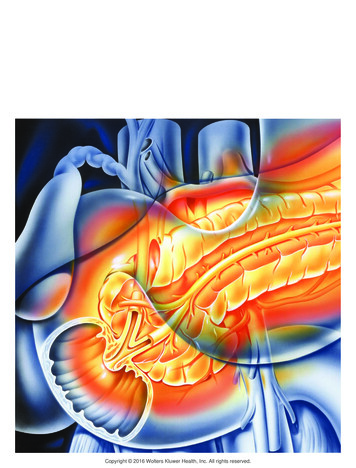
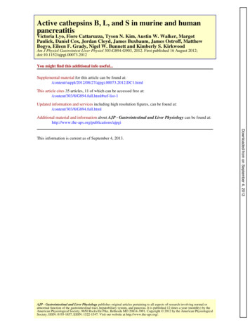
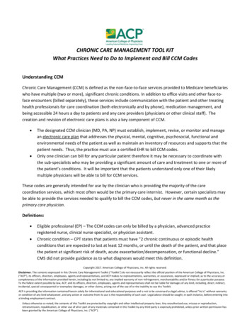
Chronic Pancreatitis:IntroductionAuthors: Anthony N. Kalloo, MD; Lynn Norwitz, BS; Charles J. Yeo, MDChronic pancreatitis is a relatively rare disorder occurring in about 20 per 100,000 population. The disease is progressive withpersistent inflammation leading to damage and/or destruction of the pancreas. Endocrine and exocrine functional impairment resultsfrom the irreversible pancreatic injury.The pancreas is located deep in the retroperitoneal space of the upper part of the abdomen (Figure 1). It is almost completelycovered by the stomach and duodenum. This elongated gland (12–20 cm in the adult) has a lobe-like structure. Variation in shapeand exact body location is common. In most people, the larger part of the gland's head is located to the right of the spine or directlyover the spinal column and extends to the spleen. The pancreas has both exocrine and endocrine functions. In its exocrine capacity,the acinar cells produce digestive juices, which are secreted into the intestine and are essential in the breakdown and metabolism ofproteins, fats and carbohydrates. In its endocrine function capacity, the pancreas also produces insulin and glucagon, which aresecreted into the blood to regulate glucose levels.Figure 1. Location of thepancreas in the body.What is Chronic Pancreatitis?Chronic pancreatitis is characterized by inflammatory changes of the pancreas involving some or all of the following: fibrosis, calcification, pancreatic ductalinflammation, and pancreatic stone formation (Figure 2).Although autopsies indicate that there is a 0.5–5% incidence of pancreatitis, the true prevalence is unknown.In recent years, there have been several attempts to classify chronic pancreatitis, but these have met with difficulty for several reasons. Classification based onhistological appearance (obstructive versus calcific pancreatitis) has not proven to be useful. There is ambiguity in nomenclature, a lack of consensus amonginvestigators, and finally, histology is not always available. Efforts to classify this disorder based on etiology appear to be a more practical approach.SymptomsThe clinical presentation of chronic pancreatitis is usually abdominal pain, ranging from a sudden acute abdominal catastrophe to mild episodes of deep epigastricpain and possible vomiting.Chronic pancreatitis may produce constant, dull, unremitting abdominal pain, epigastric tenderness, weight loss, steatorrhea and glucose intolerance. Diarrhea maybe chronic, with as many as six or more bowel movements per day. The diarrhea is caused by fat malabsorption, which results in bulky, foul-smelling stools that mayappear oily and float (steatorrhea). With the head of the gland on the right side, lying within the curve of the duodenum at the second lumbar vertebra (L2) level of thespine (Figure 3), the pain of chronic pancreatitis often radiates to the back, although it may radiate to both upper and lower quadrants. Sitting up and leaning forwardmay help to relieve or reduce the discomfort.Figure 3. Typical posture to reduce pancreatic-type pain. Copyright 2001-2013 All Rights Reserved.600 North Wolfe Street, Baltimore, Maryland 21287
Chronic Pancreatitis:AnatomyThe pancreas lies behind the peritoneum of the posterior abdominal wall and is oblique in orientation. The head of the pancreas is on the right side and lies within the"C" curve of the duodenum at the second vertebral level (L2). The tip of the pancreas extends across the abdominal cavity almost to the spleen. Collecting ductsempty digestive juices into the pancreatic duct, which runs from the head to the tail of the organ. The pancreatic duct empties into the duodenum at the duodenalpapilla, alongside the common bile duct (Figure 4).Figure 4. Normal anatomy.The duct of Wirsung is the main pancreatic duct extending from the tail of the organ to the major duodenal papilla, or ampulla of Vater. The widest part of the duct is inthe head of the pancreas (4 mm), tapering to 2 mm at the tail in adults. The duct of Wirsung is close, and almost parallel, to the distal common bile duct beforecombining to form a common duct channel prior to approaching the duodenum. In approximately 70% of people, an accessory pancreatic duct of Santorini (dorsalpancreatic duct) is present. This duct may communicate with the main pancreatic duct. The degree of communication of the dorsal and ventral duct varies frompatient to patient (Figure 5A).Figure 5. A, Anatomy of the major and minor papilla; B, sphincter of Oddi; C, endoscopic view.Smooth circular muscle surrounding the end of the common bile duct (biliary sphincter) and main pancreatic duct (pancreatic sphincter) merge at the level of theampulla of Vater and is called the sphincter of Oddi (Figure 5A). This musculature is embryologically, anatomically, and physiologically different from the surroundingsmooth musculature of the duodenum. The normal appearance through the endoscope includes the major and minor papilla. The major papilla extends 1 cm into theduodenum with an orifice diameter of 1 mm. The minor papilla is 20–30 mm proximal and medial to the major papilla. Its orifice is tiny and may be difficult to identify(Figure 5B). Dysfunction of the pancreatic sphincter may result in unexplained abdominal pain or pancreatitis. The sphincter of Oddi is a dynamic structure thatrelaxes (Figure 6A) and contracts (Figure 6B) to change the dimensions of the ampulla of Vater.Figure 6. A, B, Function of the sphincter of Oddi.
Regions of the PancreasThe pancreas may be divided into five major regions—the head, neck, body, tail, and uncinate process (Figure 7). The distal end of the common bile duct can befound behind the upper border of the head of the pancreas. This duct courses through the posterior aspect of the pancreatic head before passing through the head toreach the ampulla of Vater (major papilla). The uncinate process is the segment of pancreatic tissue that extends from the posterior of the head. The neck of thepancreas, a part of the gland 3–4 cm wide, joins the head and body. The pancreatic body lies against the aorta and posterior parietes, and anteriorly contacts theantrum of the stomach.Figure 7. Regions of the pancreas. Copyright 2001-2013 All Rights Reserved.600 North Wolfe Street, Baltimore, Maryland 21287
Chronic Pancreatitis:CausesAlcoholThe most common cause of chronic pancreatitis in Western societies is alcohol. Alcohol consumption has been implicated in approximately 70% of cases as a majorcause of this disease. Developing between 30 and 40 years of age, this chronic pancreatitis is more common in men than in women. A direct relationship existsbetween the daily consumption of alcohol and the risk of development of chronic pancreatitis. Although the length of time required to produce symptoms is unknown,there is clearly a correlation between the quantity and duration of alcohol consumption and the risk of developing chronic pancreatitis. It is estimated that alcoholintake greater than 20g per day over a period of 6–12 years produces changes consistent with chronic pancreatitis. There are several theories regarding themechanism by which alcohol produces chronic pancreatitis; however, the exact mechanism is unknown.Pancreas DivisumThe most common congenital anomaly of the pancreas, pancreas divisum, occurs in approximately 10% of the population and results from incomplete or absentfusion of the dorsal and dorsal ducts during embryological development. In pancreas divisum, the ventral duct of Wirsung empties into the duodenum through themajor papilla, but drains only a small portion of the pancreas (ventral portion). Other regions of the pancreas, including the tail, body, neck, and the remainder of thehead, drain secretions into the duodenum through the minor papilla via the dorsal duct of Santorini (hence the term dominant dorsal duct syndrome) (Figure 8).Recent clinical trials have supported the concept that obstruction of the minor papilla may cause acute pancreatitis or chronic pancreatitis in a subgroup of patientswith pancreas divisum. Endoscopic or surgical therapy directed to the minor papilla has been effective in treating these patients.Figure 8. Anatomy of pancreas divisum.Tropical PancreatitisTropical pancreatitis is found predominantly throughout Asia, Africa and other tropical locales. Men and women are affected with equal frequency without any knownetiological factors. Young people (mean age at onset, 12–15 years) may be affected. Although the etiology is speculative, malnutrition may play an important role in itspathogenesis. Patients usually develop recurrent abdominal pain in childhood and diabetes mellitus later in life. Pancreatic stone formation is present in the majorityof these patients.HyperparathyroidismChronic pancreatitis occurs in untreated hyperparathyroidism. Hypercalcemia is thought to be the mechanism by which hyperparathyroidism causes chronicpancreatitis. Hypercalcemia causes an increase in calcium secretion by the pancreas. In the animal model, hypercalcemia causes stimulation of the acinar cell,increases calcium secretion, and alters the diffusion barrier between the pancreatic duct lumen and the interstitial space.TraumaTrauma to the back or abdomen may produce pancreatic injury (Figure 9A) leading to chronic pancreatitis. Trauma may result in inflammation and the formation ofpseudocysts or strictures (Figure 9B). Many cases of pancreatic injury are associated with ductal disruption.Figure 9. A, B, Pancreatic injury from trauma.Obstructive pancreatitisChronic pancreatitis is also associated with obstruction of the pancreatic duct secondary to strictures related to pancreatic inflammation, or benign or malignant
tumors. Sphincter of Oddi dysfunction involving the pancreatic or ampullary sphincter of the duct is thought to be another cause. Pathological findings in this type ofpancreatitis include the absence of intraductal calculi or plugs, and uniform dilation of the duct distal to the obstruction site.Idiopathic Chronic PancreatitisIdiopathic chronic pancreatitis is the major form of nonalcoholic disease in Europe and North America, occurring in 10–40% of those with chronic pancreatitis. Thisform of pancreatitis affects juveniles, with an onset of symptoms at a median age of 18. The senile type appears to peak at 60 years of age. Arteriosclerosis has beensuggested as a cause of senile chronic pancreatitis, although firm evidence implicating vascular insufficiency is lacking.Cystic FibrosisThere is an association between patients with pancreatitis and mutation of the cystic fibrosis transmembrane conductance regulator (CFTR) gene. In this subset ofpatients, there is no evidence of cystic fibrosis lung disease. It is possible that some patients who carry the diagnosis of idiopathic pancreatitis may well havepancreatitis as a result of this genetic mutation.Hereditary Chronic PancreatitisHereditary chronic pancreatitis appears in childhood at a mean age of 10–12 years. This form of pancreatitis affects familial groups and involves a small number ofrelated individuals. Hereditary chronic pancreatitis is transmitted through an autosomal dominant gene of incomplete penetrance, and the incidence is relatively equalin both sexes. The majority of these patients express one of two mutations (which are R122H or N29I) in the cationic trypsinogen gene (PRSS1 gene). This defectprevents deactivation of trypsinogen resulting in autodigestion. Hereditary chronic pancreatitis is characterized by recurrent attacks of abdominal pain. Diabetesdevelops in approximately 20% of these patients 8–10 years after the onset of pain. Hereditary chronic pancreatitis has about a 40% lifetime risk of pancreatic cancerwith patients in the fifth to seventh decades of life having the highest risk. Copyright 2001-2013 All Rights Reserved.600 North Wolfe Street, Baltimore, Maryland 21287
Chronic Pancreatitis:DiagnosisOverviewOver the years, numerous tests have been developed to diagnose chronic pancreatitis; however, their sensitivity and specificity are poor. To date, historicalinformation, serum enzymes, exocrine function, and radiographic studies seem to be the most reliable indicators of the disease. Biochemical measurements ofpancreatic function are helpful.Biochemical MeasurementsIsoamylase, lipase, trypsin, and elastase levels may be low, normal, or elevated in patients with chronic pancreatitis. In early or mild cases of chronic pancreatitis, it isdifficult to make a definitive diagnosis based on serum enzyme levels alone.The secretin stimulation test is the most sensitive test to diagnose early pancreatic disease in patients who have developed malabsorption problems.The bentiromide test is inexpensive, convenient, and easily available for diagnosis of advanced disease. This test, however, has a low sensitivity for diagnosing earlyor mild chronic pancreatitis. Essentially a urine test, it requires normal renal function, adequate diuresis, and proper absorption in the intestines. Para-aminobenzoicacid (PABA) is the result of the synthetic tripeptide bentiromide, cleaved by pancreatic chymotrypsin, in the duodenum and excreted in the urine (Figure 10A). Patientsconsistently excrete less PABA with chronic pancreatitis because of impaired chymotrypsin secretion by the pancreas (Figure 10B).Figure 10. Bentiromide test; A, para-aminobenzoic acid (PABA) excreted in urine; B, chronicpancreatitis — little or no PABA in the urine.The quantitative measurement of fecal fat is diagnostic in determining malabsorption. In this test, a known quantity of dietary fat is consumed. Normally 7% or less ofthe ingested fat is detectable in the stool. In chronic pancreatitis, a two-stage test is more sensitive and specific. The test uses fecal collection with and without theuse of pancreatic enzyme replacement to differentiate steatorrhea secondary to chronic pancreatitis from other causes. Steatorrhea due to chronic pancreatitis ariseswhen 90% of pancreatic exocrine function has been lost.Plasma cholecystokinin (CCK) may be elevated in chronic pancreatitis patients compared with those with normal pancreatic function.Tests of pancreatic exocrine function may directly measure enzyme or bicarbonate secretions, or indirectly demonstrate malabsorption of a compound that requirespancreatic digestion for normal absorption. None of the methods targeted at exocrine function are absolutely accurate in terms of assessing exocrine secretion. Inaddition, none of these secretion assays appears to be able to differentiate chronic pancreatitis from carcinoma of the pancreas.Radiological TestingPlain Abdominal FilmA plain film of the abdomen is usually the first diagnostic test used to establish a diagnosis of chronic pancreatitis. Diffuse, speckled calcification of the gland maysuffice as a positive finding (Figure 11).Figure 11. Abdominal x-ray showing diffuse calcification.Transabdominal UltrasoundTransabdominal ultrasound is a simple, noninvasive, and relatively inexpensive imaging technique. Findings of a dilated pancreatic duct (greater than 4 mm),calcification, and large cavities (greater than 1 cm) are associated with chronic pancreatitis. With a 70% sensitivity and 90% specificity, a satisfactory ultrasoundexamination negates the need for additional confirmatory testing.
Computed Tomography (CT)More sensitive than transabdominal ultrasound, CT (computed tomography) scanning can demonstrate duct dilation, cystic lesions, and calcification (Figure 12). Thistechnique is useful in discriminating chronic pancreatitis from pancreatic carcinoma.Figure 12. CT scan demonstrating chronic pancreatitis.Magnetic Resonance Cholangiopancreatography (MRCP)Magnetic resonance cholangiopancreatography (MRCP) represents a major advance in the demonstration of pancreatic ductal anatomy. MRCP yields satisfactorypancreatograms in patients with chronic pancreatitis in whom a CT scan showed no abnormalities. No ductal or intravenous injection of contrast medium is necessary,and the patient is not exposed to irradiation. MRCP is derived from an enhanced MRI and may be adjusted to optimally visualize the biliary and pancreatic ducts(Figure 13). Dynamic secretin magnetic resonance pancreatography (DSMRP) has further advanced pancreatic visualization. DSMRP may improve the clinician'sability to detect early chronic pancreatitis. Further studies are needed to fully assess this novel approach.Figure 13. MRCP demonstrating chronic pancreatitis.Endoscopic DiagnosisGastrointestinal endoscopy allows the physician to visualize and biopsy the mucosa of the upper gastrointestinal tract. Endoscopy permits visualization of theesophagus, stomach and duodenum. The enteroscope allows visualization of at least 50% of the small intestine, including most of the jejunum and different degreesof the ileum. During these procedures, the patient may be given a pharyngeal topical anesthetic that helps to prevent gagging. Pain medication and a sedative mayalso be administered before the procedure. The patient is placed in the left lateral position (Figure 14).Figure 14. Room set-up and patient positioning for ERCP.An endoscope is a thin, flexible, lighted tube that is passed through the mouth and pharynx and into the esophagus. The endoscope transmits an image of theesophagus, stomach and duodenum to a monitor, which is visible to the physician. The endoscopy room is equipped with an x-ray machine and monitor screen, whichare used to help identify bile and pancreatic ducts. The endoscope introduces air into the stomach, expanding the folds of tissue and enhancing the examination ofthe stomach.
Figure 15. A, B, Position of the scope in the duodenum for ERCP.Endoscopic Retrograde Cholangiopancreatography (ERCP)ERCP is an endoscopic technique for visualization of the bile and pancreatic ducts. During this procedure, the physician inserts a side-viewing endoscope (Figure 16)in the duodenum facing the major papilla (Figure 15B). The side-viewing scope (duodenoscope) is specially designed to facilitate placement of endoscopicaccessories into the bile and pancreatic ducts. The endoscopic accessories may be passed through the biopsy channel (Figure 16) into the ducts. A catheter is usedto inject dye into both pancreatic and biliary ducts to obtain x-ray images using fluoroscopy (Figure 14). During the procedure, the physician is able to see two sets ofimages: the endoscopic image of the duodenum and major papilla, and the fluoroscopic image of the bile and pancreatic ducts.Figure 16. Side-viewing endoscope.The endoscope is designed to be held in the left hand, with the thumb operating up and down angulation. The index finger operates the suction and air/wateroperations. The right hand is responsible for advancing, withdrawing and torquing the insertion tube. The right hand also operates left and right angulation of theendoscope and passes accessories through the instrument.A variety of instruments can be utilized through the endoscope (Figure 15B). Electrosurgical devices, such as snares, biopsy forceps, heater probes; BICAP devicesfor polyp removal and cauterization, dilation balloons, stents, catheters, and esophageal prostheses can be used. Lithotripsy devices, injection devices, brushes,forceps, scissors, and magnetic extraction devices may also be inserted through the endoscope. Cameras may be attached for photo-documentation and dualexaminer viewing. Video cameras may also be attached for full-color motion picture viewing during endoscopic procedures or for later review.ERCP is a sensitive and specific diagnostic tool in chronic pancreatitis. ERCP shows details of the pancreatic ductal anatomy, including strictures, ductal rupture andpseudocysts (Figure 17).Remarkable advances have been made in endoscopy over the last 25 years. Video technology has made gastrointestinal endoscopy easier for the endoscopist andsafer for the patient, and it facilitates a greater transfer of clinical information. The future holds the promise for even better devices and technology.Figure 17. A, B, ERCP of normal pancreatic and biliary ducts.The changes seen on ERCP (Figure 18A) are often inadequate to be of diagnostic value in the patient with chronic pancreatitis. Mild pancreatitis may present withminimal dilation of the main pancreatic duct and some clubbing of the side branches of the duct (Figure 18B).
Figure 18. A, B, ERCP demonstrating mild chronic pancreatitis.The patient with moderately-staged chronic pancreatitis shows moderate dilation of the main pancreatic duct (1.5 times the normal size) on endoscopic retrogradecholangiopancreatography (Figure 19A). This is accompanied by moderate clubbing of the side branches of the main pancreatic duct (Figure 19B).Figure 19. A, B, ERCP demonstrating moderate chronic pancreatitis.A characteristic "chain of lakes" appearance of the main pancreatic duct can be noted on ERCP in patients with severe chronic pancreatitis (Figure 20A). The mainpancreatic duct is enlarged (greater than 1.5 times) with increased tortuosity. There is severe clubbing and dilation of the side branches. Stone formation andocclusion of the pancreatic duct may occur in this stage of the disease (Figure 20B). On surgical examination of the organ, the gland is hard and grainy, and may beyellowish-gray.Figure 20. A, B. ERCP demonstrating severe chronic pancreatitis.Endoscopic Ultrasonography (EUS)Endoscopic ultrasonography is the most sensitive imaging tool for the diagnosis of chronic pancreatitis, and has been proven to be more accurate than the CT scan.Endoscopic ultrasound is a highly technical, low-risk diagnostic procedure that utilizes high-frequency ultrasound during endoscopy to evaluate and diagnosedigestive tract disorders. EUS allows imaging of the pancreas at close proximity with high resolution. Hence, it may detect changes consistent with chronicpancreatitis in the patient in whom ERCP and other tests are normal. An EUS scope is advanced within the gastrointestinal tract against, or in close proximity to, thepancreas. From a position in the stomach or duodenum, the endoscope allows visualization of the pancreas and adjacent structures (Figure 21C).
Figure 21. A, Radial and B, linear array EUS scopes; C, in position to scan pancreas; D, correspondingEUS image.There are two types of EUS scopes: radial scanning (Figure 21A) and linear array (Figure 21B). The radial type scans in a plane perpendicular to the axis of thescope (Figure 21A) to produce 360º images similar to a CT "slice" (Figure 21C and D). The transducer appears as a "bull's-eye" within the image (Figure 22).Figure 22. EUS image of a normal pancreas.The high resolution of the image allows clear differentiation between normal (Figure 23) and diseased ducts (Figure 24). The linear array type (Figure 23) scans in aplane parallel to the axis of the scope. It has the advantage of allowing visualization of the needle while performing a procedure (see Figure 30, EUS celiac plexusblock). EUS allows the endoscopist to perform fine needle aspiration of lesions to differentiate malignancies from focal chronic pancreatitis.Figure 23, 24. Comparison of EUS of normal pancreas and EUS of chronic pancreatitis. Copyright 2001-2013 All Rights Reserved.600 North Wolfe Street, Baltimore, Maryland 21287
Chronic Pancreatitis:TherapyOverviewChronic pancreatitis patients require supportive measures. The initial stage in management of patients with chronic pancreatitis should include assessment of theetiology and severity of the disease, because both of these factors affect the mode of treatment. Treatment is generally directed toward control of pain, correction ofproblems related to pancreatic exocrine and endocrine insufficiency, and the correction of associated biliary tract and gastrointestinal tract pathology .Abdominal pain is a difficult symptom to treat in chronic pancreatitis. Because pain is a subjective sensation, there is no objective parameter for measurement ormeans to monitor its occurrence.Medical TherapyAlcohol and Cigarette SmokingAvoidance of alcohol ingestion decreases the frequency and the severity of abdominal pain. Cigarette smoking has been correlated with intraductal calcifications inchronic pancreatitis patients. Also, both alcohol and cigarette smoking correlated significantly with number of pain relapses. Patients with chronic pancreatitis shouldbe advised to avoid cigarettes and alcohol.AnalgesicsNon-narcotic analgesics (salicylates, acetaminophen, ibuprofen and nonsteroidal analgesics) should be used initially for pain control. These drugs should be usedbefore meals to prevent postprandial exacerbation of pain. Dosage should be individualized, beginning with the lowest effective dose. With increased severity of pain,dosing frequency and strength should be increased. Episodes of severe abdominal pain may require limited use of narcotic analgesics such as acetaminophen withcodeine. Opiate analgesics are required in severe cases of chronic pancreatitis. The pain of chronic pancreatitis is usually intermittent and postprandial, but whenpain becomes persistent, affecting the patient's lifestyle, effective pain management becomes the most crucial part of treatment.Enzyme TherapyThe therapeutic goal of pancreatic enzyme therapy is to control diarrhea and help the patient to gain body weight. Many physicians advocate the use of pancreaticenzymes with acid suppression to inhibit pancreatic secretion and possibly decrease pancreatic intraductal pressure, and lessening pain. Enzyme therapy in chronicpancreatitis is critical for management of malabsorption problems. Diarrhea symptoms significantly improve with oral pancreatic enzyme therapy (with at least24,000–32,000 units of lipase), but complete correction of steatorrhea is sometimes difficult to achieve, even with large amounts of enzyme supplementation. Theclinical usefulness of pancreatic enzymes may be assessed by the patient's weight, ideally gaining two pounds each week and stabilizing at 10% below ideal bodyweight.Treatment of MalnutritionA result of maldigestion and malabsorption of fats, carbohydrates and proteins, protein energy malnutrition is a frequent abnormality in patients with chronicpancreatitis. Therapy for protein energy malnutrition requires correction of malabsorption, and administration of high-protein, high-calorie diets. In severelymalnourished chronic pancreatitis patients, total parenteral nutrition may be the preferred treatment. The pancreas is nutrition-sensitive; consequently, malnutritionmay lead to atrophy or fibrosis. Medium-chain triglyceride preparations are a good source of lipid calories for this group of patients. However, nausea and unpleasanttaste frequently limit its use.Surgical TherapyThe progression of chronic pancreatitis is not always predictable, but typically the disease can be characterized by intractable abdominal pain, a state of exhaustionresulting from lack of food and water, chronic depression, and often chemical dependency. Although the malabsorption and diabetes mellitus associated with chronicpancreatitis can be treated medically, intractable pain ultimately becomes a major surgical indication in approximately one-third of patients. There is controversy overthe role and timing of surgery in management of the patient with chronic pancreatitis. Early intervention is recommended to prevent irreversible functional impairmentof the pancreas. Because the surgery is not uniformly successful and there is a significant recurrence of symptoms, others advocate expectant therapy.There is no single surgical procedure uniformly recommended for all patients with chronic pancreatitis. The surgical procedure is selected according to the severity ofpain, ductal morphology, the extent of parenchymal disease, and the overall condition of the patient. The goal of surgery in chronic pancreatitis patients is to relieveintractable pain while preserving endocrine and exocrine functions of the pancreas. The results of surgical procedures are inconsistent in their ability to control pain.The Puestow ProcedureThe longitudinal pancreaticojejunostomy, or Puestow's procedure, is the prototypic drainage procedure for patients with marked dilation of the main pancreatic duct(greater than 7–8 mm). An 8–10-cm segment of the pancreatic duct is unroofed and intraductal concretions removed (Figure 25A). The jejunum is divided (Figure25B) and the opened pancreatic duct is anastomosed to the jejunum (Figure 25C). This allows adequate drainage to enter the jejunum. A jejunojejunostomyreconnects the jejunum to restore continuity of the gastrointestinal tract (Figure 25D). This procedure is successful in relieving pain in 70–80% of patients in the shortterm. Pancreatic function remains unchanged because there has not been resection of the gland. It is a safe and effective surgery with low morbidity and mortality.
Figure 25. The Puestow procedure.Whipple ProcedurePancreatoduodenectomy, or Whipple resection, has been recommended for treatment of chronic pancreatitis primarily involving the head of the pancreas. Theprocedure has a mortality rate of less than 5% and 25–30% morbidity. The procedure is indicated for patients who have failed previous duct drainage procedures,those with multiple, small pseudocysts located in the head of the pancreas and/or uncinate portions of the gland, those with symptomatic gastric or biliary obstructionassociated with extensive fibrosis or multiple pseudocysts, and those with hemorrhage from inflammatory aneurysms involving major peripancreatic vessels. Standardpancreaticoduodenectomy involves resection of the head of the pancreas, duodenum, gallbladder, distal common bile duct and antrum. In chronic pancreatitis,preservation of the antrum and proximal 1–2 cm of duodenum is a necessary modification in preserving the pylorus and minimizing severe endocrine insufficiency.Pain relief is achieved in 60–80% o
Patients usually develop recurrent abdominal pain in childhood and diabetes mellitus later in life. Pancreatic stone formation is present in the majority of these patients. Hyperparathyroidism Chronic pancreatitis occurs in untreated hyperparathyroidism. Hypercalcemia is thought to be