Transcription
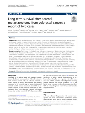
Cadegiani and Kater BMC Endocrine Disorders (2016) 16:48DOI 10.1186/s12902-016-0128-4RESEARCH ARTICLEOpen AccessAdrenal fatigue does not exist: a systematicreviewFlavio A. Cadegiani and Claudio E. Kater*AbstractBackground: The term “adrenal fatigue” (“AF”) has been used by some doctors, healthcare providers, and thegeneral media to describe an alleged condition caused by chronic exposure to stressful situations. Despite this,“AF” has not been recognized by any Endocrinology society, who claim there is no hard evidence for the existence.The aim of this systematic review is to verify whether there is substantiation for “AF”.Methods: A systematic search was performed at PUBMED, MEDLINE (Ebsco) and Cochrane databases, from thebeginning of the data until April 22nd, 2016. Searched key words were: “adrenal” “fatigue”, “adrenal” “burnout”,“adrenal” “exhaustion”, “hypoadrenia”, “burnout” “cortisol”, “fatigue” “cortisol”, “clinical” “burnout”, “cortisol” “vitalility”, “adrenal” “vitality”, and “cortisol” “exhaustion”. Eligibility criteria were: (1) articles written in English, (2)cortisol profile and fatigue or energy status as the primary outcome, (3) performed tests for evaluating the adrenalaxis, (4) absence of influence of corticosteroid therapy, and (5) absence of confounding diseases. Type of questionnaireto distinct fatigued subjects, population studied, tests performed of selected studies were analyzed.Results: From 3,470 articles found, 58 studies fulfilled the criteria: 33 were carried in healthy individuals, and 25 insymptomatic patients. The most assessed exams were “Direct Awakening Cortisol” (n 29), “Cortisol AwakeningResponse” (n 27) and “Salivary Cortisol Rhythm” (n 26).Discussion: We found an almost systematic finding of conflicting results derived from most of the studies methodsutilized, regardless of the validation and the quality of performed tests. Some limitations of the review include: (1)heterogeneity of the study design; (2) the descriptive nature of most studies; (3) the poor quality assessment of fatigue;(4) the use of an unsubstantiated methodology in terms of cortisol assessment (not endorsed by endocrinologists);(5) false premises leading to an incorrect sequence of research direction; and, (6) inappropriate/invalid conclusionsregarding causality and association between different information.Conclusion: This systematic review proves that there is no substantiation that “adrenal fatigue” is an actual medicalcondition. Therefore, adrenal fatigue is still a myth.Keywords: Adrenal depletion, Adrenal fatigue, Cortisol, Adrenal insufficiency, Burnout, FatigueAbbreviations: 24 h UFC, 24-h urinary free cortisol; ACTH, Adrenocorticotropic hormone; ADAS, Abbreviateddyadic adjustment scale; AF, Adrenal fatigue; AUC, Estimated cortisol release (area under the curve); BFI, Brieffatigue inventory; CAR, Cortisol awakening response; CFQ, Chalder fatigue questionnaire; CFS, Chronic fatiguesyndrome; CST, Cosyntropin stimulation test; DAC, Direct awakening cortisol; DCSRD, Diagnosis criteria ofstress-related exhaustion disorder; DHEA-S, Dehydroepiandrosterone sulfate; DST, Dexamethasone suppression test;FAQ, Fatigue assessment questionnaire; FMG, Fibromyalgia; FSE, Fatigue severity scale; H/B, Healthy/Burnout;(Continued on next page)* Correspondence: kater@unifesp.brFrom the Adrenal and Hypertension Unit, Division of Endocrinology andMetabolism, Department of Medicine, Escola Paulista de Medicina,Universidade Federal de São Paulo (EPM/UNIFESP), R. Pedro de Toledo 781–13th floor, 04039-032 São Paulo, SP, Brazil 2016 The Author(s). Open Access This article is distributed under the terms of the Creative Commons Attribution 4.0International License (http://creativecommons.org/licenses/by/4.0/), which permits unrestricted use, distribution, andreproduction in any medium, provided you give appropriate credit to the original author(s) and the source, provide a link tothe Creative Commons license, and indicate if changes were made. The Creative Commons Public Domain Dedication o/1.0/) applies to the data made available in this article, unless otherwise stated.
Cadegiani and Kater BMC Endocrine Disorders (2016) 16:48Page 2 of 16(Continued from previous page)HRFS, HIV-related fatigue scale; Maastricht, Maastricht vital exhaustion questionnaire; Maslach, Maslach burnoutinventory; MFI, Multidimensional fatigue inventory; MP, Memory performance; MSC, morning serum cortisol(& salivary); MST, Mental stress tests; NC-WHO, Neurasthenia criteria; NRWS, Need for recovery from work scale;NSC, Night salivary cortisol; POMS, Profile of mood states; SCR, Salivary cortisol rhythm; SEQ, Exclusion withstress-energy questionnaire; SF-36, Short form health servey 36; SMBQ, Shirom-melamed burnout questionnaire;SOFI, Swedish occupational fatigue inventory; UFC, 24 h Urinary free cortisolBackgroundThe term “adrenal fatigue” (“AF”) has been used bysome doctors, healthcare providers, and the generalmedia to describe an alleged condition caused bychronic exposure to stressful situations. According tothis theory, chronic stress could potentially lead to“overuse” of the adrenal glands, eventually resulting intheir functional failure. In a recent search on Google(April 22, 2016), “adrenal fatigue” provided 640,000 results, and the association of the two words exhibited1,540,000 findings. Despite this, “adrenal fatigue” hasnot been recognized by any endocrinology societies todate, who claim there is no evidence for the existenceof this syndrome [1].Conversely, some medical societies, although unrecognizedby American Board of Medical Specialties and Association of American Medical Colleges [2, 3], claim thatadrenal fatigue is a real and underdiagnosed disease[4, 5]. According to these societies, to screen for “AF”in patients, a questionnaire developed by Dr. Wilson,who is reportedly the first person to describe this supposed syndrome, is recommended to be used [6]. Inaddition, patients suspected of “AF” are now beingtested for serum basal cortisol levels and salivary cortisol rhythm. Those who present impaired results fromthese tests are then treated with corticosteroids, regardless of the etiology. As a result, corticosteroids(mainly hydrocortisone) are probably being prescribedto a large number of patients, as at least 24,000 healthproviders [7] are instructed by one medical society(The American Academy of Anti-Aging Medicine – A4M)to prescribe corticosteroids in these cases.Arguments for corticosteroid use as a treatment for“claimed AF” include: [1] the immediate and significantimprovement seen in patients who are prescribed corticosteroid, and [2] the long and extensive clinical symptomatology of this alleged disease, which shows a slowdepletion before clinical and severe hypocortisolism ensues [4–6]. Moreover, others claim that endocrinologistsuse much too strict diagnostic criteria before prescribingcorticosteroids, and thus, many sufferers would not bereceiving adequate treatment [4, 6]. However, there arelogical counterarguments to routine corticosteroid usein these patients. First, corticosteroids promote a senseof wellbeing (usually temporary), regardless of thepatient’s condition. Second, even at low and physiological doses, corticosteroids increase the risk for severaldisorders, such as psychiatric disorders [8–11], osteoporosis [12], myopathy [13], glaucoma [14], metabolic disorders [14, 15], sleep disturbances [16] and cardiovasculardiseases [17, 18].Therefore, is “adrenal fatigue” an actual disorder? Isfatigue related to depleted adrenal function? Does fatigued healthy subjects present relative adrenal failure?Is adrenal involved in the pathophysiology of fatigue indiseases? Which tests were performed in order to establish markers or triggers? The aim of this systematicreview was to determine the correlation between adrenal status and fatigue states, including the recentlydescribed “burnout” or “burnout syndrome”, and otherfatigue-related diseases. The primary objective was toevaluate the methodology for fatigue status assessment,including cortisol tests, and to examine the results ofstudies involving cortisol and fatigue correlation.MethodsSearch strategiesThe PRISMA protocol for systematic reviews was utilized for this study design. A systematic search wasconducted through the electronic PUBMED, MEDLINE(Ebsco), and COCHRANE databases, from the beginning of the data until April 22, 2016. The search strategy included the following keywords: (1) “adrenal fatigue”; (2) “adrenal burnout”; (3) “adrenal exhaustion”; (4) “adrenal” “fatigue”; (5) “hypoadrenia”; (6)“cortisol” “fatigue”; (7) “cortisol” “burnout”; (8)“clinical” “burnout”; (9) “cortisol” “vitality”; (10) “adrenal” “vitality”; and (11) “cortisol” “exhaustion”,where “a b” means “a” and “b” together in the exactexpression, and “a” ”b” means that both words neededto be contained in the article, but not necessarily together. Although the terms “adrenal fatigue” and “adrenal” “fatigue” were searched, as articles found usingthe first criteria were also found using the second criteria, further analysis were performed for the exactexpression that matched with the disease. We alsoanalyzed articles mentioned within identified studieswhenever the alleged disorder, or a similar situation,were described (such as cortisol profile and exhaustionor fatigued patients).
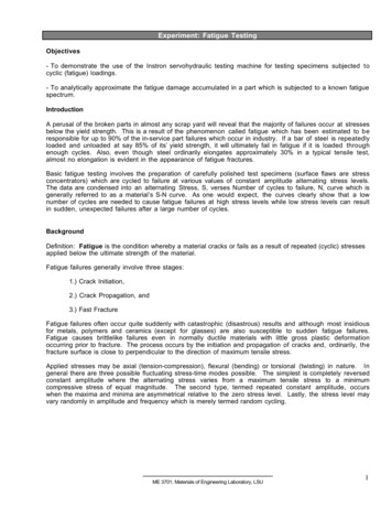
Cadegiani and Kater BMC Endocrine Disorders (2016) 16:48Data extractionAll studies were evaluated by the two reviewers (F.A.C.and C.E.K.) after removal of duplicate articles, accordingto: (1) authorship, (2) journal, (3) publication date, (4)studied population, (5) definition of “fatigue”, “exhaustion”, and “burnout”, (6) study design and methods, (7)analysis methods to assess adrenal axis, (8) results, (9)conclusions, and (10) study variables and bias.Inclusion and exclusion criteriaInclusion criteria were: (1) whole article written in English,(2) cortisol profile and fatigue or energy status as the primary outcome, (3) specific tests performed for evaluatingthe adrenal axis, (4) absence of corticosteroid therapy, (5)absence of confounding diseases that would lead to animpaired cortisol status caused by the disorder itself(such as depression, alcoholism, and morbid obesity).Studies with a merely description about adrenal axisimpairment with no tests performed were excluded.Quality assessmentThe abstract of each of identified study was analyzed byone of the authors (F.A.C.), and was excluded if it did notmeet the eligibility criteria. The studies that fulfilled theinclusion criteria were entirely evaluated regarding the rationale, method design, primary outcome, assessment offatigue, statistical analysis, results, discussion, and conclusions, in order to improve data quality. Those studies thatpresented any bias in the methodology, results, or interpretation of the exposed data, which could be reflected inthe analysis of the study as a whole, were also excluded.Statistical analysisEach of the studied populations, each of the questionnaires and each of the tests performed were quantified,Fig. 1 Study selectionPage 3 of 16whereas tests results were analyzed in terms of percentage of type of responses for each of the tests performed.Results were analyzed in general and according to theunderlying disease.ResultsStudy selectionIn total, 3,470 articles were identified. A summary of thestudy selection is shown in Fig. 1. The search for “adrenal” “burnout” yielded 56 studies; “adrenal” “exhaustion” yielded 446 articles; “adrenal” “fatigue”yielded 1,353 articles; “fatigue” “cortisol” yielded 1,128articles; “cortisol” “burnout” yielded 102 articles; “cortisol” “vitality” yielded 37 articles; “adrenal” “vitality”yielded 53 articles; “hypoadrenia” yielded 9 articles articles (“hypoadrenocorticism” yielded 1,302 articles but isused to refer to hypocortisolism in animals, and therefore, was not included here); and “cortisol” “exhaustion” yielded 286 articles. Twelve studies were excludedbecause they were written in languages other thanEnglish, 1,989 were excluded because there was norelation with the purpose of the systematic review,whereas 905 articles of interest were duplicates. Ofthe 564 remaining studies, 504 had only descriptivecharacteristics or contained results already presentedin another study (in which tests were performed), andtherefore were excluded. Two studies were excludedbecause despite of the correlation between cortisolprofile and burnout or multiple sclerosis, they did notperform correlation between fatigue and cortisol, butother aspects, as depression and pain [19, 20]. For thesystematic review, we analyzed all the included andnot excluded studies, which represent a total of 58 articles (1.67 % of the original search) (Table 1).
Cadegiani and Kater BMC Endocrine Disorders (2016) 16:48Page 4 of 16Table 1 Summary of selected studiesFirst Author &ReferenceYear ofNumber of PopulationPublication PatientsQuestionnaire CCNlSchmidt [69](part 1)2016265BreastCancerFAQDAC, CAR, SCR, NSC NSC, AUC, Nl CAR,Nl DAC, SCRPts. mostly on chemotherapy(ChTx); schemes not specified.No controls, only correlationbetween fatigue levels and tests.ChTx related to worse fatigue.Initial tests performed during,w/ or w/o ChTx.Schmidt [69](part 2)2016265BreastCancerFAQDAC, CAR, SCR, NSC AUC, CAR, NSC, SCR, Nl DACSecond test of the study,performed after 14 weeksof procedures.Sjors [25]2015220H/BSMBQDAC, CAR CAR, DACResults normalized afteradjustment of anti-depressiveuse; SCR results not providedOosterholt[28]201591H/BMaslachDAC, CAR, SCR,AUC, CAR 60 minNl SCR, CAR, Nl AUC,Nl CAR 60 min, DACControl of variations did notchange resultsDe Vente , MSTNl MSC, Nl MSTMST: Nl (women), reduced (men);MSC: Nl (men), reduced (women).Mental arithmetic and publicspeech stressors also performedLennartsson[24]201556H/BDCSRD;exclusionw/ SEQDHEA-S; ACTH;ACTH and DHEA-Spost-TSST (TSSTstress test performed) DHEA-S, post-TSSTDHEA-S, Nl ACTH,Nl post-TSST ACTHTao [23]2015171H/BMaslachMSC, ACTH ACTH, MSCJonsson [29]201551H/BSMBQMSC, MSTNl MSC, MSTLennartsson[27]201556H/BDCSRDand SMBQMSC, MST, ACTH,ACTH post-TSST(TSST stresstest performed)Nl MSC, Nl MST,Nl ACTH, Nlpost-TSST ACTHSevere BO: lower ACTH, cortisolresponse to TSST vs controls,whereas low BO: higher ACTH,cortisol responses vs controlsSchmaling[26]201562HealthyADASAUC, SCR AUC, SCR31 couples studied, one of whichhad chronic fatigue (but not , CAR, SCR, NSCNl DAC, Nl CAR,Nl NSC, Nl SCR**Colocar como SAUDÁVEL –porque é só lombalgia (increasedCAR in Low back pain)Powell [67]201576MultipleSclerosisCFQDAC, CAR, SCR DAC, CAR,Nl SCRSleep disorders excluded;adjusted for depressive symptoms;NSC not published. Multiplesclerosis had increased awakeningcortisol and decreased CARCruz [70]201543BreastcancerBFI and CFQMSC, DHEA-SNl MSC, Nl DHEA-SPts undergoing ChTx chDAC, CAR, SCR, NSC DAC, CAR, SCR, NSCOnly BO; Groups of severe distressor depression not includedAggarwal [30]2014227HealthyCFQMSC, NSC, 0.25 mgDST, DHEA-SNl MSC, Nl NSC,Nl 0.25 mg DST, DHEA-SEvaluation of chronic, widespreadpain, chronic orofacial pain,chronic fatigue (but not CFS),irritable bowel syndromeTell D [71]2014130BreastcancerMFIDAC, CAR, SCR, NSC CAR, SCR, DAC, NSCPost-surgery breast cancer,regardless of ChTx. Notadjusted to sleeping patternsWolfram [32]201353H/BMaslachMSC, 1 mcg CST,DST/CRH test (1.5 mgDST 100 mcg CRH)Nl MSC, post-1 mcgACTH, Nl ACTH andcortisol DST-CRHHigh over-commitment presentblunted serum and salivarycortisol and ACTH responsesto DST-CRH testKlaassen [33]201327HealthyMP (Beck &Luine, 2010)and StressTasks(Wang, 2006)MSC, post-stressNl MSC, Nl post-stressA complex test sequence wasperformed but not reproduced
Cadegiani and Kater BMC Endocrine Disorders (2016) 16:48Page 5 of 16Table 1 Summary of selected studies (Continued)Eek [34]2012581HealthySOFI-20DAC, CAR, NSC,SCR, MSCNl DAC, Nl CAR, NlNSC, Nl SCR, Nl MSCWomen: reduced awakening,increased CAR, increased SCR;Men: increased awakening andreduced CAR – when fatiguedSjors [35]2012247H/BDCSRDand SMBQDAC, 15 min CARNl DAC, Nl 15 minCARRahman [54]201130CFSPreviousMSC, SCR, NSCDx – NoquestionnaireNl MSC, Nl SCR,Nl NSCMoya-Albiol[37]201064H/BMaslachDAC, CARNl DAC, Nl CARKumari [38]20094,299HealthySF-36DAC, CAR, NSC, SCR DAC, CAR, NSC, SCRAdjusted for WC, BMI, sleepduration, CVD medication,depressive symptoms, smoking,alcohol intake provides Nlawakening but lower SCROsterberg[39]2009304H/BMaslachDAC, CAR, NSC, SCRNl DAC, Nl CAR, NSC, SCR0.5 mg DST was not comparedto controlsWingenfeld[41]2009279H/BMaslach andMaastrichtAUC, SCRNl AUC, Nl SCRDAC and CAR not done;conclusions different from results.For AUC, Low BO: Nl, moderate:increased, severe: decreasedRydstedt [40]200976HealthyNRWSDAC, NSCNl DAC, Nl NSCPapadopoulos 2009[55]38CFSCFQ andSF-36MSC, AUC, morningAUC, 0,5 mg DST MSC, AUC, MAUC,Nl 0.5 mg DSTData on absolute cortisol levelsat each point not published.DST reduction evaluated bypercent reduction.Bay [72]200975Posttraumaticbrain injuryPOMSAUCNl AUCCorrelation between brain injuryrelated fatigue level and cortisolAUC. Basal and NSC results notreported; SCR not evaluated.Sudhaus [73]200943ChronicLombalgiaMFIDAC, CAR, MAUC CAR, Nl DAC, Nl MAUC(correlation betweenfatigue levels amonglow back pain subjects)Lindeberg [36] 200878HealthySF-36DAC, CAR, NSC, SCRNl DAC, CAR,Nl NSC, SCRSertoz [42]200872H/BMaslachBasal and post1.0 mcg DST cortisolNl basal cortisol and1.0 mg DSTBellingrath[43]2008101H/BMaslach andMaastrichtDAC, CAR, NSC, SCR,0.25 mg DSTNl DAC, Nl CAR, Nl NSC,Nl SCR, 0.25 mg DSTNater [57]2008185CFSSF-36and MFIDAC, CAR, MAUCNl DAC, Nl CAR, MAUCTorres-Harding 2008[56]108CFSFSEAUC, SCRNl AUC, Nl SCRMultiple psychological testsperformed. Data on NSC,basal and CAR not published.Sonnenschein[45]200742H/BMaslachCAR, 0.5 mg DST,DHEA-SNl CAR, Nl 0.5 mg DST,Nl DHEA-SAdjusted for depression, sleepquality. Awakening levels andeach level graphics not availableHarris [44]200744HealthySF-36DAC, CAR, NSC, SCRNl DAC, Nl CAR, Nl NSC,Nl SCROther aspects also correlated:complains, job stress anddemand, QOL and coping.Adjusted for coffee and tobacco.Langelaan [46] 200655H/BMaslachDAC, CAR, 0.5 mgDST, DHEA-SNl DAC, Nl CAR, Nl 0.5 mg Engaged work also compared andDST, Nl DHEA-Shad stronger suppression in DSTMommersteeg 2006[47]109HealthyNC-WHODAC, CAR, 0.5 mgDST, SCR, AUC, NSCNl DAC, Nl CAR, Nl NSC, NlSCR, Nl 0.5 mg DST, Nl AUCBarroso [74]40HIVHRFSMSC, NSC MSC, NSC2006Jerjes [58]200680CFSCFQUFC, TCM UFC, Nl TCMGrossi [48]200564H/BSMBQDAC, CAR DAC, CARGroups were high x moderate x lowBO score; correlation was significant
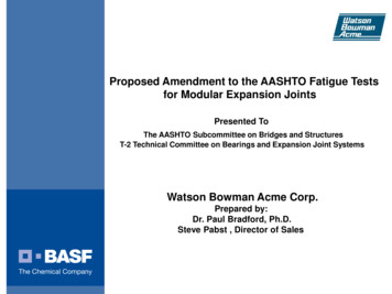
Cadegiani and Kater BMC Endocrine Disorders (2016) 16:48Page 6 of 16Table 1 Summary of selected studies (Continued)Segal [59]200540CFSNoMSC, 1 mcg CSTquestionnaire MSC, 1 mcg CSTJerjes [60]200535CFSCFQMSC, SCR, NSC, AUC MSC, SCR, AUC ,Nl NSCBower [75]200529BreastcancerSF-36DAC, AUC, SCR, NSC AUC, SCR, Nl DAC, NSCPost-ChTx (regardless of time)complete cancer remission andexclusion of other disordersMcLean [76]200555FybromialgiaSF-36DAC, 60 min CAR,SCR, AUC, NSCNl DAC, Nl 60 min CAR,Nl SCR, Nl AUC, Nl NSC(correlation betweenfatigue levels amongFMG subjects)FMG subjects presented NlDAC and CAR, as controls.Roberts [62]200492CFSCFQ andSF-36DAC, CAR, MAUCNl DAC, CAR, MAUCCrofford [61]200472CFS/FMGPOMSACTH, MSC, SCR,NSC, AUCNl ACTH, Nl SCR,Nl NSC, AUC, Nl MSCTests performed in: CFS, FMG andCFS FMG; FMG w/o fatigue hadNl AUC and increased BMC levelsMoch [50]200316H/BMaslachUFC, DHEA-S,ACTH, MSC UFC, Nl DHEA-S,Nl ACTH, MSCOnly women; longitudinalevaluation – Nl initial cortisol.De Vente [49]200345H/BMaslachDAC, MSC, post-TSST DAC, MSC, Nl post-TSSTGaab [63]200242CFSMFIDAC, CAR, SCR,0.5 mg DST 0.5 mg DST, Nl AUC,Nl CAR, Nl DAC, Nl SCR,Nl NSCCAR also performed at 15,45 and 60 min.Dekkers [77]200053Rheumatoid MFIArthritisDAC, CAR, SCR, AUCNl AUC, Nl SCR Nl, DAC, CAR5/25 subjects with RA takingprednisone (5–10 mg/d); RSsubjects had smaller SCR,increased AM cortisol anddecreased CAR. 15 and 45 minCAR also performed.Melamed [51]1999111H/BSMBQ andMaastrichtMSC and 4 PMcortisol MSC, Nl 4 PM cortisolPruessner [52]199966HealthyMaslachDAC, CAR,0.5 mg DST DAC, CAR, 0.5 mg DST15 min and 60 min CARalso performedStrickland [65]199874CFSNotspecified/detailedMSC, NSC NSC, Nl MSCAdjusted for depressionYoung [66]199845CFSNC-WHOUFC, SCR, MSC, AUCNl UFC, Nl SCR,Nl MSC, Nl AUCScott [64]199828CFSNot specified MSC, ACTH, 100 mcg(not detailed) CRH cortisolstimulationNl MSC, Nl ACTH, CRHstim test: cortisol, ACTHRaikkonen [53] 199622HealthyNot assessed Cortisol/ACTH ratio; CST, Nl DST, Nl OGTT,Nl MSC, Nl ACTH1 mcg CST, DST(non specified),OGTT, MSC, ACTH,cortisol/ACTH ratioDHEA-S collected only in CFS.No questionnaires used.Full article not assessed – notin PUBMED or other databaseQuestionnaires: SMBQ shirom-melamed burnout questionnaire, BFI brief fatigue inventory, CFQ chalder fatigue questionnaire, Maslach maslach burnout inventory,SF-36 short form health survey 36, NC-WHO neurasthenia criteria, DCSRD diagnosis criteria of stress-related exhaustion disorder, SEQ stress-energy questionnaire,ADAS, abbreviated dyadic adjustment scale, MFI multidimensional fatigue inventory, FAQ fatigue assessment questionnaire, MP memory performance,POMS profile of mood states, Stress Tasks, FSE fatigue severity scale, SOFI Swedish occupational fatigue inventory, Maastricht Maastricht vital exhaustionquestionnaire, NRWS need for recovery from work scale, HRFS HIV-related fatigue scale, WC waist circumferenceOther abbreviations: CFS chronic fatigue syndrome, H/B healthy/burnout, 24 h-UFC 24-h urinary free cortisol, FMG fibromyalgia; : Increased or elevated; : Decreased or reduced; : Unchanged; Nl: NormalStudy characteristicsAmong the 58 studies included, 33 (56.9 % of the selectedstudies) were performed in healthy subjects [21–53], sincewe considered “burnout” not an actual disorder but instead a stressful condition presented by some groups ofhealth workers. Despite the several studies describing cortisol impairment in Chronic Fatigue Syndrome (CFS), only13 (22.4 %) studies performed an actual assessment ofthe hypothalamic–pituitary–adrenal (HPA) axis [54–66].Twelve studies (20.7 %) were found in which tests for cortisol profiling were performed for other diseases [67–77].However, for analysis purposes, one study [69] was dividedinto two studies as it performed two distinct protocols atdifferent moments. Among these, five were done performed in patients with a diagnosis of breast cancer whohad undergone or were undergoing chemotherapy. One
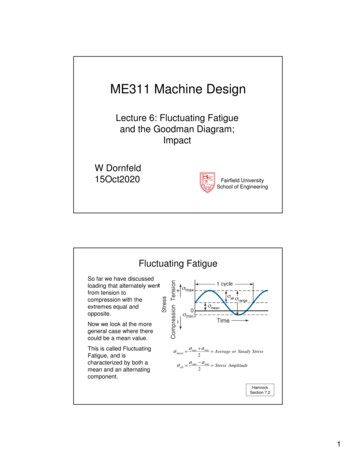
Cadegiani and Kater BMC Endocrine Disorders (2016) 16:48study tested patients with fibromyalgia, two studiescompared patients with chronic lower pain, one withrheumatoid arthritis, one with post brain injury, twowith multiple sclerosis, and one involved patients withhuman immunodeficiency virus (HIV) and CFS. Onestudy evaluated both patients with fibromyalgia and patients with CFS in different groups.The median number of tested subjects in the 58 studieswas 72 (range: 16–4,299). The median numbers ofparticipants in articles involving healthy individuals,patients with CFS, or patients with other diseaseswere 76 (16–4,299), 45 (28–185), and 65 (29–1150),respectively. The largest number of healthy subjectsincluded groups of workers whose cortisol resultswere compared to exhaustion and fatigue status, in anattempt to discriminate correlations between cortisoland energy levels. One study involving 4,299 individuals was responsible for more subjects than the sumof all the other studies.Methods used to evaluate fatigue in the general studypopulationSome authors utilized more than one method to compare the different patients and were included in multiplegroups. A summary of all the methods used to assess fatigue, and their results, is shown in Table 2. Among the58 studies, 27 (46.6 %) utilized the Cortisol AwakeningResponse (CAR) to assess the HPA axis. This method isbased on previous studies [77–81] that indicate cortisollevels rise by 50 % on average within 30 min of wakingas a physiological response to stay alert, with a bluntedCAR resulting in fatigue symptoms. For the CAR, salivary cortisol is collected immediately on waking (t 0)and again 30 min later (t 30), and the difference (deltacortisol) between the two measurements are analyzed.Among the 27 studies that employed CAR, fourteen(51.9 %) showed a normal response, nine (23.3 %) had adiminished delta cortisol, and four (14.8 %) demonstrated an increased delta cortisol.Another method that became widely used to evaluateexhaustion/burnout/fatigue states is the salivary cortisolrhythm (SCR), which evaluates the change in cortisollevels between morning, afternoon, and late night. Atotal of 26 studies evaluated SCR (44.8 %). Some heterogeneity in the method was found between studies, but ingeneral, salivary cortisol was collected at 8 AM, 4 PM,and 10–11 PM. While the SCR is considered as anotherfatigue marker [82, 83], like the CAR, there is no justification for considering this as an etiology for “adrenal fatigue”. Sixteen (61.5 %) studies showed no differencebetween fatigued and control patients, whereas seven(26.9 %) demonstrated an impaired decrease in the circadian SCR. The remaining three (11.6 %) studies disclosed a more pronounced decrease in cortisol level.Page 7 of 16The direct awakening cortisol (DAC) level, collected atthe exact moment of waking, was used in 29 studies(50.0 %). Unlike CAR, DAC reflects sleep quality rather than being a possible identifying factor of fatigue[84–86], even though a poor quality sleep plays an important role in the fatigue process [87–89]. In studiesthat employed DAC, inconsistent results were observed: normal results were found in nineteen (65.5 %)studies, elevated levels were shown in four (13.8 %),and reduced levels in six (20.7 %).The DAC, CAR and SCR methods were by far themost commonly elected ones for examining the correlation between cortisol profile and fatigue status.However, a few other studies analyzed other aspects ofcortisol release.The dexamethasone (Dex) suppression test (DST) wasalso used in nine (15.3 %) studies. The DST identifiesautonomous hypercortisolism, as cortisol production isnormally suppressed by Dex. DSTs have also been usedto investigate hypocortisolism, based on the supposedassumption that it promotes “oversuppression” of cortisol in low cortisol states, indicating that lower levels ofcortisol would disclose a more prolonged suppressionthan controls [55, 90–93], although many studies do notshow correlation between DST and fatigue [47, 55, 94, 95].In six studies, a lower Dex dose (0.5 mg) was used in anattempt to improve the test sensitivity. Among these, fourstudies (66.7 %) showed the same results for both groups,whereas in two others (33.3 %), the test resulted in lowerand prolonged suppression of cortisol levels in fatiguedsubjects. Moreover, an even lower Dex dose (0.25 mg) wasperformed in two studies and resulted in reduced cortisolin one study and normal levels in the group with exhaustion. In one study, Dex dose was not specified, but levelswere not different among exhausted and control groups.As a whole, the DST was used in nine studies, and nosignificant differences were observed between fatiguedand non-fatigued groups in six of these studies(66.7 %), whereas reduced levels were observed inthree studies (33.3 %).Adrenocorticotropic hormone (ACTH) is a pituitarypeptide hormone that stimulates cortisol productionby the adrenocortical zona fasciculata. Elevated ACTHoccurs early in primary adrenal insufficiency, whereasinappropriate (normal) ACTH levels in the presence oflow serum cortisol are found in secondary adrenal failure. Although, normal ACTH levels with normal cortisol levels does not exclude the possibility of relativeadrenocortical failure. Six (10.3 %) studies employedthe morning ACTH levels to compare fatigued andnon-fatigued patients; no significant differences forACTH, as well as for cortisol, were found in five studies (83.3 %), meanwhile one showed elevated ACTHlevels in burnout patients (16.7 %).
Cadegiani and Kater BMC Endocrine Disorders (2016) 16:48Page 8 of 16Table 2 Assessed methods and results of all selected studies (N 58)Procedure (*)Number of studies (% of total)Not different (%)Decreased (%)Increased (%)DAC29 (50.0 %)19 (65.5 %)6 (20.7 %)4 (13.8 %)CAR27 (46.6 %)14 (51.9 %)9 (33.3 %)4 (14.8 %)SCR26 (44.8 %)16 (61.5 %)7 (26.9 %)3 (11.5 %)MSC22 (37.9 %)14 (63.6 %)4 (18.2 %)4 (18.2 %)NSC22 (37.9 %)13 (59.1 %)3 (13.6 %)6 (27.3 %)AUC13 (22.4 %)8 (61.5 %)3 (23.1 %)2 (15.4 %)DST9 (15.5 %)6 (66.7 %)3 (33.3 %)-DHEA-S6 (10.3 %)4 (66.7 %)2 (33.3 %)-ACTH6 (10.3 %)5 (83.3 %)-1 (16.7 %)MST5 (8.6 %)4 (80.0 %)1 (20.0 %)-UFC3 (5.2 %)1 (33.3 %)2 (66.7 %)-CST3 (5.2 %)-2 (66.7 %)1 (33.3 %)MAUC3 (5.2 %)-2 (66.7 %)1 (33.3 %)CAR 60 min2 (3.4 %)2 (100 %)--ACTH MST2 (3.4 %)2 (100 %)--4 PM cortisol11--DST CRH cortisol11--DST CRH ACTH11--CAR 15 min11--TCM11--DHEA-S MST1-1-CRST cortisol1-1-CRST ACTH1-1-OGTT cortisol1--1Cortisol/ACTH ratio1--1Legends: (*): DAC direct awakening cortisol, CAR cortisol awakening response, SCR salivary cortisol rhythm, MSC morning serum (& salivary) cortisol, NSC nightsalivary cortisol, AUC area under-the-curve (Estimated Cortisol Release), DST dexamethasone suppression test, DHEA-S dehydroepiandrosterone sulfate, ACTHadrenocorticotropic hormone, MST mental stress test, UFC 24 h-urinary free cortisol, CST cosyntropin stimulation test, MAUC morning area under-the-curve(morning estimated cortisol release), CRH corticotropin releasing hormone, TCM total urinary cortisol metab
2016 257 H/B Maslach HCC Nl Schmidt [69] (part 1) 2016 265 Breast Cancer FAQ DAC, CAR, SCR, NSC NSC, AUC, Nl CAR, Nl DAC, SCR Pts. mostly on chemotherapy (ChTx); schemes not specified. No controls, only correlation between fatigue levels and tests. ChTx related to worse fatigue. In