Transcription
HOSPITAL CONSULTIN PARTNERSHIP WITH THE SOCIETY FOR DERMATOLOGY HOSPITALISTSUpdate on CalciphylaxisEtiopathogenesis, Diagnosis,and ManagementpyUrmi Khanna, MD; Arturo Dominguez, MD; Jesse Keller, MD; Daniela Kroshinsky, MD, MPH;Alex G. Ortega-Loayza, MD; Lindsay Strowd, MD; Robert G. Micheletti, MDcomanagement. In this article, we provide an update on calciphylaxisetiopathogenesis, diagnosis, and management, and we highlight thechallenges faced in managing this potentially fatal condition.Cutis. 2018;102:395-400.CCU T ISDo aintain a high index of suspicion for calciphylaxisMin patients with end-stage renal disease on chronicdialysis presenting with severely painful livedoidplaques or retiform purpura, particularly in fat-richbody sites. Skin biopsies may be limited by biopsy site, inadequate biopsy depth, missed areas of microcalcification, and absence of definitive histologic criteria.Special calcium stains and review by an experienceddermatopathologist may lower the rate of falsenegative biopsies. In cases where the most likely clinical diagnosis iscalciphylaxis, treatment should be initiated even ifdefinitive histopathology findings are lacking. Treatment should be multimodal, including elimination of risk factors, intravenous sodium thiosulfate,agents addressing calcium-phosphate metabolism,and surgical debridement, if indicated.no tPRACTICE POINTSCalciphylaxis is a rare painful skin condition classically seen inpatients with end-stage renal disease (ESRD), particularly those onchronic dialysis; however, it also has been increasingly reported inpatients with normal renal function. Calciphylaxis is associated withhigh mortality rates, and excruciating pain and nonhealing ulcersoften lead to recurrent hospitalizations and infectious complications.It is critical for dermatologists to recognize the clinical features ofcalciphylaxis to ensure accurate and timely diagnosis and properalciphylaxis, also known as calcific uremic arteriolopathy, is a painful skin condition classically seenin patients with end-stage renal disease (ESRD),particularly those on chronic dialysis.1,2 It also has increasingly been reported in patients with normal renal function and calcium and phosphate homeostasis.3,4 Effectivediagnosis and management of calciphylaxis remains challenging for physicians.2,5 The condition is characterizedby tissue ischemia caused by calcification of cutaneousarteriolar vessels. As a result, calciphylaxis is associatedwith high mortality rates, ranging from 60% to 80%.5,6Excruciating pain and nonhealing ulcers often lead torecurrent hospitalizations and infectious complications,7and poor nutritional status, chronic pain, depression, andNEW PODCASTDr. Robert G. Micheletti discusses calciphylaxisdiagnosis and management with Cutis Editor-in-ChiefVincent A. DeLeo, MD, in a new episode of the “Peerto Peer” podcast https://apple.com/2yNkYN6Dr. Khanna is from the Department of Dermatology, Cleveland Clinic, Ohio. Dr. Dominguez is from the Department of Dermatology, University ofTexas Southwestern Medical Center, Dallas. Drs. Keller and Ortega-Loayza are from the Department of Dermatology, Oregon Health & ScienceUniversity, Portland. Dr. Kroshinsky is from the Department of Dermatology, Massachusetts General Hospital, Boston. Dr. Strowd is from theDepartment of Dermatology, Wake Forest University School of Medicine, Winston-Salem, North Carolina. Dr. Micheletti is from the Departments ofDermatology and Medicine, University of Pennsylvania, Philadelphia.The authors report no conflict of interest.Correspondence: Robert G. Micheletti, MD CUTISVOL. 102 NO. 6 I DECEMBER 2018 395Copyright Cutis 2018. No part of this publication may be reproduced, stored, or transmitted without the prior written permission of the Publisher.
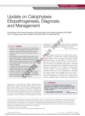
HOSPITAL CONSULTEtiology and PathogenesisCalciphylaxis is most commonly seen on the legs, abdomen, and buttocks.2 Patients with ESRD commonlydevelop proximal lesions affecting adipose-rich sites andhave a poor prognosis. Distal lesions are more common inpatients with nonuremic calciphylaxis, and mortality ratesare lower in this population.2Early lesions present as painful skin nodules or indurated plaques that often are rock-hard or firm to palpationwith overlying mottling or a livedoid pattern (Figure, A).Early lesions progress from livedo reticularis to livedoracemosa and then to retiform purpura (Figure, B).Purpuric lesions later evolve into black eschars (Figure, C),then to necrotic, ulcerated, malodorous plaques or nodules in later stages of the disease (Figure, D). Lesions alsomay develop a gangrenous sclerotic appearance.2,5Although most patients with calciphylaxis have ESRD,nonuremic patients also can develop the disease. Thosewith calciphylaxis who do not have renal dysfunction frequently have other risk factors for the disease and oftenDonoCalciphylaxis is thought to have a multifactorial etiologywith the exact cause or trigger unknown.7 A long list ofrisk factors and triggers is associated with the condition(Table 1). Calciphylaxis primarily affects small arteries(40–600 μm in diameter) that become calcified due to animbalance between inhibitors and promoters of calcification.2,11 Fetuin-A and matrix Gla protein inhibit vascularcalcification and are downregulated in calciphylaxis.12,13Dysfunctional calcium, phosphate, and parathyroidClinical FeaturespyCalciphylaxis is considered a rare dermatosis with anestimated annual incidence of 1% to 4% in ESRD patientson dialysis. Recent data suggest that incidence of calciphylaxis is rising,5,7,9 which may stem from an increaseduse of calcium-based phosphate binders, an actual risein disease incidence, and/or increased recognition ofthe disease.5 It is difficult to estimate the exact diseaseburden of calciphylaxis because the diagnostic criteriaare not well defined, often leading to missed or delayeddiagnosis.3,10 Furthermore, there is no centralized registryfor calciphylaxis cases.3coEpidemiologyhormone regulatory pathways provide an increased substrate for the process of calcification, which causes endothelial damage and microthrombosis, resulting in tissueischemia and infarction.14,15 Notably, there is growinginterest in the role of vitamin K in the pathogenesis ofcalciphylaxis. Vitamin K inhibits vascular calcification,possibly by increasing the circulating levels of carboxylated matrix Gla protein.16tinsomnia can further complicate recovery and lead topoor quality of life.8We provide an update on calciphylaxis etiopathogenesis,diagnosis, and management. We also highlight some challenges faced in managing this potentially fatal condition.Calciphylaxis Risk Factors and TriggersISTABLE 1.TAutoimmune disease: systemic lupus erythematosusCUCoagulation disorders: antithrombin III deficiency, lupus anticoagulant, protein C deficiencyDisturbed calcium/phosphate homeostasis: ESRD, hypercalcemia, hyperphosphatemia, secondary hyperparathyroidismEnvironmental factors: exposure to heavy metals (eg, aluminum) and UV lightEthnicity: whiteGender: femaleGenetic polymorphisms: CD73 (rs4431401, rs9444348), FGF23 and vitamin D receptor (rs7310492, rs11063118, rs13312747,and rs17882106)HypoalbuminemiaMalignancy: Metastatic cancer (eg, colon, lungs), POEMS syndromeMedications: Calcium-containing medications (eg, phosphate binders), corticosteroids, iron supplements, recombinant humanparathyroid hormone, warfarin, vitamins D and EMetabolic diseases: diabetes, ESRD (especially dependence on dialysis for 2 years, recent transition onto dialysis, or insufficientdialysis [ie, missed sessions])Nutritional disorders: gastric bypass surgery, malnutrition, obesitySkin trauma: skin biopsy, subcutaneous insulin or heparin injection, other direct trauma to the skinAbbreviations: ESRD, end-stage renal disease; POEMS, polyneuropathy, organomegaly, endocrinopathy, monoclonal gammopathy,skin changes.396 I CUTIS WWW.MDEDGE.COM/CUTISCopyright Cutis 2018. No part of this publication may be reproduced, stored, or transmitted without the prior written permission of the Publisher.
HOSPITAL CONSULTBonotcopyADDCTISEarly lesions of calciphylaxis often appear as indurated plaques with overlying mottling or livedoid pattern (A) that progress to retiform purpura (B).Purpuric lesions then evolve into black eschars (C). In later stages, necrotic, ulcerated, malodorous plaques or nodules are present (D).CUreport another notable health problem in the weeks ormonths prior to presentation.4 More than half of patientswith calciphylaxis become bedridden or require use of awheelchair.17 Pain is characteristically severe throughoutthe course of the disease; it may even precede the appearance of the skin lesions.18 Because the pain is associatedwith ischemia, it tends to be relatively refractory to treatment with opioids. Rare extracutaneous vascular calcifications may lead to visual impairment, gastrointestinal tractbleeding, and myopathy.5,9,19,20DiagnosisConsidering the high morbidity and mortality associatedwith calciphylaxis, it is important to provide accurate andtimely diagnosis; however, there currently are no validateddiagnostic criteria for calciphylaxis. Careful correlation ofclinical and histologic findings is required. Calciphylaxisbiopsies have demonstrated medial calcification and proliferation of the intima of small- to medium-sized arteries.21 Lobular and septal panniculitis and extravascularsoft-tissue calcification, particularly stippled calcificationof the eccrine sweat glands, also has been seen.2,22 SpecialWWW.MDEDGE.COM/CUTIScalcium stains (eg, von Kossa, Alizarin red) increasethe sensitivity of biopsy by highlighting subtle areas ofintravascular and extravascular calcification.5,23 Sufficientsampling of subcutaneous tissue and specimen evaluation by an experienced dermatopathologist are necessaryto ensure proper interpretation of the histologic findings.Despite these measures, skin biopsies may be nondiagnostic or falsely negative; therefore, when there ishigh clinical suspicion, it may be appropriate to moveforward with a presumptive diagnosis of calciphylaxiseven if the histologic findings are nondiagnostic.1,9,24It also is worth noting that localized progression andulceration may occur following skin biopsy, such thatbiopsy may even be contraindicated in certain cases(eg, penile calciphylaxis).Standard laboratory workup for calciphylaxis includesevaluation for associated risk factors as well as exclusionof other conditions in the differential diagnosis (Table 2).Blood tests to evaluate for risk factors include liver andrenal function tests, a complete metabolic panel, parathyroid hormone level, and serum albumin level.5 Elevatedcalcium and phosphate levels may signal disturbedVOL. 102 NO. 6I DECEMBER 2018 397Copyright Cutis 2018. No part of this publication may be reproduced, stored, or transmitted without the prior written permission of the Publisher.
HOSPITAL CONSULTCalciphylaxis DifferentialDiagnosesTABLE 3.TABLE 2.Calciphylaxis Treatment OptionsMedical TreatmentsAntiphospholipid syndrome (or other hypercoagulable state)Anticoagulants (excluding warfarin)Atherosclerosis (peripheral vascular disease)Becaplermin (recombinant platelet-derived growth factor)Calcinosis cutisBisphosphonates (eg, pamidronate, alendronate)*CellulitisCinacalcet (in the setting of elevated parathyroid hormone)aCholesterol embolismDiscontinue medications (if possible) that may contribute tocalciphylaxis (eg, warfarin, steroids)aHematomaIntralesional sodium thiosulfateInfectious ulcerIntravenous sodium thiosulfateaLivedoid vasculopathyPentoxifyllinePyoderma gangrenosumProstaglandinsSmall- to medium-vessel vasculitispyNon–calcium-containing phosphate binders (eg, sevelamer)aMartorell hypertensive ischemic ulcerVitamin K supplementationcoVenous stasis ulcerSurgical TreatmentsDebridement (in appropriate candidates)ParathyroidectomyRenal transplantationMiscellaneousHyperbaric oxygen therapyLocal wound careaPain managementaTherapies most frequently employed by the authors.aCUTISDonocalcium and phosphate homeostasis but are neithersensitive nor specific for the diagnosis.25 Complete bloodcell count, blood cultures, thorough hypercoagulabilityworkup (including but not limited to antiphospholipidantibodies, proteins C and S, factor V Leiden, antithrombin III, homocysteine, methylenetetrahydrofolate reductase mutation, and cryoglobulins), rheumatoid factor,antineutrophil cytoplasmic antibodies, and antinuclearantibody testing may be relevant to help identify contributing factors or mimickers of calciphylaxis.5 Variousimaging modalities also have been used to evaluate forthe presence of soft-tissue calcification in areas of suspected calciphylaxis, including radiography, mammography, computed tomography, ultrasonography, nuclearbone scintigraphy, and spectroscopy.2,26,27 Unfortunately,there currently is no standardized reproducible imagingmodality for reliable diagnosis of calciphylaxis. Ultimately,histologic and radiographic findings should always beinterpreted in the context of relevant clinical findings.2,9tDermal regenerative templatePreventionReduction of the net calcium phosphorus product mayhelp reduce the risk of calciphylaxis in ESRD patients,which can be accomplished by using non–calciumphosphate binders, adequate dialysis, and restrictinguse of vitamin D and vitamin K antagonists.2,5 There arelimited data regarding the benefits of using bisphosphonates and cinacalcet in ESRD patients on dialysis toprevent calciphylaxis.28,29ManagementManagement of calciphylaxis is multifactorial. Besidesdermatology and nephrology, specialists in pain398 I CUTIS management, wound care, plastic surgery, and nutritionare critical partners in management.1,5,9,30 Nephrologistscan help optimize calcium and phosphate balance andensure adequate dialysis. Pain specialists can aid in creating aggressive multiagent pain regimens that target theneuropathic/ischemic and physical aspects of calciphylaxis pain. When appropriate, nutrition specialists canhelp establish high-protein, low-phosphorus diets, andwound specialists can provide access to advanced wounddressings and adjunctive hyperbaric oxygen therapy.Plastic surgeons can provide conservative debridementprocedures in a subset of patients, usually those withdistal stable disease.The limited understanding of the etiopathogenesisof calciphylaxis and the lack of data on its management are reflected in the limited treatment options forthe disease (Table 3).2,5,9 There are no formal algorithmsfor the treatment of calciphylaxis. Therapeutic trials arescarce, and most of the current treatment recommendations are based on small retrospective reports or caseseries. Sodium thiosulfate has been the most widely usedWWW.MDEDGE.COM/CUTISCopyright Cutis 2018. No part of this publication may be reproduced, stored, or transmitted without the prior written permission of the Publisher.
HOSPITAL CONSULTTABLE 4.Clinical Trials for the Treatment of -blind,placebo-controlledclinical trialCALISTA trial: evaluate the safety and efficacy of intravenous sodiumthiosulfate injection for treatment of acute calciphylaxis–associated pain inhemodialysis patientsIINCT02790073Multicenter, openlabel clinical trialEvaluate the effect of SNF472 (hexasodium phytate) in addition tostandard of care for promoting wound healing and therapeutic responsein hemodialysis patients with calciphylaxisINCT01289626Single center, openlabel clinical trialEvaluate the efficacy of lanthanum carbonate for remission of skin lesionsand reduction of phosphate levels in ESRD patients with calciphylaxisNANCT02278692Single center,randomized, parallelassignment trialEvaluate the effects of oral vitamin K supplementation on anticalcificationfactor (carboxylated matrix Gla protein) levels and clinical outcomes al studyObservational follow-up study of patients from the ST-001 CALISTA studyNANCT03146793Single centerobservational,retrospectivecohort studyEvaluate whether early administration of sodium thiosulfate reducesmortality in dialysis patients with calciphylaxisnotcopyMethodsDoAbbreviations: ESRD, end-stage renal disease; NA, not applicable.CUTIStreatment option since 2004, when its use in calciphylaxiswas first reported.31 Sodium thiosulfate chelates calciumand is thought to have antioxidant and vasodilatory properties.32 There are a few promising clinical trials and largescale studies (Table 4) that aim to evaluate the efficacyof existing treatments (eg, sodium thiosulfate) as well asnovel treatment options such as lanthanum carbonate,SNF472 (hexasodium phytate), and vitamin K.33-36PrognosisCalciphylaxis is a potentially fatal condition with a poorprognosis and a median survival rate of approximately1 year following the appearance of skin lesions.37-39Patients with proximal lesions and those on peritonealdialysis (as opposed to hemodialysis) have a worse prognosis.40 Mortality rates are estimated to be 30% at 6 months,50% at 12 months, and 80% at 2 years, with sepsis secondary to infection of cutaneous ulcers being the leading causeof death.37-39 The impact of calciphylaxis on patient qualityof life and activities of daily living is severe.8,17to help formulate evidence-based diagnostic criteria.Radiographic and histologic studies, as well as othertools for early and accurate diagnosis of calciphylaxis,should be studied for feasibility, accuracy, and reproducibility. The incidence of nonuremic calciphylaxis pointstoward pathogenic pathways besides those based onthe bone-mineral axis. Basic science research directedat improving understanding of the pathophysiology ofcalciphylaxis would be helpful in devising new treatmentstrategies targeting these pathways. Establishment of acollaborative, multi-institutional calciphylaxis workinggroup would enable experts to formulate therapeuticguidelines based on current evidence. Such a group couldfacilitate initiation of large prospective studies to establishthe efficacy of existing and new treatment modalities forcalciphylaxis. A working group within the Society forDermatology Hospitalists has been tasked with addressing these issues and is currently establishing a multicenter calciphylaxis database.REFERENCESFuture DirectionsMulti-institution cohort studies and collaborative registries are needed to provide updated informationrelated to the epidemiology, diagnosis, treatment, morbidity, and mortality associated with calciphylaxis andWWW.MDEDGE.COM/CUTIS1.2.3.Nigwekar SU, Kroshinsky D, Nazarian RM, et al. Calciphylaxis: riskfactors, diagnosis, and treatment. Am J Kidney Dis. 2015;66:133-146.Nigwekar SU, Thadhani RI, Brandenburg VM. Calciphylaxis. N Engl JMed. 2018;378:1704-1714.Davis JM. The relationship between obesity and calciphylaxis: a reviewof the literature. Ostomy Wound Manage. 2016;62:12-18.VOL. 102 NO. 6I DECEMBER 2018 399Copyright Cutis 2018. No part of this publication may be reproduced, stored, or transmitted without the prior written permission of the Publisher.
HOSPITAL CONSULT13.14.15.16.17.18.19.20.21.22.400 I CUTIS 26.27.28.29.30.31.32.33.py25.co11.12.24.Cassius C, Moguelet P, Monfort JB, et al. Calciphylaxis in haemodialysedpatients: diagnostic value of calcifications in cutaneous biopsy. Br JDermatol. 2018;178:292-293.Sreedhar A, Sheikh HA, Scagliotti CJ, et al. Advanced-stage calciphylaxis: think before you punch. Cleve Clin J Med. 2016;83:562-564.Brandenburg VM, Kramann R, Rothe H, et al. Calcific uraemic arteriolopathy (calciphylaxis): data from a large nation-wide registry. NephrolDial Transplant. 2017;32:126-132.Paul S, Rabito CA, Vedak P, et al. The role of bone scintigraphy in thediagnosis of calciphylaxis. JAMA Dermatol. 2017;153:101-103.Shmidt E, Murthy NS, Knudsen JM, et al. Net-like pattern of calcification on plain soft-tissue radiographs in patients with calciphylaxis. J AmAcad Dermatol. 2012;67:1296-1301.EVOLVE Trial Investigators; Chertow GM, Block GA, Correa-RotterR, et al. Effect of cinacalcet on cardiovascular disease in patients undergoing dialysis. N Engl J Med. 2012;367:2482-2494.Rogers NM, Teubner DJO, Coates PT. Calcific uremicarteriolopathy: advances in pathogenesis and treatment. Semin Dial.2007;20:150-157.Nigwekar SU. Multidisciplinary approach to calcific uremic arteriolopathy. Curr Opin Nephrol Hypertens. 2015;24:531-537.Cicone JS, Petronis JB, Embert CD, et al. Successful treatmentof calciphylaxis with intravenous sodium thiosulfate. Am J KidneyDis. 2004;43:1104-1108.Chen NX, O’Neill K, Akl NK, et al. Adipocyte induced arterial calcification is prevented with sodium thiosulfate. Biochem Biophys Res Commun.2014;449:151-156.Chan MR, Ghandour F, Murali NS, et al. Pilot study of the effect of lanthanum carbonate in patients with calciphylaxis: a Wisconsin Networkfor Health Research (WiNHR) study. J Nephrol Ther. 2014;4:1000162.Perelló J, Gómez M, Ferrer MD, et al. SNF472, a novel inhibitor ofvascular calcification, could be administered during hemodialysis toattain potentially therapeutic phytate levels. J Nephrol. 2018;31:287-296.Christiadi D, Singer RF. Calciphylaxis in a dialysis patient successfullytreated with high-dose vitamin K supplementation. Clin KidneyJ. 2018;11:528-529.Caluwe R, Vandecasteele S, Van Vlem B, et al. Vitamin K2 supplementation in haemodialysis patients: a randomized dose-finding study.Nephrol Dial Transplant. 2014;29:1385-1390.McCarthy JT, El-Azhary RA, Patzelt MT, et al. Survival, risk factors, andeffect of treatment in 101 patients with calciphylaxis. Mayo Clin Proc.2016;91:1384-1394.Fine A, Zacharias J. Calciphylaxis is usually non-ulcerating: risk factors,outcome and therapy. Kidney Int. 2002;61:2210-2217.Nigwekar SU, Zhao S, Wenger J, et al. A nationally representativestudy of calcific uremic arteriolopathy risk factors. J Am Soc Nephrol.2016;27:3421-3429.Zhang Y, Corapi KM, Luongo M, et al. Calciphylaxis in peritonealdialysis patients: a single center cohort study. Int J Nephrol Renovasc Dis.2016;9:235-241.t10.23.no9.o8.D7.IS6.T5.Bajaj R, Courbebaisse M, Kroshinsky D, et al. Calciphylaxis in patientswith normal renal function: a case series and systematic review.Mayo Clin Proc. 2018;93:1202-1212.Hafner J, Keusch G, Wahl C, et al. Uremic small-artery diseasewith medial calcification and intimal hyperplasia (so-called calciphylaxis): a complication of chronic renal failure and benefit from parathyroidectomy. J Am Acad Dermatol. 1995;33:954-962.Jeong HS, Dominguez AR. Calciphylaxis: controversies in pathogenesis,diagnosis and treatment. Am J Med Sci. 2016;351:217-227.Westphal SG, Plumb T. Calciphylaxis. In: StatPearls. Treasure Island,FL: StatPearls Publishing; 2018. https://www.ncbi.nlm.nih.gov/books/NBK519020. Accessed November 12, 2018.Riemer CA, El-Azhary RA, Wu KL, et al. Underreported use of palliativecare and patient-reported outcome measures to address reduced qualityof life in patients with calciphylaxis: a systematic review. Br J Dermatol.2017;177:1510-1518.Nigwekar SU. Calciphylaxis. Curr Opin Nephrol Hypertens. 2017;26:276-281.Fine A, Fontaine B. Calciphylaxis: the beginning of the end? Perit DialInt. 2008;28:268-270.Lin WT, Chao CM. Tumoral calcinosis in renal failure. QJM. 2014;107:387.Schafer C, Heiss A, Schwarz A, et al. The serum protein alpha 2Heremans-Schmid glycoprotein/fetuin-A is a systemically acting inhibitor of ectopic calcification. J Clin Invest. 2003;112:357-366.Luo G, Ducy P, McKee MD, et al. Spontaneous calcification ofarteries and cartilage in mice lacking matrix GLA protein. Nature.1997;386:78-81.Bleyer AJ, Choi M, Igwemezie B, et al. A case control study of proximalcalciphylaxis. Am J Kidney Dis. 1998;32:376-383.Ahmed S, O’Neill KD, Hood AF, et al. Calciphylaxis is associated withhyperphosphatemia and increased osteopontin expression by vascularsmooth muscle cells. Am J Kidney Dis. 2001;37:267-276.Nigwekar SU, Bloch DB, Nazarian RM, et al. Vitamin K-dependentcarboxylation of matrix gla protein influences the risk of calciphylaxis.J Am Soc Nephrol. 2017;28:1717-1722.Weenig RH, Sewell LD, Davis MD, et al. Calciphylaxis: natural history,risk factor analysis, and outcome. J Am Acad Dermatol. 2007;56:569-579.Polizzotto MN, Bryan T, Ashby MA, et al. Symptomatic management ofcalciphylaxis: a case series and review of the literature. J Pain SymptomManage. 2006;32:186-190.Gupta N, Haq KF, Mahajan S, et al. Gastrointestinal bleeding secondaryto calciphylaxis. Am J Case Rep. 2015;16:818-822.Edelstein CL, Wickham MK, Kirby PA. Systemic calciphylaxis presentingas a painful, proximal myopathy. Postgrad Med J. 1992;68:209-211.Mochel MC, Arakari RY, Wang G, et al. Cutaneous calciphylaxis: aretrospective histopathologic evaluation. Am J Dermatopathol.2013;35:582-586.Chen TY, Lehman JS, Gibson LE, et al. Histopathology of calciphylaxis: cohort study with clinical correlations. Am J Dermatopathol. E.COM/CUTISCopyright Cutis 2018. No part of this publication may be reproduced, stored, or transmitted without the prior written permission of the Publisher.
Dr. Strowd is from the Department of Dermatology, Wake Forest University School of Medicine, Winston-Salem, North Carolina. Dr. Micheletti is from the Departments of Dermatology and Medicine, University of Pennsylvania, Philadelphia. The authors report no conflict of interest. Correspondence: Robert G. Micheletti