Transcription
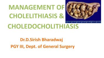
MANAGEMENT OFCHOLELITHIASIS &CHOLEDOCHOLITHIASISDr.D.Sirish BharadwajPGY III, Dept. of General Surgery
DEFINITION The presence of gallstones in thegallbladder is calledcholelithiasis. The presence of gallstones inthe common bile duct iscalled choledocholithiasis
Effects of GallstonesIn the gallbladder: Asymptomatic stonesBiliary colic with periodicityAcute cholecystitisChronic cholecystitisEmpyema gallbladderPerforation causing biliary peritonitis or pericholecystitic abscessMucocele of the gallbladderLimey gallbladderGallstone ileus leading to small bowel obstructionCarcinoma gallbladder
In the common bile duct(CBD): Obstructive jaundiceCholangitisPancreatitisMirizzi syndrome
Current discussion: Chronic cholecystitisAcute calculous cholecystitisCholedocholothiasisAysmptomatic gallstones
CLINICALPRESENTATION
Chronic cholecystitis Biliary colic a common misnomer because the pain is not colicky The pain is described as a bandlike tightness of theupper abdomen in the epigastrium or right upper quadrant constant pain that builds in intensity, and can radiate to the back,interscapular region, Attacks usually last for more than 1 hour but subsides by 24 hours;
may be associated with nausea and vomiting. Bloating and belching – 50% Intolerance to fatty meals Flatulent dyspepsia The physical examination usually normal if they are pain-free. During an episode of biliary colic, mild right upper quadranttenderness may be present.
Acute calculous cholecystitis Right upper quadrant pain, similar in severity but much longer induration than pain from previous episodes of biliary colic fever, nausea, and vomiting.On physical exam, right upper quadrant tenderness and guarding are usually presentinferior to the right costal margin, distinguishing the episode fromsimple biliary colic. When inflammation spreads to the peritoneum, patients develop morediffuse tenderness, guarding and rigidity. A mass, the gallbladder and adherent omentum, is occasionallypalpable,
Murphy's sign, inspiratory arrest with deep palpation in theright upper quadrant, may also be present.
A mild leukocytosis is usually present (12,000-14,000cells/mm3). mild elevations in: serum bilirubin ( 4 mg/dL),alkaline phosphatase,transaminases,amylase may be present.
CholedocholithiasisCommon bile duct stones may be silent and are often discoveredincidentally. In these patients, biliary obstruction is transient, and laboratorytests may be normal.Clinical features suspicious for biliary obstruction(obstructive/surgicaljaundice) due to common bile duct stones: biliary colic, jaundice, pruritis Pale/clay coloured stools, and dark of the urine.
cont. Vitamin deficiency Obstruction of bile flow also interferes with absorption of the fatsoluble vitamins A,D,E, and K Fever and chills may be presentCharcot’s triad - seen in Choledocholithiasis with ascending cholangitis : Fever, Rt. Upper quadrant pain, jaundiceIf untreated, may progress to septic shock – Rey old’s pentad: Charcot’s triad hypote sio e tal status cha ges
BLOOD INV: TLC increased Serum bilirubin ( 3.0 mg/dL), serum aminotransferases raised alkaline phosphatase & gamma glutamyl transpeptidase areelevated (more than AST/ALT)URINALYSIS: Urine Bilirubin is increased. Urobilinogen is negative.
Aysmptomatic gallstones 10% of males and 20% of females Gallstones detected incidentally while performing USGabdomen for some other reasons.
MANAGEMENT
INVESTIGATIONS Laboratory investigations– LFT’s– Hepatitis B&C viral serology– Urine Imaging– Non-invasive/ minimally invasive– Invasive
Imaging Non-invasive / minimally invasive––––––Abdominal X-rayOral cholecystogramUltrasonography (US)Magnetic Resonance Cholangio-Pancreatography (MRCP)CholescintigraphyComputerized Tomography (CT) Invasive––––Endoscopic Retrograde Cholangio-Pancreatography (ERCP)Endoscopic UltrasoundPercutaneous Transhepatic Cholangiography (PTC)Intraoperative cholangiography
X-ray abdomen X-rays: 15% stones are radiopaque, May show Mercedes-Benz sign porcelain GB may be seen. Air in biliary tree(Pneumobilia) emphysematous GB wall.
Ultrasonography AccurateQuickInexpensiveEasily availableOperator dependentSuboptimal scan
Ultrasonography Biliary calculiSize of GBThickness of GB wallInflammation around GB - Pericholecystic fluid, sonographicMurphy’s. Size of CBD (Normal CBD diameter 6-8mm)
Ultrasonography Intra and extra-hepatic biliary dilatation and level ofobstruction. 70-95 % sensitive. 80-100 % specific.
MRCP Non-invasive Contrast not required. Demonstrates– Ductal obstruction– Visualizes stones– Strictures– Other intra & extra ductal abnormalities
Cholescintigraphy Technitium 99 (Tc99-IDA chelate complex). HIDA/ PIPIDA/ DISIDA scan. Gallbladder visualized within 30min to 1 hour in absence ofdisease. Not visualized in Acute cholecystitis 97 % sensitive and 94 % specific. Diagnose obstruction.
Bile leaks (detect andquantify). CBD obstruction appears asnon visualization of smallintestine. No external radiationexposure to the patient. Less helpful when thepatient is fasting for morethan 5 days, with a 40%false-positive rate.HIDA
CT Scan Not useful in benign biliary disease. Useful when Carcinoma gallbladder is suspected Gall stones often not visualized. Cholecystitis is underdiagnosed. Higher dose of radiation.
Endoscopic Ultrasound Procedure: USG probe passed through an upper GI endoscope and kept inpylorus/duodenum area High frequency used - 20-40Mhz Evaluates Pancreato-biliary system. Detection of microlithiasis Choledocholithiasis Evaluation of benign and malignant strictures. Detects regional lymphnodes Relationship to vascular structures.
Endoscopic UltrasoundAdvantages: High resolution imaging. Less invasive. No exposure to radiation. Aspiration of a cyst or FNAC.Disadvantages: Higher operator dependency. Cost and availability. Visualization is limited to 8 cm.
ERCPMore of a therapeutic than diagnostic technique.
ERCP – Diagnostic Gold standard of imaging for biliary tree. Detects stones or malignant strictures Identifies the cause and level of obstruction
Percutaneous Transhepatic Cholangiography (PTC)
Percutaneous Transhepatic Cholangiography (PTC) More of a palliative technique.Bile ducts are cannulated directly.Demonstrates areas of stricture/obstruction.Effective in pts with a dilated biliary ductal systemIndications: When ERCP fails or is not possible. Stenting for biliary drainage. Prior to biliary drainage procedure.
Contraindications: bleeding tendency, Unfit for surgery, Hydatid Cysts, Ascites, CLD (chronic liver disease)
TREATMENT
ASYMPTOMATIC CHOLELITHIASISProphylactic cholecystectomy considered in high risk pool: Elderly diabeticsThose who do not have immediate access to hospitalThose from CA gallbladder belt – UP & BiharPt with hemolytic anemias such as Sickle cell anemiaPorcelain gall bladderLarge gallstones ( 2.5cms)Long common channel of bile and pancreatic ductsbariatric surgeryPrior to transplantation – life threatening infection in theimmunocompromised.
Acute calculous cholecystitis Bed restpatient placed on NPO to allow GI tract and gallbladder to rest.NG tube placed on low suction.Fluids are given IV in order to replace lost fluids from NG tube suction.Anticholinergics such as Bentyl (dicyclomine hydrochloride)to decreaseGB and biliary tree tone. (20mg IM q4-6). Tramadol 50mg IV/TID Antiemetics (Ondansetron). Antibiotics (Cefotaxim 1g IV/BD, Metronidazole 500mg IV/TID, Amikacin500mg IV/BD) need to cover Ecoli(39%), Klebsiella(54%),Enterobacter(34%), enterococci, group D strep.
Management Laparoscopic cholecystectomy is the definitive treatment forpatients with acute cholecystitis. Early cholecystectomy performed within 2 to 3 days ofpresentation preferred over interval or delayed cholecystectomy that isperformed 6 to 10 weeks after initial medical therapy. About 20% of patients fail initial medical therapy and requiresurgery during the initial admission
Occasionally, the inflammatory process obscures the structures in thetriangle of Calot, precluding safe dissection and ligation of the cysticduct. In these patients, partial cholecystectomy,cauterization of the remaining gallbladder mucosa &drainage avoid injury to the common bile duct.
In patients considered too unstable to tolerate a laparotomy,percutaneous cholecystostomy under local anesthesia can beperformed to drain the gallbladder. This procedure leaves the gallbladder in place, which may be asource of ongoing sepsis. Drainage and IV antibiotics, followed by interval laparoscopiccholecystectomy, can then be performed after 3 to 6 months toallow the patient to recover and the acute inflammation to resolve.
Chronic Cholecystitis Observation & dietary/lifestyle changes for pts with very mildsymptoms Elective laparoscopic cholecystectomy with CBD explorationin pts with severe/recurrent symptoms Diabetic patients should have a cholecystectomy promptlybecause they are at higher risk for acute cholecystitis or evengangrenous cholecystitis. Pregnant women with symptomatic gallstones who failexpectant management with dietary modification can safelyundergo surgery during the second trimester
SUPPORTIVE OR DIETARY MANAGEMENT Low fat dietPowdered supplements high in protein and carbohydratesCooked fruitsRice or tapiocaLean meatsSmashed potatoesNon gas forming vegetables
The following to be avoided EggsCreamPorkFried foods, cheese and rich dressingsGas forming vegetables - LegumesAlcohol
Non operative treatment: For pts where Lap chole contraindicated Generally unsuccessful and used rarely Dissolution with oral bile salt therapy (Ursodeoxycholic acid,Chenodeoxycholic acid) Contact dissolution – cannulation of GB & infusion of organic solvent MTBE ESWL - generally combined with oral dissolution treatment to helpdissolve the fragmented pieces of the original gallstone. Intracorporeal lithotripsy
Extracorporeal shock wave lithotripsy (ESWL) For solitary stones that are less than 2 centimeters in diameter. The patient sits in a tub of water. High-energy, ultrasound shock waves are directed through theabdominal wall toward the stones. The shock waves travel through the soft tissues of the body and breakup the stones. The stone fragments are then usually small enough to be passedthrough the bile duct and into the intestines.
CHOLEDOCHOLITHIASISTreatment:ERCP sphincterotomy with a balloon sweep and extraction of thestone followed by Laparoscopic cholecystectomy in the sameadmission.
ERCP - TherapeuticIndications: if expertise in laparoscopic common bile duct exploration is notavailable. worsening cholangitis, ampullary stone impaction, biliary pancreatitis, multiple comorbidities, and cirrhosis
Various applications: Endoscopic sphincterotomy/papillotomy Removal of stones Insertion of stents Dilation of strictures Extraction of worms
Contra-indications of ERCP Acute PancreatitisPancreatic PseudocystPrevious Pancreato-duodenectomyCoagulation disordersRecent Myocardial InfarctionH/o contrast dye anaphylaxisNot fit for surgery
If stones are present in the commonbile duct, an endoscopicsphincterotomy must be performed toremove them BEFORE acholecystectomy is done. A number of variousinstruments are insertedthrough the endoscope in orderto "cut" or stretch the sphincter. Once this is done, additionalinstruments are passed thatenable the removal of stonesand the stretching of narrowedregions of the ducts. Drains (stents) can also be usedto prevent a narrowed area fromrapidly returning to itspreviously narrowed state.
Complications of ERCP PancreatitisDuodenal perforationBleedingCholangitisDye related allergic reactions
REASONS FOR FAILURE OF ERCP multiple stones,intrahepatic stones,impacted stones,difficulty with cannulation,duodenal diverticula,biliary stricture
LAPAROSCOPICCHOLECYSTECTOMY
CBD stones identified but not removed during cholecystectomy– ERCP for stone extraction.
OPEN CHOLECYSTECTOMY performed as a conversion from an attempted laparoscopiccholecystectomy (4-35%) or when Laparoscopic facility is not availableIndications for Open Cholecystectomy: Poor pulmonary or cardiac reserve Suspected or known gallbladder cancer Cirrhosis and portal hypertension Third-trimester pregnancy Combined procedure
IntraoperativeCholangiography
An intraoperative cholangiogram at the time of cholecystectomy willdocument the presence of common bile duct stones.Indications: Elevated preoperative liver enzymes (AST, ALT, ALP, bilirubin) Unclear anatomy during laparoscopic dissection Suspicion of intraoperative injury to biliary tract Dilated common bile duct on preoperative imaging Gallstone pancreatitis without endoscopic clearance of common bileduct Jaundice Large common bile duct and small stones Unsuccessful preoperative endoscopic retrogradecholangiopancreatography for choledocholithiasis
CBD EXPLORATION
LAPAROSCOPIC CBD EXPLORATION Laparoscopic common bile duct exploration through:(choledochoscope) cystic duct or with formal choledochotomy allows the stones to be retrievedduring the same procedure. If the expertise and instrumentation for laparoscopic common bileduct exploration are not available: a drain should be placed and left adjacent next to the cystic duct & ERCP with stone extraction is performed the following day.
Open Common Bile Duct Exploration An open common bile duct exploration should be performed ifendoscopic intervention is not available or not feasiblebecause of anatomic restrictions or expertise. If a choledochotomy is performed, a T tube is left in place. The purpose of the T tube is to provide access to the bili
Chronic Cholecystitis Observation & dietary/lifestyle changes for pts with very mild symptoms Elective laparoscopic cholecystectomy with CBD exploration in pts with severe/recurrent symptoms Diabetic patients should have a cholecystectomy promptly because they are at higher risk for acute cholecystitis or even gangrenous cholecystitis . Pregnant women with symptomatic gallstones who fail .