Transcription
Radiation OncologyBioMed CentralOpen AccessStudy protocolRecommendations for implementing stereotactic radiotherapy inperipheral stage IA non-small cell lung cancer: report from theQuality Assurance Working Party of the randomised phase IIIROSEL studyCoen W Hurkmans*1, Johan P Cuijpers2, Frank J Lagerwaard2,Joachim Widder3, Uulke A van der Heide4, Danny Schuring1 andSuresh Senan2Address: 1Department of Radiation Therapy, Catharina Hospital, Eindhoven, The Netherlands, 2Department of Radiation Oncology, VUUniversity Medical Center, Amsterdam, The Netherlands, 3Department of Radiation Oncology, University Medical Center Groningen, Groningen,The Netherlands and 4Department of Radiation Oncology, University Medical Center Utrecht, Utrecht, The NetherlandsEmail: Coen W Hurkmans* - coen.hurkmans@catharina-ziekenhuis.nl; Johan P Cuijpers - jp.cuijpers@vumc.nl;Frank J Lagerwaard - fj.lagerwaard@vumc.nl; Joachim Widder - j.widder@rt.umcg.nl; Uulke A van der Heide - u.a.vanderheide@umcutrecht.nl;Danny Schuring - danny.schuring@catharina-ziekenhuis.nl; Suresh Senan - s.senan@vumc.nl* Corresponding authorPublished: 12 January 2009Radiation Oncology 2009, 4:1doi:10.1186/1748-717X-4-1Received: 24 September 2008Accepted: 12 January 2009This article is available from: http://www.ro-journal.com/content/4/1/1 2009 Hurkmans et al; licensee BioMed Central Ltd.This is an Open Access article distributed under the terms of the Creative Commons Attribution License (http://creativecommons.org/licenses/by/2.0),which permits unrestricted use, distribution, and reproduction in any medium, provided the original work is properly cited.AbstractBackground: A phase III multi-centre randomised trial (ROSEL) has been initiated to establish therole of stereotactic radiotherapy in patients with operable stage IA lung cancer. Due to rapidchanges in radiotherapy technology and evolving techniques for image-guided delivery, guidelineshad to be developed in order to ensure uniformity in implementation of stereotactic radiotherapyin this multi-centre study.Methods/Design: A Quality Assurance Working Party was formed by radiation oncologists andclinical physicists from both academic as well as non-academic hospitals that had alreadyimplemented stereotactic radiotherapy for lung cancer. A literature survey was conducted andconsensus meetings were held in which both the knowledge from the literature and clinicalexperience were pooled. In addition, a planning study was performed in 26 stage I patients, of which22 were stage 1A, in order to develop and evaluate the planning guidelines. Plans were optimisedaccording to parameters adopted from RTOG trials using both an algorithm with a simplehomogeneity correction (Type A) and a more advanced algorithm (Type B). Dose conformityrequirements were then formulated based on these results.Conclusion: Based on current literature and expert experience, guidelines were formulated forthis phase III study of stereotactic radiotherapy versus surgery. These guidelines can serve tofacilitate the design of future multi-centre clinical trials of stereotactic radiotherapy in other patientgroups and aid a more uniform implementation of this technique outside clinical trials.Page 1 of 14(page number not for citation purposes)
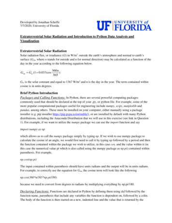
Radiation Oncology 2009, 4:1BackgroundUntil recently, conventionally fractionated high-doseradiation therapy was the preferred treatment in patientswith stage I NSCLC who were unfit to undergo surgery ordeclined surgery. This is, however, a poor alternative tosurgery in operable patients as the mean reported crudelocal recurrence rates are as high as 40% (range 6–70%),resulting in a three year overall and cause-specific survivalof only 34 and 39%, respectively [1].Recently, stereotactic radiotherapy has gained much interest in the treatment of medically inoperable patients withstage I lung cancer, as local control rates are dramaticallyimproved with this technique compared to conventionalfractionation. In studies where schedules with a biologically effective dose (BED) larger than 100 Gy are used, typical local control rates are approximately 90%. The largestseries were reported from Japan [2,3], United States [4]and the Netherlands [5], comprising experience in over750 patients. Onishi et al. [6] retrospectively described theresults of 257 patients treated in 14 Japanese centres usinga number of different fractionation schedules and deliveryapproaches. This Japanese study also included nearly 100patients who refused surgery, and the 5-year overall survival rate of 70.8% observed after a BED of 100 Gy amongthose patients is at least equivalent to the outcome aftersurgery [7-9]. Currently, several phase II trials have startedin operable lung cancer patients [10] (RTOG 0618 andJCOG 0403), however, to date no prospective multi-centre randomized studies have been performed to comparestereotactic radiotherapy with surgery in patients withoperable lung h direct and indirect). In case of surgery, a lobectomyshould be carried out, but limited resections are acceptable. Careful radiological follow-up is performed withinthe trial in patients treated by SRT, as salvage surgery ormediastinal radiation therapy might still be possible incase of clinical, radiological or histological evidence oflocal or hilar disease progression.Accreditation and dosimetry guidelines have been previously developed for trials of stereotactic radiotherapy suchas RTOG 0236 and JCOG 0403 [12-14]. However, a reassessment was considered necessary because a new patientgroup was being treated with stereotactic radiotherapy,namely patients who were fit to undergo both primaryand salvage surgery. As a result, normal tissue dose-constraints had to be more stringently defined in order tominimize the risk of increased complications after salvagesurgery. Furthermore, IGRT technology from differentvendors has been rapidly adopted at various Dutch centres, which had to be taken into account. The resultingguidelines include both minimum requirements thatmust be met by each participating centre as well as recommendations for possible further improvements. They arepresented here in order to facilitate the implementation offuture multi-centre studies, to stimulate and improve theimplementation of stereotactic techniques in clinical practice and to improve the comparability of results.MethodsA randomized phase III trial of Radiosurgery Or Surgeryfor operable Early stage (stage 1A) non-small cell Lungcancer (ROSEL, ClinicalTrials.gov ID NCT00687986)has been opened for accrual in August 2008. The study isinitiated by the VU medical centre Amsterdam and theDutch Lung Cancer Research Group. The primary studyobjectives are to compare local and regional control, quality of life and treatment costs at 2 and 5 years in patientswho are randomized to either surgery or radiosurgery(Figure 1). Treatment costs are a primary end-point, as thecosts associated with surgery for stage IA in The Netherlands are far higher than the present costs of stereotacticradiotherapy [11]. These costs are expected to be evenmore if the costs of post-operative revalidation and loss ofeconomic activity are taken into account. However,patients treated with stereotactic radiotherapy could incurcosts for salvage treatment if a higher incidence of local orregional recurrences is detected. Therefore, treatment costswere considered to be a relevant end-point.A ROSEL Quality Assurance Working Party was formed byradiation oncologists and medical physicists from bothacademic as well as non-academic hospitals that hadalready implemented stereotactic radiotherapy for lungcancer. Several working party meetings were organised inwhich both the knowledge from literature and clinicalexperience were shared and amalgamated. In support ofthese meetings, a literature search was conducted bysearching MEDLINE with different key words and theirpermutations such as stereotactic radiotherapy, stage Ilung cancer, treatment planning, CT scan, patient positioning and tumour mobility. Abstract books of theASTRO, ASCO, AAPM and ESTRO/ECCO from 2004 to2008 were reviewed. It was recognized that there was littledata available in the literature about the influence of different planning algorithms on the planning of stereotacticradiotherapy. Therefore, an additional planning study wasperformed in 22 stage IA and 4 stage 1B non-small celllung cancer patients in order to develop and evaluate theplanning guidelines differentiated according to type ofdose calculation algorithm used. Patient characteristicsand treatment planning details have been reported previously [15].Secondary objectives include overall survival, pulmonaryfunction tests, quality adjusted life years and total costsIn brief, a four-dimensional (4D)-CT was reconstructed inten equally spaced time bins using respiratory phase bin-Page 2 of 14(page number not for citation purposes)
Radiation Oncology 2009, 4:1http://www.ro-journal.com/content/4/1/1Figure studyROSEL1designROSEL study design.Page 3 of 14(page number not for citation purposes)
Radiation Oncology 2009, 4:1ning for each patient. From these phases, a maximumintensity projection (MIP) was reconstructed [16]. Thedatasets were then imported in the Pinnacle3 treatmentplanning system (Philips Medical Systems, Wisconsin).Using the MIP dataset, an experienced radiation oncologist delineated the internal target volume (ITV). Organs atrisk were delineated on an average-density CT reconstruction. The PTV was created by expanding the ITV with a 3mm margin. The treatment plans consisted of 9 equallyspaced coplanar 6 MV beams which were not allowed toenter through the oesophagus, heart, spinal cord or contralateral lung. The plans were inversely optimized usingthe direct aperture optimization module of the Pinnacle3treatment planning system with the same objectives asused in the ROSEL trial. Three different plans were created; using an advanced (type A) dose calculation algorithm, a less advanced (type B) algorithm and a planassuming all tissues within the body to have unit density,in accordance with the RTOG study 0236 and 0618 protocols [17,18].In order to facilitate the clinical use of these recommendations, we divided the process of implementing high-doseradiotherapy into the following headings: CT scanningand patient positioning, target volume definition, organsat risk definition, Dose calculation algorithms and fractionation, dose prescription, coverage and constraints,treatment planning and treatment execution.Patient positioning and CT scanningThe patient should be scanned in the treatment positionwhich should be supine with both arms raised above thehead using an arm-rest or other fixation device. Positionswhich are less comfortable for the patient should beavoided so as to prevent the likelihood of uncontrolledmovement during scanning or treatment. Four-dimensional (4D) CT scanning is strongly recommended inorder to account for an individualised assessment andincorporation of tumour motion [19-21]. Preferably 10but no less than 6 breathing phases should be reconstructed in order to determine the tumour movement fortreatment planning. Using 10 phases, it was found thatgenerally the full amplitude of motion can be captured[22]. Within the ROSEL trial, acquisition of a slow-CTscan or multiple (at least 3) rapid planning scans coveringthe entire range of tumour motion is also allowed, as 4DCT scanners are not widely available yet. However, targetvolume delineation might be more difficult as the images,and thus also the tumour volume, of slow-CT scans areblurred [23,24]. All centres participating in the ROSELstudy will most likely be able to implement 4D-CT scanning in the near future. Generally, intravenous contrast isnot necessary for planning CT scans for early stage lungcancer, but contrast-enhanced CT images may still be usedfor dose calculations. Although the effect of (IV) contrast on dose calculations for lung patients is notspecifically studied, the influence of IV contrast in headand neck intensity modulated radiotherapy plans wasproven to be insignificant [25]. The slice spacing betweenreconstructed CT images should be 3 mm over the complete tumour trajectory and 5 mm elsewhere. The scanshould encompass the entire lung volume in order to calculate meaningful lung dose-volume parameters.Target volume definitionThe gross tumour volume (GTV) will generally be contoured using CT pulmonary windows; however, soft tissuewindows may be used to avoid inclusion of adjacent vessels or chest wall structures within the GTV. The correctness of the GTV delineation should be checked in axial,sagittal and coronal views. The clinical target volume(CTV) is assumed to be identical to the GTV, i.e. with nomargin for microscopic disease added, which appears tobe justified by the high local control rates observed inpatients undergoing careful post-treatment follow-up[26]. This approach has also been accepted in the ASTROACR recommendations on stereotactic radiotherapy [27].For PTV definition, two main treatment planning and execution techniques can be distinguished; planning andirradiation based on the internal target volume (ITV) concept or the time-averaged mean position of the tumour.PTV based on the ITV conceptFor 4D CT scans, the ITV can be derived from the union ofGTV delineations on all breathing phases or alternatively,from contouring on a maximum intensity projection(MIP) CT-dataset [28,29]. The appropriateness of theMIP-delineation should at least be confirmed by a visualinspection of the projected ITV contours on the CT-datasets of the end-inspiration and end-expiration phase binsusing axial, sagittal and coronal views. In addition to theMIP contouring, the GTV should also be contoured in asingle phase (preferably the end-expiration phase,because this is the most stable tumour position and thephase with the least breathing artefacts) in all patients inorder to determine the GTV size. For checking the ITV contour based on the MIP it is not necessary to delineate theend-inspiration and end-expiration phase bins (visualassessment suffices). Alternatively, the ITV may be constructed by the union of all delineations of the GTV in allbreathing phases. If only 3D CT data is available, the ITVshould be based on either multiple slow CT-scans covering the whole tumour trajectory or an additional marginof 3–5 mm in all directions around the CTV determinedon a single slow CT-scan [30]. The ITV to PTV margin isprimarily meant to take into account patient set-up uncertainties. However, small intra-fractional variations in thetumour motion and mean position may be present. Alsointer-fractional variations may be present, but these mightPage 4 of 14(page number not for citation purposes)
Radiation Oncology 2009, 4:1be corrected for using tumour based image guided position verification and correction [31]. In addition, smalldelineation uncertainties will exist. Thus, a minimum of 3mm ITV to PTV margin is required in all dimensions, evenif a set-up error of 3 mm can be guaranteed. On the otherhand, the ITV to PTV margin should not exceed 5 mm, asthis would unnecessarily enlarge treated volumes. In casean institution would need to apply a larger margin, e.g.because of their set-up accuracy, it is advised to firstimprove its (set-up) technique (see also paragraph abouttreatment execution).PTV based on the mean tumour positionAs an alternative to the ITV concept, planning and irradiation based on the time-averaged mean position of thetumour has been developed [32]. In contrast to the ITV toPTV margin discussed previously, the CTV to PTV marginneeded here should take the tumour motion into account.However, similar to the reasoning given for the ITV to PTVmargin, a minimum margin of 3 mm should be used forthe incorporation of the other uncertainties.Organs at risk definitionDose volume criteria for organs at risk (OAR) given in anext paragraph are all constraints to the highest dosesreceived by the OAR. As a consequence, the impact of differences in delineation protocols between institutions isnot expected to be high, as these differences are likely tobe primarily of influence on the delineations located outside the high dose region. However, in order to supportfuture normal tissue complication probability (NTCP)modelling studies, the OAR delineation guidelines as usedin the ROSEL protocol are given below.When 4D-CT scans are used for treatment planning, thecritical OAR should be contoured on the relevant reference reconstruction (i.e. the scan used for dose calculations, see also paragraph about treatment planning). Thiscan generally be performed without taking into accountpotential mobility of these organs, as current experience isbased on this type of delineations. However, extremes ofmotion of organs such as the oesophagus may influencethe choice of beam arrangements in case of 'peripheral'lesions located in the proximity of the mediastinum [33].Also, patient set-up corrections due to tumour shifts leadto a change in the dose given to the OAR. To avoid excessive doses to OAR, it is recommended to evaluate theimpact of such shifts on the OAR dose during treatmentplanning. This might be accomplished by using Planningorgan at Risk Volumes (PRV) [34].The spinal cord and oesophagus should be contouredstarting at least 10 cm above the superior extent of the PTVand continuing on every CT slice to at least 10 cm belowthe inferior extent of the PTV. For patients with ted in the mid- or lower zones of the lungs, the pericardium and/or heart should be contoured as a singlestructure. The superior aspect (or base) for purposes ofcontouring will begin at the level of the inferior aspect ofthe aortic arch (aorto-pulmonary window) and extendinferiorly to the apex of the heart.The defined ipsilateral brachial plexus originates from thespinal nerves exiting the neural foramen on the involvedside from around C5 to T2 [35,36].For peripheral tumours in the upper lobes, the majortrunks of the brachial plexus should be contoured, usingthe subclavian and axillary vessels as surrogates. This neurovascular complex will be contoured starting proximallyat the bifurcation of the brachiocephalic trunk into thejugular/subclavian veins (or carotid/subclavian arteries)and following along the route of the subclavian vein tothe axillary vein ending after the neurovascular structurescross the 2nd rib.The trachea and proximal bronchial tree are contoured astwo separate structures using mediastinal windows on CTto correspond to the mucosa, submucosa and cartilagerings and airway channels associated with these structures.For this purpose, the trachea will be divided into two sections: the proximal trachea and the distal 2 cm of trachea.The proximal trachea will be contoured as one structure,and the distal 2 cm of trachea will be included in the structure identified as proximal bronchial tree (main carina,right and left main bronchi, right and left upper lobebronchi, intermedius bronchus, right middle lobe bronchus, lingular bronchus, right and left lower lobe bronchi).Delineation of the chest wall has not been regularly performed. Little is known about chest wall morbidity in relation to dose in stereotactic radiotherapy, and thereforedelineation is not mandatory within the ROSEL trial [37].However, it is recommended to delineate the chest wall incase of tumours in close proximity to the chest wall. Thiswill aid the development of NTCP models concerningchest wall toxicity.Dose calculation algorithms and fractionationA number of different dose fractionation schedules havebeen reported for lung SRT [38,39], but the optimal dosefractionation schedule may vary with tumour stage andlocation. Although no randomized studies comparing different fractionation schedules have been conducted forstage I tumours, most of the clinical experience is basedon schedules with 3 fractions of 20 Gy. In RTOG study0236, RTOG study 0618 and in the ROSEL study, this fractionation scheme is used. In all studies, eligibility forinclusion was limited to lesions located 2 cm distal toPage 5 of 14(page number not for citation purposes)
Radiation Oncology 2009, 4:1the hilar structures. Within the ROSEL study, a more conservative fractionation scheme of 5 fractions of 12 Gy isalso allowed for patients with a tumour with broad contact to the thoracic wall or adjacent to the heart or mediastinum. Lung function is not considered to affect thescheduling or fractionation. The largest clinical experiencepublished thus far did not exclude any patient on the basisof poor lung function [26], and did not observe excessivelung toxicity when 'risk-adapted' SRT schemes were usedThis is supported by 2 recent reviews [40,41]. A report byTimmerman [42] which suggested that toxicity rates werehigh for central tumors treated with SRT has been criticized on the grounds of the toxicity definitions used [43].However, it is recognized that differences between calculation algorithms in the various treatment planning systems may be as high as 30% in individual cases [15].These differences are largest for lung tumour treatmentplans, and generally increase with decreasing field size,which is especially relevant in stereotactic radiotherapy ofstage 1A lung tumours. Thus, depending on the treatmentplanning algorithm used, one should actually use an alternative nominal fraction dose to deliver the same actualdose to the patient. Unfortunately, extensive data comparing all the calculation algorithms that are likely to be usedin the ROSEL study are not available. For the nominaldose fractionation schedules allowed within the ROSELtrial two main type of algorithms are distinguished[15,44]. Type A models: Models primarily based on electronicpath length (EPL) scaling for inhomogeneity corrections.Changes in lateral transport of electrons are not (well)modelled. The algorithms in this group are e.g. Eclipse/ModBatho and Eclipse/ETAR from Varian, OMP/PB andPlato/ETAR from Nucletron, PrecisePLAN from Elekta, Iplan Dose/PB from BrainLAB, and XiO/Convolution fromCMS. Type B models: Models that in an approximate way consider changes in lateral electron transport. The models inthis group are e.g. Pinnacle/CC from Philips Medical Systems, Eclipse/AAA from Varian, OMP/CC from Nucletron,I-Plan-dose with XVMC Monte-Carlo algorithm fromBrainLAB and XiO/Superposition from CMS.As a guideline, the fractionation schedule(s) and doseconstraints one wants to implement should be adapted tothe dose algorithm used. For example, within the ROSELtrial, it was decided that for type A models, a standard fractionation schedule of 3 fractions of 20 Gy or 3 fractions of18 Gy and a conservative fractionation schedule of 5 fractions of 12 Gy or 5 fractions of 11 Gy could be allowed.For type B models, the standard fractionation should be 3fractions of 18 Gy and the conservative /1should be 5 fractions of 12 Gy or 5 fractions of 11 Gy. A 3fractions of 20 Gy schedule is not allowed in combinationwith type B models in the ROSEL trial, as this might leadto dose levels being approximately 10% higher than thedose levels with which extensive experience has beengained in the VU Medical Centre Amsterdam, using a typeA algorithm. These higher dose levels might lead toincreased morbidity. The fractionation of 5 times 12 Gy isstill allowed with type B models since the errors of type Aalgorithms in calculating dose to the thoracic wall, heartor mediastinum are expected to be less significant.Although this also would lead to approximately 10%higher dose levels, the biologically effective dose for thePTV will still be well below the BED of the 3 fractionsschedule. There are no indications in the literature thatthis would lead to an unacceptable level of morbidity. It ishighly recommended to include dose algorithm specificsin future reports about stereotactic radiotherapy for lungtumours. If a more accurate algorithm becomes availableto the authors of such articles, one should also considerthe publication of the recalculated data. These data can beused to improve our dose-effect models, which aid the further improvement of stereotactic radiotherapy.Dose prescription, coverage and constraintsIn line with current multi-institutional trials and multiplesingle-centre experiences, the dose prescription should bebased on 95% of the target volume (PTV) receiving at leastthe nominal fraction dose (e.g., 20 Gy per fraction 60 Gytotal), and 99% of the target volume (PTV) receiving aminimum of 90% of the fraction dose. The dose maximum within the PTV should preferably not be less than110% or exceed 140% of the prescribed dose, similar tothe criteria formulated in RTOG protocol 0618 [18]. Thelocation of the treatment plan normalization point,which is in fact only influencing the display of the dosedistribution, can be left to the institutions preference.RTOG trial 0236 defined a set of parameters to quantifythe conformity of the dose and PTV coverage. The sameparameters were used in RTOG trial 0618 and are usedhere. However, the ROSEL trial requires the use of inhomogeneity corrections, whereas this is not allowed withinthe RTOG trials. Consequently, the dose conformityrequirements in the ROSEL study differ from the RTOGrecommendations. Moreover, a distinction in these valuesis made between type A and B algorithms, because of thesignificant differences in calculation results between them(Table 1).From Figure 2 it is clear that using a type B algorithm, it ismore difficult to conform the planned dose to the PTVthan using a type A algorithm, especially for a small PTV.This is caused by the increased influence of lateral scatterdisequilibrium for smaller PTV, which is modelled betterPage 6 of 14(page number not for citation purposes)
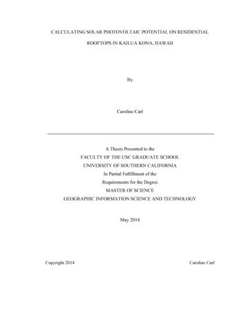
Radiation Oncology 2009, 4:1http://www.ro-journal.com/content/4/1/1Table 1: Dose conformity requirements and definition of protocol deviations. R100% and R50% ratio of respectively the 100% and 50%Prescription Isodose Volume to the PTV. D2 cm dose maximum at 2 cm from the PTV as percentage of the prescribed dose. V20 Gy Percent of lung receiving 20 Gy or more (both lungs minus GTV).R50%R100%None 1.15 1.15 0None 8 7 6 1.25 1.15 0DeviationMinor8–107–86–6.5None 55 65 65DeviationMinor55–6065–7065–75V20 Gy (%)NoneNone 12 9 6DeviationMinor12–149–116–8using a type B algorithm. Thus, a less strict conformityrequirement was formulated. The difference between typeB and type A or unit density calculations is even more pronounced for the R50% values (Figure 3). Also for the doseat 2 cm from the PTV (Figure 4) and the percentage of thelung receiving more than 20 Gy (Figure 5), it is clear thata type B algorithm will result in higher values, due to thefact that the change in lateral scattering in lung tissue istaken into account much better. Again, the conformityrequirements for type B algorithms were relaxed for theseparameters. However, relaxation of these requirementsdoes not result in an actual inferior patient treatment. Onthe contrary, because these more advanced algorithmsprovide a better description of the actual dose distribution, the user has a greater opportunity to optimize thedose distribution to the stated requirements. Therefore,the use of these more advanced algorithms is stronglyencouraged. Please note that the figures presented here arebased on the treatment plans generated without recalculation with a more advanced algorithm, thus representingtreatment planning clinical practice within the ROSELtrial, while in the article of Schuring and Hurkmans theresults were presented after recalculation, thus quantifying the actual delivered dose differences arising from theuse of different algorithms [15]. To emphasize theimprovement that can be achieved using a more advancedalgorithm over a type A algorithm or a unit density calculation, the dose to the PTV after recalculation is given inFigure 6 (reprinted with permission from Schuring andHurkmans [15]. The figure clearly shows that The EPLplans (Type A algorithm) consistently overestimate thedose to the PTV, resulting in an average D95 of 48 Gy, 20%None 65 70 70DeviationMinor65–7570–8070–80PTV (cc)DeviationMinor 4 6 8Type B models (more advanced algorithms)R50%D2 cm (%)R100%NoneType A models (standard algorithms)D2 cm (%)4–66–88–10V20 Gy (%)None 5 6 100–2020–40 40PTV (cc)DeviationMinor5–86–1010–150–2020–40 40lower than the prescribed value. The overestimation of thedose increased with decreasing PTV size, although largevariations are observed between individual patients. Forthe unit density calculations the recalculated D95 rangedbetween as much as 63 and 42 Gy for individual patients.Dose-volume constraints for OAR within the ROSEL protocol are given in Table 2 and differ from the ones used inRTOG 0236 and 0618 (for lung constraints, see previousTable 1). A reassessment was considered necessarybecause a new patient group will be treated with stereotactic radiotherapy within the ROSEL trial, namely patientswho are fit to undergo both primary and salvage surgery.As a result, normal tissue dose-constraints have to bemore stringently defined in order to minimize the risk ofincreased complications after salvage surgery. Additionally, new constraints were formulated to be used for the 5fraction scheme. Furthermore, the constraints are basedon 1 cc volumes (except for the spinal cord), to prevent anexcessive dependency on the calculation grid size in theevaluation of these parameters. Skin dose, with the constraint that no point within the skin should receive a dosehigher than 24 Gy as dictated in RTOG 0618 is notincluded in Table 2, as dose calculations within thisregion are often not very accurate and
lung cancer, treatment planning, CT scan, patient posi-tioning and tumour mobility. Abstract books of the ASTRO, ASCO, AAPM and ESTRO/ECCO from 2004 to 2008 were reviewed. It was recognized that there was little data available in the literature about the influence of dif-ferent planning