Transcription
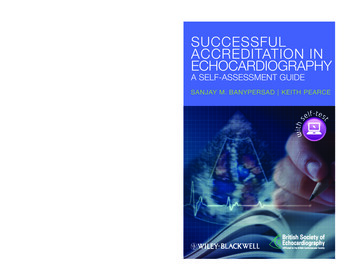
A SELF-ASSESSMENT GUIDES A N J AY M . B A N Y P E R S A DKEITH PEARCEMBChB, BMedSci (Hons), MRCP (UK),Cardiology SpR, The Heart Hospital, London, UKPrincipal Cardiac Physiologist,Wythenshawe Hospital, Manchester, UKSitting an accreditation examination is adaunting prospect for many trainee echocardiographers. And with an increasing drive for theaccreditation of echocardiography laboratoriesand individual echocardiographers, there is anincreasing need for an all-encompassing revisionaid for those seeking accreditation.T I T L E S O F R E L AT E D I N T E R E S TThe editors of this unique book have producedthe only echocardiography revision aid basedon the syllabus and format of the BritishSociety of Echocardiography (BSE) nationalechocardiography accreditation examinationand similar examinations administered acrossEurope. Written by BSE accredited members,fully endorsed by the BSE, and with a forewordby BSE past-President, Dr. Simon Ray, thisindispensable guide provides a valuable insightinto how echocardiography accreditationexams are aphy in Pediatric andCongenital Heart DiseaseLai, ISBN 978-1405174015This book is accompanied by acompanion website:www.accreditationechocardiography.comThe website includes: 89 interactive Multiple-Choice Questions 193 VideoclipsCover design: Fortiori DesignCover images: iStockBANYPERSAD PEARCECrucially, to support students with the morechallenging video section of the exam, acompanion website provides video cases, andwith clear and concisely-structured explanationsto all questions, this is an essential tool foranyone preparing to sit an echocardiographyexamination.Practical Handbook of Echocardiography:101 Case StudiesSun, ISBN 978-1-4051-9556-0SU CCESSFU L ACCRED ITATIO N IN ECHOCARDIOGRAPHYSUCCESSFULACCREDITATION INECHOCARDIOGRAPHYSUCCESSFULACCREDITATION INECHOCARDIOGRAPHYA SELF-ASSESSMENT GUIDESANJAY M. BANYPERSAD KEITH PEARCE
Banypersad bindex.indd 20411/19/2011 2:36:55 PM
Successful Accreditationin EchocardiographyBanypersad ffirs.indd i12/2/2011 3:52:29 PM
COMPANION WEBSITEThis book is accompanied by a companion website:www.accreditationechocardiography.comThe website includes: 89 interactive Multiple-Choice Questions193 VideoclipsBanypersad ffirs.indd ii12/2/2011 3:52:29 PM
SuccessfulAccreditation inEchocardiographyA Self-Assessment GuideSanjay M. Banypersad MBChB,BMedSci (Hons), MRCP (UK)Cardiology SpRThe Heart HospitalLondonUKKeith PearcePrincipal Cardiac PhysiologistWythenshawe HospitalManchesterUKEndorsed by the British Societyof EchocardiographyA John Wiley & Sons, Ltd., PublicationBanypersad ffirs.indd iii12/2/2011 3:52:29 PM
This edition first published 2012 2012 by John Wiley & Sons, Ltd.Wiley-Blackwell is an imprint of John Wiley & Sons, formed by the merger of Wiley’s global Scientific,Technical and Medical business with Blackwell Publishing.Registered Office:John Wiley & Sons, Ltd, The Atrium, Southern Gate, Chichester, West Sussex, PO19 8SQ, UKEditorial Offices:9600 Garsington Road, Oxford, OX4 2DQ, UK111 River Street, Hoboken, NJ 07030-5774, USAFor details of our global editorial offices, for customer services and for information about howto apply for permission to reuse the copyright material in this book please see our websiteat www.wiley.com/wiley-blackwellThe right of the author to be identified as the author of this work has been asserted in accordancewith the UK Copyright, Designs and Patents Act 1988.All rights reserved. No part of this publication may be reproduced, stored in a retrieval system,or transmitted, in any form or by any means, electronic, mechanical, photocopying, recordingor otherwise, except as permitted by the UK Copyright, Designs and Patents Act 1988, withoutthe prior permission of the publisher.Designations used by companies to distinguish their products are often claimed as trademarks.All brand names and product names used in this book are trade names, service marks, trademarksor registered trademarks of their respective owners. The publisher is not associated with any productor vendor mentioned in this book. This publication is designed to provide accurate and authoritativeinformation in regard to the subject matter covered. It is sold on the understanding that the publisheris not engaged in rendering professional services. If professional advice or other expert assistance isrequired, the services of a competent professional should be sought.The contents of this work are intended to further general scientific research, understanding, anddiscussion only and are not intended and should not be relied upon as recommending or promotinga specific method, diagnosis, or treatment by physicians for any particular patient. The publisherand the author make no representations or warranties with respect to the accuracy or completenessof the contents of this work and specifically disclaim all warranties, including without limitationany implied warranties of fitness for a particular purpose. In view of ongoing research, equipmentmodifications, changes in governmental regulations, and the constant flow of information relatingto the use of medicines, equipment, and devices, the reader is urged to review and evaluate theinformation provided in the package insert or instructions for each medicine, equipment, or device for,among other things, any changes in the instructions or indication of usage and for added warnings andprecautions. Readers should consult with a specialist where appropriate. The fact that an organizationor Website is referred to in this work as a citation and/or a potential source of further information doesnot mean that the author or the publisher endorses the information the organization or Website mayprovide or recommendations it may make. Further, readers should be aware that Internet Websiteslisted in this work may have changed or disappeared between when this work was written and whenit is read. No warranty may be created or extended by any promotional statements for this work.Neither the publisher nor the author shall be liable for any damages arising herefrom.Library of Congress Cataloging-in-Publication DataBanypersad, Sanjay M.Successful accreditation in echocardiography : a self-assessment guide / Sanjay M. Banypersad,Keith Pearce.p. ; cm.Includes index.ISBN-13: 978-0-4706-5692-1 (pbk. : alk. paper)ISBN-10: 0-470-65692-1 (pbk. : alk. paper)I. Pearce, Keith (Keith A.) II. Title.[DNLM: 1. Echocardiography–Examination Questions. WG 18.2]LC classification not assigned616.1′2307543076–dc232011029720A catalogue record for this book is available from the British Library.Wiley also publishes its books in a variety of electronic formats. Some content that appears in printmay not be available in electronic books.Set in 9.25/12pt Meridien by SPi Publisher Services, Pondicherry, India12012Banypersad ffirs.indd iv12/2/2011 3:52:30 PM
ContentsForeword, viiPreface, viiiAcknowledgements, ixAbbreviations, x1 Basic Physics and AnatomyQuestions, 1Answers, 62 The Aortic ValveQuestions, 14Answers, 193 Left Ventricular AssessmentQuestions, 27Answers, 344 The Mitral ValveQuestions, 44Answers, 495 Right Ventricular AssessmentQuestions, 57Answers, 626 Prosthetic Valves and EndocarditisQuestions, 70Answers, 757 Pericardial Disease and Cardiac MassesQuestions, 82Answers, 878 Adult Congenital Heart DiseaseQuestions, 94Answers, 99vBanypersad ftoc.indd v11/19/2011 2:32:15 PM
CONTENTS9 Video QuestionsCase 1, 106Case 2, 109Case 3, 111Case 4, 115Case 5, 119Case 6, 122Case 7, 125Case 8, 130Case 9, 133Case 10, 137Case 11, 140Case 12, 145Case 13, 149Case 14, 152Case 15, 161Case 16, 168Case 17, 172Case 18, 175Case 19, 177Case 20, 181Video Answers, 186Index, 196COMPANION WEBSITEThis book is accompanied by a companion website:www.accreditationechocardiography.comThe website includes: 89 interactive Multiple-Choice Questions193 VideoclipsviBanypersad ftoc.indd vi11/19/2011 2:32:15 PM
ForewordEchocardiography is a mainstay of cardiac diagnostics and remains byfar the most commonly performed imaging examination in cardiologypractice. The development of easily portable and hand held machineshas enhanced its use in bedside diagnosis and emergency assessmentwhile real time 3-D imaging, tissue Doppler and speckle tracking provide a sophisticated insight into myocardial structure and function. Intandem with the development of technology has come the recognitionthat echocardiography is only as good as the individual performing theexamination and that the training, accreditation and continuing education of echocardiographers is essential to the effective functioning ofa clinical service. Moreover there is an increasing drive for the accreditation of echocardiography laboratories and individual accreditationof echocardiographers is a central part of this process.Sitting an accreditation examination is a daunting prospect formany trainee echocardiographers. There are numerous textbooks onechocardiography covering the range from basic to advanced imagingbut few that provide specific preparation for examinations. In thisbook Sanjay Banypersad, Keith Pearce and their colleagues have setout to provide a revision aid based broadly on the current syllabus ofthe British Society for Echocardiography. Writing unambiguous multiple choice questions and selecting video cases relevant to clinicalpractice is far from easy and the authors and text reviewers have madestrenuous efforts to ensure the accuracy and relevance of the content.No book of this type is sufficient on its own to provide all theinformation required for individual accreditation but used in conjunction with one of the comprehensive echocardiography textsavailable it should be very useful to those preparing for examinationsor simply wanting to refresh their knowledge.Simon Ray, BSc, MD, FRCP, FACC, FESCConsultant CardiologistHonorary Professor of CardiologyUniversity Hospitals of South ManchesterManchester Academic Health Sciences CentreManchester, UKviiBanypersad fbetw.indd vii11/22/2011 3:13:11 PM
PrefaceThere has been a vast expansion in the field of cardiac imaging inrecent years. Coronary CT is now part of NICE guidance for low-riskischaemic heart disease and cardiac MRI is increasingly favoured forcertain pathologies. Echocardiography remains however of paramount importance in the cardiological assessment of patients. Itsfundamental advantage lies in being widely available, cost-effective andeasily portable without any appreciable reduction in picture quality.This has meant not only an increase in the number of studies beingperformed per year, but also in the specialty of the operator performingthe studies. Emergency physicians and anaesthetists are now wellversed in the application of echocardiography to critically ill patientsin the resuscitation department, ICU or operating theatres.It is important therefore that adherence to a quality standard issafeguarded to ensure that the patient receives a uniformly highstandard of examination. There are a number of accreditationprocesses worldwide and this book is designed to broadly mimic thelayout of the British Society of Echocardiography Transthoracicaccreditation process, which currently comprises a written MCQpaper and a video section. This book has 8 chapters derived fromthe current syllabus and each chapter consists of 20 MCQ stylequestions each with 5 ‘True/False’ stems, except the LV Assessmentchapter which has 30 questions. Chapter 9 is comprised of 20 videocases each consisting of 4 or 5 questions with the option to pick one‘best-fit’ answer from the given stems.It is my hope that all candidates sitting a board exam or accreditation will find this book an invaluable revision aid and that thosenot sitting for accreditation will still nevertheless find it useful fortheir continued professional development.Sanjay M. BanypersadviiiBanypersad fpref.indd viii11/22/2011 3:14:01 PM
AcknowledgementsWe would like to extend our gratitude to the following people fortheir time and effort spent in addition to their clinical duties, inorder to peer-review all the material in this book.Dr Simon Ray, Consultant Cardiologist, University HospitalsSouth Manchester NHS Foundation Trust, Wythenshawe Hospital,Southmoor Road, Manchester, UK.Dr Nik Abidin, Consultant Cardiologist, Salford Royal NHS Foundation Trust, Salford Royal Hospital, Stott Lane, Salford, UK.Miss Jane Lynch, Expert Cardiac Physiologist, University HospitalsSouth Manchester NHS Foundation Trust, Wythenshawe Hospital,Southmoor Road, Manchester, UK.Dr Anna Herrey, Consultant in Cardiology, The Heart Hospital,16–18 Westmoreland Street, London, UK.Dr Ansuman Saha, Consultant Cardiologist, East Surrey Hospital,Canada Avenue, Redhill, Surrey, UK.Dr Richard Bogle, Consultant Cardiologist, Epsom and St. HelierUniversity Hospital NHS Trust, Wrythe Lane, Carshalton, Surrey, UK.Dr Anita MacNab, Consultant Cardiologist, University HospitalsSouth Manchester NHS Foundation Trust, Wythenshawe Hospital,Southmoor Road, Manchester, UK.Dr Bruce Irwin, SpR in Cardiology, University Hospitals SouthManchester NHS Foundation Trust, Wythenshawe Hospital,Southmoor Road, Manchester, UK.We are also grateful to all the echocardiographers and technicians inthe echocardiography department at Wythenshawe Hospital and tothe University Hospitals South Manchester NHS Foundation Trustfor their permission to use the images and video files.Sanjay M. Banypersad would also like to add a final vote of thanksto his parents and younger brother, Vishal, for their constant wordsof support and encouragement throughout.ixBanypersad flast.indd ix11/22/2011 3:13:41 PM
CHCMHOCM5-HydroxytryptamineAmerican College of Cardiologyadult congenital heart diseaseAmerican Heart Associationatrial fibrillationaortic regurgitationarrhythmogenic right ventricular cardiomyopathyaortic stenosisatrial septal defectaortic valveaortic valve replacementatrioventricular septal defectsblood pressurebody surface areaBritish Society of Echocardiographycoronary artery diseasecardiac resynchronisation therapycross-sectional areacomputed tomographycontinuous wavedecibeldilated cardiomyopathychange in pressuredobutamine stress echocardiogramchange in timechange in volumeelectrocardiogramnot strictly an abbreviation – refers to anterior mitralleaflet movement on M-mode in the active and passivephase of transmitral flowejection fractionE-point septal separationEuropean Society of Cardiologyhypertrophic cardiomyopathyhypertrophic obstructive cardiomyopathyxBanypersad flast.indd x11/22/2011 3:13:41 PM
A BBREVIATION SAPPMPRPRFPSPVPWRARBBBRCARCMheart rateintensive care unitintravenousinferior vena cavaIsovolumetric contraction timeIsovolumetric relaxation timeinterventricular septum in diastolejugular venous pressureleft atriumleft anterior descendingleft bundle branch blockleft ventricleleft ventricular assist deviceleft ventricular end-diastolic dimensionleft ventricular end-diastolic pressureleft ventricular end-systolic dimensionleft ventricular hypertrophyleft ventricular inflow tractleft ventricular outflow tractmyocardial infarctionmitral stenosismitral regurgitationmagnetic resonance imagingmitral valvemitral valve prolapsemitral valve replacementNational Institute for Health and Clinical Excellencepulmonary arterypatent ductus arteriosuspulmonary embolismpatent foramen ovaleproximal isovelocity surface areapermanent pacemakerpulmonary regurgitationpulse-resonance frequencypulmonary stenosispulmonary valvepulsed waveright atriumright bundle branch blockright coronary arteryrestrictive cardiomyopathyxiBanypersad flast.indd xi11/22/2011 3:13:41 PM
A B B R E V I AT I DVTIregurgitant orifice arearight ventricleright ventricular hypertrophyright ventricular outflow tractregional wall motion abnormalitysystemic lupus erythematosusstroke volumesuperior vena cavasystemic vascular resistancetricuspid annular plane systolic excursiontuberculosistransoesophageal echocardiographytricuspid regurgitationtransthoracic echocardiographytricuspid valvevelocityventricular septal defectvelocity time integralxiiBanypersad flast.indd xii11/22/2011 3:13:41 PM
1Basic Physicsand AnatomyQUESTIONSFor each question below, decide whether the answers provided aretrue or false.1 The following is true of ultrasound waves:a. Propagate through medium like lightb. Are part of the electromagnetic spectrumc. Loudness is measured in decibelsd. The decibel scale shows a linear relationship with amplituderatioe. Can be reflected but not refracted2 The following are true of ultrasound waves during 2D echo:a. The optimal image is formed when the medium isperpendicular to the ultrasound beamb. The narrowest part of the beam (the focal zone) can be variedc. Side lobes are artefacts only found with phased-arraytransducersd. Structures smaller in diameter than the wavelength of theultrasound beam may cause scattering of the beame. Travel faster in blood than in bone3 During standard TTE:a. Dropout occurs when there is parallel alignment of the beamwith the tissueb. At a higher frequency, the ultrasound beam has a higherpenetration depthc. Doppler studies are based on scattering of the ultrasoundbeam by red blood cellsSuccessful Accreditation in Echocardiography: A Self-Assessment Guide,First Edition. Sanjay M. Banypersad and Keith Pearce. 2012 John Wiley & Sons, Ltd. Published 2012 by John Wiley & Sons, Ltd.1Banypersad c01.indd 111/22/2011 3:09:12 PM
B A S I C P H Y S I C S AND ANAT OMY: QUE S T IONSd. The transmitted ultrasound waves are attenuated withincreasing mismatch in acoustic impedancee. Axial resolution degrades more than lateral resolution whenthe depth is increased4 The following are true of image resolution and artefacts:a. M-mode has excellent temporal resolutionb. Prosthetic valves cause acoustic shadowing as well asreverberationsc. Tissue harmonic imaging improves endocardial borderdefinition but has no effect on valvesd. High PRF can cause uncertainty due to range ambiguitye. Low aliasing velocities with colour Doppler can overestimateregurgitation5 During echocardiography, the following can be changed by theoperator:a. Impedanceb. Focusc. Amplituded. Wavelengthe. PRF6 Regarding the use of tissue Doppler imaging:a. It can be used to calculate myocardial tissue velocitiesb. It can give information on segmental LV functionc. Unlike transmitral E and A velocities are, tissue Dopplerimaging-derived E’ and A’ waves are not preload dependentd. Gives a more accurate assessment of IVRT than transmitralDopplere. The heart’s movement in the chest cavity can be a limitationof the technique7 When using M-mode to assess LV ejection fraction:a. May be inaccurate if the beam is obliqueb. Results may not be indicative of overall function in ischaemicheart diseasec. End-systolic dimensions are usually measured on the R waveof the ECGd. A fractional shortening of 30% can be normale. The result is more accurate than EF derived using theSimpson’s method2Banypersad c01.indd 211/22/2011 3:09:13 PM
B AS IC P HYSICS A N D A N ATOMY: QU ESTION S8 Regarding PW Doppler, the following are true:a. Is subject to the Nyquist limitb. Has two dedicated crystals for sending and receivingc. Can measure velocities at varying depthd. Is used in tissue Doppler imaginge. More than one sample volume can be assessed at a time9 Regarding continuous-wave Doppler, the following are true:a. Transmits and receives an impulse in sequence.b. Is useful in assessing mid-cavity step-ups in gradientc. Often aliases at high velocitiesd. Is limited in that it cannot separate individual velocitiesalong the length of a beame. Is useful when assessing peak aortic velocity10 For a 5 MHz transducer at an angle of 60 to blood flow, theDoppler frequency shift is 10 kHz. The following are true:a. The wavelength is approximately 0.3 mmb. The maximum depth is 2–3 cmc. The blood velocity is approximately 3 m/sd. Lowering the transducer frequency to 1 MHz increasesmaximum depth to 20 cme. Optimal accuracy occurs with the Doppler cursorperpendicular to the direction of flow11 In standard 2D echocardiography of a patient lying in the leftlateral position:a. The atrial septum is best visualised in the apical 4-chamberviewb. In the apical 4-chamber view, tilting the ultrasound beamposteriorly reveals the 5-chamber viewc. In the parasternal long-axis view, tilting the beam inferomedially reveals the RV inflowd. In the parasternal long axis view, the normal LA is 4.5 cmin mene. Coronary arteries can sometimes be seen in the parasternalshort-axis view12 Regarding the parasternal short-axis view:a. The most posterior of the aortic valve cusps is thenon-coronary cuspb. The mitral valve leaflets are clearly seen3Banypersad c01.indd 311/22/2011 3:09:13 PM
B A S I C P H Y S I C S AND ANAT OMY: QUE S T IONSc. It is a useful view for detecting PV abnormalitiesd. It is a useful view for calculating PA pressuree. Eccentric jets of regurgitant aortic or mitral valves can beclearly demonstrated13 In the apical 4-chamber view:a. The right ventricular wall is thinner than that of the LVb. A septal ‘knuckle’ is often seen in elderly peoplec. The Chiari network may be seen in the LAd. Rotating to the apical 3-chamber view reveals theinferior walle. Rotating to the apical 2-chamber view shows theaortic valve14 Regarding spectral Doppler signals:a. The normal mitral E wave is greater than the A wave inyoung peopleb. Peak aortic velocity of 2 m/s can be normal with someprosthetic valvesc. In AF, an average of at least five consecutive signals shouldbe takend. CW Doppler is usually needed for high velocities to avoidaliasinge. A fast sweep-speed is required to assess for respiratoryvariation15 The following relationships between structures is true in theparasternal long axis:a. The left coronary cusp of the aortic valve is anteriorb. A fibrous band separates the anterior mitral valve leaflet andthe aortic rootc. In the RV inflow view, the anterior and posterior tricuspidvalve leaflets are seend. The moderator band can be seen in the RVe. The nodules of Arantius are features of the mitral valve16 The following parameters would not affect frame rate:a. Increasing the depthb. Increasing the sector sizec. Increasing the line densityd. Increasing the transmit frequencye. Decreasing the sector size4Banypersad c01.indd 411/22/2011 3:09:13 PM
B AS IC P HYSICS A N D A N ATOMY: QU ESTION S17 The type of filter used for tissue Doppler imaging is a:a. High-pass filterb. Band-pass filterc. Low-pass filterd. Reject filtere. Notch filter18 Dobutamine stress echo:a. Cannot be used to detect myocardial viabilityb. Can be used to diagnose CADc. Is more sensitive and specific than exercise stress testingd. Can be used to predict anaesthetic risk for major surgerye. Is usually performed using agitated saline contrast19 Harmonic imaging:a. Was developed to improve endocardial definitionb. Uses a transmit frequency equal to the receive frequencyc. Enhances the detection of transpulmonary contrastd. Makes valvular structures appear thickere. Should not be used when making Doppler recordings20 The following statements are true:a. Absorption is the transfer of ultrasound energy to the tissueduring propagationb. Acoustic impedance is the product of tissue density and thepropagation velocity through itc. Shifting the zero velocity baseline may eliminate aliasing inthe pulsed-wave Doppler moded. Shadowing results in the presence of echoes directly behinda strong echo reflectore. A longitudinal wave is a cyclic disturbance in which theenergy propagation is parallel to the direction of particlemotion5Banypersad c01.indd 511/22/2011 3:09:13 PM
Basic Physicsand AnatomyANSWERS1 a. Fb. Fc. Td. Fe. FVisible light is part of the electromagnetic spectrum and is propagatedas a transverse waves. Sound is not part of the electromagneticspectrum and is propagated as longitudinal waves, with oscillationsparallel to the direction of propagation. Loudness is measured indecibels and the scale shows a logarithmic relationship to amplituderatio i.e. dB 20 log (V/R) (where V represents acoustic pressureand R is a reference value). Ultrasound waves can be both reflectedand refracted, the latter being responsible for false images inaberrant locations.2 a. Tb. Tc. Fd. Te. FReflection of ultrasound waves (and therefore imaging) is optimalwhen the tissue interface is perpendicular with the ultrasound beam.The normal ultrasound beam from a transducer of diameter D,travels through an aperture and has an initial columnar near zone;beyond this, there is divergence of the beam, according to sin θ 1.22λ/D, which causes image degradation. However, the transducerface can be altered to become, for example, more concave, changingthe position of the narrowest point of the beam so that imageresolution is greater – this is the focal zone and it is variable. Sidelobes are beams dispersed laterally to the main beam leading toimage artefact and are common to all transducers; grating lobesare specific to phased-array transducers. Scattering is caused by6Banypersad c01.indd 611/22/2011 3:09:13 PM
B AS IC P HY SICS A N D A N ATOMY: A N SWERSstructures smaller than the wavelength of the ultrasound beam.Structures larger in wavelength cause reflection or refraction. Thepropagation velocity in bone is double that of blood.3 a. Tb. Fc. Td. Te. FParallel alignment causes very little of the ultrasound beam to bereflected back to the transducer, causing image dropout; this istypically seen of the atrial septum in apical 4-chamber view.A higher frequency produces higher image resolution but decreasespenetration depth. The wavelength of ultrasound is 0.2–1 mm,whereas that of a red cell is about 7–10 μm, hence as stated above,red blood cells are effective scatterers and form the principle ofDoppler flow studies. Air has high acoustic impedance, so anyair between the transducer and the body causes a significantacoustic impedance mismatch and therefore attenuation of thetransmitted beam; attenuation can also affect the reflected beam.Axial resolution is relatively unchanged with increasing depthbecause the beam remains parallel to the tissues. However, lateralresolution decreases because beam width increases due todivergence.4 a. Tb. Tc. Fd. Te. TM-mode does have excellent temporal resolution and is often usedto assess high-speed motion such as mitral valve leaflet fluttering.Prosthetic valves can cause reverberation and acoustic shadowingbeyond the valve image. Harmonics improve border definition butalso make valves appear thicker, thus standard imaging shouldalways be used in conjunction with harmonics. High PRF is useful todetect very high velocities, but range ambiguity means that thedepth at which that velocity occurs could be located at any one ofseveral points along the insonating beam. Low aliasing velocitiescause distinct colour changes at lower velocities than normal, makingthe degree of regurgitation seem higher than it actually is.7Banypersad c01.indd 711/22/2011 3:09:13 PM
B A S I C P H Y S I C S AND ANAT OMY: ANS WE R S5 a. Fb. Tc. Td. Fe. TImpedance is a property of the tissue itself. Wavelength is usuallyfixed, and since velocity is constant through a given medium, PRFcan be altered to produce varying depth. Amplitude is alteredthrough gain and the focus can be varied as explained above (seeanswer to Question 4)6 a. Tb. Tc. Td. Fe. TTissue Doppler imaging can assess myocardial tissue velocities andindeed, the myocardial velocity gradient between 2 positions on theventricle; it can therefore be very useful for assessing segmentalmotion and function. Because myocardial velocities rather thanblood flow velocities are measured, they are less preload dependent.However, IVRT is best measured with conventional PW Doppler asmyocardial movement does not necessarily correlate with valveopening and closure.7 a. Tb. Tc. Fd. Te. FM-mode has excellent time resolution and endocardial bordermotion is well imaged. A very oblique beam will overestimate cavitysize and underestimate function as displacement is at an angle to theinsonating beam. Maximal displacement measurement will occurwhen the beam is perpendicular to the chamber. Regional wallmotion abnormalities are common is ischaemic heart disease and alarge apical infarct with preserved basal segments would overestimateLV function with M-mode. End-diastolic dimensions are measuredon the R wave and the normal range for fractional shortening is25–45%. Simpson’s method is a more accurate measure of EF as anumber of segments across the LV cavity are included.8Banypersad c01.indd 811/22/2011 3:09:13 PM
echocardiography accreditation examination and similar examinations administered across Europe. Written by BSE accredited members, fully endorsed by the BSE, and with a foreword by BSE past-President, Dr. Simon Ray, this indispensable guide provides a valuable insight into how echo