Transcription
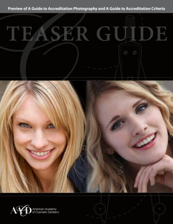
Preview of A Guide to Accreditation Photography and A Guide to Accreditation CriteriaTEASER GUIDE
Table of Contents forA Guide to Accreditation Photography: Photographic Documentationand Evaluation in Cosmetic DentistryREQUIRED VIE WS FOR CLINIC AL C ASE SUBMISSIONS . . . . . . 6HOW-TO TAKE EXCEPTIONAL DENTAL PHOTOS . . . . . . . . . . . . . . . . . 7ISSUES THAT APPLY TO ALL IMAGES . . . . . . . . . . . . . . . . . . . . . . . . . . . . . . . . . . 8FULL FACE . . . . . . . . . . . . . . . . . . . . . . . . . . . . . . . . . . . . . . . . . . . . . . . . . . . . . . . . . . . . . . . . . . . . . . . . . . . . . 9FULL SMILE . . . . . . . . . . . . . . . . . . . . . . . . . . . . . . . . . . . . . . . . . . . . . . . . . . . . . . . . . . . . . . . . . . . 10-11UPPER AND LOWER TEEETH. . . . . . . . . . . . . . . . . . . . . . . . . . . . . . . . . . . . . . . . . 12-13MAXILL ARY ANTERIOR VIE W . . . . . . . . . . . . . . . . . . . . . . . . . . . . . . . . . . . . . . . 14-15MAXILL ARY ARCH . . . . . . . . . . . . . . . . . . . . . . . . . . . . . . . . . . . . . . . . . . . . . . . . . . . . . . . . . . . . . . . 16MANDIBUL AR ARCH . . . . . . . . . . . . . . . . . . . . . . . . . . . . . . . . . . . . . . . . . . . . . . . . . . . . . . . . . . . 17ADDITIONAL PHOTOGR APHIC DOCUMENTATIONFOR DENTISTS — C ASE T YPE V. . . . . . . . . . . . . . . . . . . . . . . . . . . . . . . . . . . . . . . . . . 18ADDITIONAL PHOTOGR APHIC DOCUMENTATIONFOR L ABOR ATORY TECHNICIANS . . . . . . . . . . . . . . . . . . . . . . . . . . . . . . . . 19-20COMMON ERRORS IN DENTAL PHOTOGR APHY . . . . . . . . . . 21-23USE OF A CONTR ASTING DE VICE . . . . . . . . . . . . . . . . . . . . . . . . . . . . . . . . . . . . . . 24A Guide to Accreditation PhotographyPhotographic Documentationand Evaluation inCosmetic Dentistry22 16 11 8 5,6 4 2,8 1,4A200010005002501256030158421218www.aacd.comA Guide to Accreditation Photography
Required Views forClinical Case SubmissionsThere are 24 views required for all clinical case examinations. Of the 24 views, 12 should betaken before treatment and 12 after treatment. Additional views are required for the techniquedocumentation as well as radiographic documentation (see www.aacd.com/Accreditation foradditional information). Images may be captured in either manual or TTL mode. All intraoralimages should be captured using high f-stops to maximize depth of field.MAGNIFICATIONImages of the required views will be captured at one of three magnification ratios (1:10, 1:2, 1:1).Make any necessary magnification conversions to produce an image magnification comparable tothe images illustrated in the photography guide. Lens magnification conversion is needed for manydigital SLR cameras without full frame sensors. Settings will vary with sensor and face size. Cameraswith smaller sensors will require approximately a 1.5 times increase in the setting on the lens barrel[1:10 (1:15), 1:2 (1.3), 1:1 (1:1.5)].VIEWSNON-RETRACTED VIEWS1. Natural Full Face – frontal view – 1:10 (1:15) magnification2. Full Natural Smile – frontal view – 1:2 (1:3) magnification3. Full Natural Smile – right lateral view – 1:2 (1:3) magnification4. Full Natural Smile – left lateral view – 1:2 (1:3) magnificationRETRACTED VIEWS5. Upper and lower teeth slightly parted – frontal view – 1:2 (1:3) magnification6. Upper and lower teeth slightly parted – right lateral view – 1:2 (1:3) magnification7. Upper and lower teeth slightly parted – left lateral view – 1:2 (1:3) magnification8. Maxillary anterior in view only – frontal view – 1:1 (1:1.5) magnification9. Maxillary anterior in view only – right lateral view – 1:1 (1:1.5) magnification10. Maxillary anterior in view only – left lateral view – 1:1 (1:1.5) magnificationRETRACTED VIEWS USING A MIRROR11. Maxillary arch – occlusal view – 1:2 (1:3) magnification12. Mandibular arch – occlusal view – 1:2 (1:3) magnificationA Guide to Accreditation Photography
Upper and Lower TeethRight and Left Lateral View1:2 (1:3) MagnificationRetracted View The upper and lower teeth should be slightly parted so the incisal edges are visible. This allowsfor evaluation of incisal plane and incisal embrasures. Show as much gingiva as possible. Rotate the retractors toward the image side, while pullingthe retractors out and away from the teeth. Minimize the appearance of lips and retractors in the image. Treated teeth and adjacent tissue must be completely and clearly visible. Gingival height andcontour cannot be obscured. The vertical midline of the image should be the lateral incisor. The horizontal midline of the image should be the incisal plane, perpendicular to the verticalmidline. Reproduce natural asymmetry. Focus on the lateral incisor. Proper depth of field (high f-stop) will allow other visible teeth tobe in focus. Tongue should be positioned away from the teeth to avoid distraction. Maintain1:2 (1:3) magnification. This is not a profile (sagittal) view. The contralateral central incisor and possibly thecontralateral lateral incisor and canine should be visible, based on arch size. Remember tocenter the image on the lateral incisor. If retracted and framed properly, the contralateral cheek will obscure most of the backgroundarea.A Guide to Accreditation Photography
How To Take ExceptionalDental Photos:It’s important to capture excellent patient photos during the Accreditation process and beyond.Photos that are poorly shot can negatively impact a case even when an ideal result is achieved.To help capture excellent photos, consider using an assistant who can: Position retractors Dry the teeth as needed Hold mirrors when required Prevent mirrors from fogging.Other tips: Use a solid background for full face images. When taking occlusal views, recline the patient and ask an assistant to hold retractors.The assistant can hold an occlusal mirror against the opposite arch and prevent the mirrorfrom fogging. The photographer should stand above the patient when capturing maxillary views.When taking mandibular views, the photographer should aim below the patient’s head.Occlusal views are aided by having the patient reclined andholding retractors. An assistant holds an occlusal mirroragainst opposite arch and keeps mirror from fogging with agentle stream of dry air. Photographer stands above thepatient to capture the maxillary view and belowthe patient's head to capture the mandibular view.A Guide to Accreditation Photography
Table of Contents forA Guide to Accreditation Criteria: Contemporary Concepts in Smile DesignCHAPTER 1 – GLOBAL ESTHETICS . . . . . . . . . . . . . . . . . . . . . . . . . . . . . . . . . . . . . . . . . 6 Smile Line Incisal Edge Position Incisal Plane Midline Buccal CorridorCHAPTER 2 – MACRO ESTHETICS. . . . . . . . . . . . . . . . . . . . . . . . . . . . . . . . . . . . . . . . . . . 11 Embrasures Principles of Proportion & Central Dominance Labial Anatomy, Line Angles & Incisal Edge Emergence Profile Symmetry Axial InclinationCHAPTER 3 – MICRO ESTHETICS. . . . . . . . . . . . . . . . . . . . . . . . . . . . . . . . . . . . . . . . . . . . 18 Shade Selection Margin Placement & Design Surface Finish, Luster & Reflectivity Incisal Translucency & Surface Color CharacterizationsCHAPTER 4 – PINK ESTHETICS. . . . . . . . . . . . . . . . . . . . . . . . . . . . . . . . . . . . . . . . . . . . . . . 22 Periodontal Health Ovate Pontic Gingival Contour, Shape & PositionCHAPTER 5 – SPECIAL CONSIDER ATIONS . . . . . . . . . . . . . . . . . . . . . . . . . . . . . 25 Function & Occlusion Choice of Materials Responsible Esthetics Photography Case Selection Restorative Team: Laboratory Technician & the Restorative DentistA Guide to Accreditation CriteriaContemporary Conceptsin Smile DesignDiagnosis and Treatment Evaluation in Comprehensive Cosmetic DentistryCHAPTER 6 – ACCREDITATION C ASE T YPES. . . . . . . . . . . . . . . . . . . . . . . . . . 27REFERENCES AND RESOURCES . . . . . . . . . . . . . . . . . . . . . . . . . . . . . . . . . . . . . . . . . . . . . . 54w w w. aacd. co mA Guide to Accreditation Criteria
Global EstheticsAn assessment of dental esthetics begins, quite simply, with the smile. Figure 5 - Orientation of the midline and incisal plane to the face. Figure 6 - Midline orientation.A Guide to Accreditation Criteria
Macro EstheticsAs we begin to narrow our study of the elements of smile design beyond the components relatedto the orientation of the smile in the face and how it is enveloped by the soft tissue, our focusshifts toward the elements of macro esthetics. Macro esthetics relates to the shapes and contoursof teeth. Understanding the importance of these contours within the functional matrix will helpto ensure a predictable esthetic result. Figure 9 - The incisal embrasures should demonstrate a naturaland progressive increase in size from the central to the cuspid. A Guide to Accreditation CriteriaFigure 21 - The teeth should display a progressively increasing mesial axialinclination as you move to the posterior.
Micro EstheticsAs we continue to concentrate on the details necessary to replicate nature, our next focus ismicro esthetics.Figure 25 - The texture of a tooth is easilyidentified as the “finger print” of the toothand will significantly impact the blendingof the tooth into the smile.Pink EstheticsOur quest to study esthetics related to all elements of smile design in the absolute best health possibleincluding periodontal architecture.Figure 32 - Equal gingival zeniths from cuspid to cuspid are an acceptable relationship.A Guide to Accreditation Criteria
Accreditation Case TypesThe clinical portion of the Accreditation process requires the successful completion of five case types.These case types provide an opportunity for the dentist or laboratory technician pursuing Accreditation todemonstrate excellence in a range of disciplines that cover important aspects of cosmetic dentistry. Within eachcase type, there are particular subsets of skills that become the primary focus of the Accreditation Examinersin evaluating a case. In all case types, the Examiners are looking for the member in the Accreditation processto demonstrate a comprehensive knowledge and ability in delivering responsible esthetic care with thepatient’s best interests in mind, honoring evidenced-based accepted standards of function and health. Properdiagnosis and case selection are paramount in providing the best opportunity for success. In some case types,the outlined case requirements are more comprehensive than others. The indications for the procedures mustbe matched with the patient’s needs. The principles outlined in Contemporary Concepts in Smile Design areimportant in all case types; however, they may be weighted more in certain case types than in others. Casesinvolving a limited focus of regional treatment will obviously place less emphasis on broader aspects of smiledesign. It is important to remember in all cases submitted for Accreditation, that anything that is touched—even if it is beyond the scope of required treatment in a particular case type—will be evaluated. The followingis a description of the case types and an insider’s view of the Examiners’ Perspective for each case, as well asexamples of cases that have met the standard of excellence.A Guide to Accreditation Criteria
COMMON CRITERIA ERRORS IN ACCREDITATION CASE PRESENTATIONSIt is extremely helpful, when presenting cases for Accreditation, for members inthe process to work with a mentor. Accreditation Mentors can be identified andcontacted through the AACD Website at www.aacd.com/mentors. AccreditationMentors are active Examiners. Their eyes have been calibrated through the processand they can help members in the process avoid many frustrations or roadblocks.They volunteer their time, are passionate about what they do, and are more thanhappy to assist members in the process. Communication with mentors can best befacilitated utilizing the PowerPoint or Keynote templates that are also available onthe Website. These templates provide a vehicle to efficiently share images of cases.In many situations, it is helpful to share a case with a mentor prior to initiating anytreatment to discuss treatment planning and the suitability of cases.The current Examiners were asked to anecdotally list in order the commonflaws that they observe in presented cases. Members in the Accreditation processshould revisit their submissions prior to presentation with these criteria in mind.Understanding how Examiners see cases is obviously an asset. Although all of thecriteria have equal importance, a recent poll of Examiners highlighted these “dirtydozen” shortcomings observed in submitted Accreditation cases:CRITERION #71IS THE PERIODONTAL HEALTH OPTIMAL?There is no excuse for a prosthetic design that does not support excellent periodontalhealth. The gingiva should be pink, stippled, and firm. It should exhibit a mattesurface. The papillae should be pointed and should fill the gingival embrasures rightup to the contact area. Members in the process often underestimate the time that maybe necessary for the periodontal architecture to return to optimal health post-delivery.Examiners can not make the assumption that the tissue will improve in healthwith maturation. The only evaluation that can be made is the condition that isobserved in the photograph. The following images display increasing fault in tissuehealth of three individual cases.Moderate tissue inflammation observedproximal to the left lateral incisor.A Guide to Accreditation CriteriaModerate/severe tissue inflammationobserved at the gingival cavosurfaceof the restored teeth.Gross and catastrophic tissueinflammation.
A Guide to Accreditation PhotographyPhotographic Documentationand Evaluation inCosmetic Dentistry22 16 11 8 5,6 4 2,8 1,4A Guide to Accreditation CriteriaA200010005002501256030158Contemporary Conceptsin Smile Design42Diagnosis and Treatment Evaluation in Comprehensive Cosmetic Dentistry1218www.aacd.comw w w. aacd. co mThis is just a small sampling ofA Guide to Accreditation Photography:Photographic Documentationand Evaluation in Cosmetic DentistryandA Guide to Accreditation Criteria:Contemporary Concepts in Smile DesignTo purchase either or both guide books visitwww.aacd.com/guides.
AMERICAN ACADEMY OF COSMETIC DENTISTRY402 WEST WILSON ST.MADISON, WI 53703608.222.8583 800.543.9220FAX: 608.222.9540www.aacd.com
A Guide to Accreditation Photography Photographic Documentation and Evaluation in Cosmetic Dentistry 22 16 11 8 56 4 28 14 2000 1000 500 250 125 60 30 15 8 4 2 1 2 1 8 A www .aacd .com A Guide to Accreditation Photography Table of Contents for A Guide to Accreditation Photography: Photog