Transcription
AT T E N T I O N R E A D E R SF 05130GMS ITC cover.qxd11/4/053:05 PMPage 1We would like your comments on Inside the Cell.Please give us your opinion by filling out this postage-paid response card.1.Extent to which the publication held your interest2.Understandability3.Amount and type of information presentedJoin our Findings mailing list. For sample issues,see http://publications.nigms.nih.gov/findings/. I would like to receive Findings, a magazinethat profiles two NIGMS-supported scientists,features brief descriptions of recent clinicallyrelevant research, and includes a crosswordpuzzle based on words used in the articles.Name4.Usefulness and value of such a publicationPlease comment on whether Inside the Cell helped you learn more about:CityState1.Cell biology researchInside the CellAddressZip CodeE-mail (optional)Phone (optional)2.3.What it’s like to be a scientistThe excitement of biomedical research todayOther comments: I would like to receive a free CD-ROM containingNIGMS science education booklets on topics such ascell biology, chemistry, genetics, pharmacology, andstructural biology. These booklets are geared towarda high school and early college audience. Printcopies of the publications can be ordered mU.S. DEPARTMENT OFHEALTH AND HUMAN SERVICESNational Institutes of HealthNational Institute of General Medical SciencesNIH Publication No. 05-1051Revised September 2005http://www.nigms.nih.govU.S. DEPARTMENT OFHEALTH AND HUMAN SERVICESNational Institutes of HealthNational Institute of General Medical Sciences
F 05130GMS ITC cover.qxd11/4/053:05 PMPage 2What Is NIGMS?Discrimination ProhibitedAccessibilityThe National Institute of General Medical SciencesUnder provisions of applicable public laws enactedThis publication can be made available in(NIGMS) supports basic biomedical research onby Congress since 1964, no person in the Unitedformats that are more accessible to peoplegenes, proteins, and cells. It also funds studies onStates shall, on the grounds of race, color, nationalwith disabilities. To request this material in afundamental processes such as how cells commu origin, handicap, or age, be excluded from partici-different format, contact the NIGMS Officenicate, how our bodies use energy, and how wepation in, be denied the benefits of, or be subjectedof Communications and Public Liaison atrespond to medicines. The results of this researchto discrimination under any program or activity301-496-7301, TDD 301-402-6327; send e-mailincrease our understanding of life and lay the(or, on the basis of sex, with respect to any educa-to info@nigms.nih.gov; or write to the office atfoundation for advances in the diagnosis, treatment,tion program or activity) receiving Federal financialthe following address: 45 Center Drive MSCand prevention of disease. The Institute’s researchassistance. In addition, Executive Order 111416200, Bethesda, MD 20892-6200. If you havetraining programs produce the next generation ofprohibits discrimination on the basis of age byquestions about this publication, you can usebiomedical scientists, and NIGMS has programs tocontractors and subcontractors in the performancethe same contact information to reach the editor,encourage minorities underrepresented in biomedicalof Federal contracts, and Executive Order 11246Alisa Zapp Machalek.and behavioral science to pursue research careers.states that no federally funded contractor mayNIGMS supported the research of most of thediscriminate against any employee or applicantscientists mentioned in this booklet.for employment because of race, color, religion,sex, or national origin. Therefore, the programs ofthe National Institute of General Medical Sciencesmust be operated in compliance with these lawsAdditional Copiesand Web VersionTo order additional copies of Inside the CellDEPT OF HEALTH AND HUMAN SERVICESNATIONAL INSTITUTES OF HEALTHNATIONAL INSTITUTE OF GENERAL MEDICAL SCIENCES45 CENTER DR RM 3AN.32 MSC 6200BETHESDA MD 20892-6200OFFICIAL BUSINESSPENALTY FOR PRIVATE USE 300or other free publications available from NIGMS,go to http://publications.nigms.nih.gov/order oruse the contact information above.and Executive Orders.Inside the Cell and related material are availableonline at TIONAL INSTITUTES OF HEALTHNATIONAL INSTITUTE OF GENERAL MEDICAL SCIENCESOFFICE OF COMMUNICATIONS AND PUBLIC LIAISON45 CENTER DR RM 3AN.32 MSC 6200BETHESDA MD 20814-9692
Inside the CellU.S. DEPARTMENT OFHEALTH AND HUMAN SERVICESNational Institutes of HealthNational Institute of General Medical SciencesNIH Publication No. 05-1051Revised September 2005http: // www.nigms.nih.gov
Produced by the Office of Communications and Public LiaisonNational Institute of General Medical SciencesNational Institutes of HealthU.S. Department of Health and Human Services
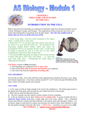
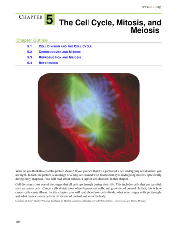
ContentsPREFACE: THE MICROSCOPIC METROPOLIS INSIDE YOU4CHAPTER 1: AN OWNER’S GUIDE TO THE CELL6Nucleus: The Cell’s Brain7Cell Membrane: Specialist in Containing and Communicating8Endoplasmic Reticulum: Protein Clothier and Lipid Factory8Golgi: Finishing, Packaging, and Mailing Centers10Lysosomes: Recycling Centers and Garbage Trucks10Mitochondria: Cellular Power Plants11Cytoskeleton: The Cell’s Skeleton and More12The Tour Ends Here14Cool Tools for Studying Cells14Science Schisms18CHAPTER 2: CELLS 101: BUSINESS BASICS20Got Energy?20Priority: Proteins21Cellular Rush Hour26The Mark of Death30CHAPTER 3: ON THE JOB: CELLULAR SPECIALTIES32Fit for the Job33All-In-One Stem Cells34You’ve Got Nerve(s)!37Nursing Baby Eggs39The Science of Senses40Cells on the Move42Big Science44CHAPTER 4: CELLULAR REPRODUCTION: MULTIPLICATION BY DIVISION46The Two Faces of Cell Division47The Cycling Cell48Mitosis: Let’s Split!50Meiosis: Sex, Heredity, and Survival52Why You’re Not Just Like Your Relatives58CHAPTER 5: THE LAST CHAPTER: CELL AGING AND DEATH60Aging: A World of Theories61Thieving Oxygen62Damage, Yes. But Aging?63Telomeres: Cellular Timekeepers64Cells That Never Die Can Kill You66Death of a Cell67Apoptosis and Mitosis: Life in Balance68Getting Rid of Troublemakers70Cell Biology: The Science of Life72GLOSSARY74
P R E FA C EBY ALISA ZAPP MACHALEKThe Microscopic Metropolis Inside YouAt this very moment, electricity is zappingthrough your brain, voracious killersIn Chapter 1, “An Owner’s Guide to the Cell,”we’ll explore some of the basic structures thatare coursing through your veins, and corrosiveallow cells to accomplish their tasks and somechemicals sizzle in bubbles from your head to yourof the ways scientists study cells. In Chapter 2,toes. In fact, your entire body is like an electrical“Cells 101: Business Basics,” we’ll focus on thecompany, chemical factory, transportation grid,functions shared by virtually all cells: making fuelcommunications network, detoxification facility,and proteins, transporting materials, and dispos hospital, and battlefield all rolled into one. Theing of wastes. In Chapter 3, “On the Job: Cellularworkers in each of these industries are your cells.Specialties,” we’ll learn how cells specialize to getCells are the smallest form of life—thetheir unique jobs done. In Chapters 4, “Cellularfunctional and structural units of all living things.Reproduction: Multiplication by Division,” andYour body contains trillions of cells, organized5, “The Last Chapter: Cell Aging and Death,”into more than 200 major types.we’ll find out how cells reproduce, age, and die.At any given time, each cell is doing thousandsMuch of the research described in this bookletof jobs. Some of these tasks are so essential for lifeis carried out by cell biologists at universitiesthat they are carried out by virtually all cells. Othersand other institutions across the nation who areare done only by cells that are highly skilled for thesupported by U.S. tax dollars, specifically thosework, whether it is covering up your insides (skindistributed by the National Institute of Generalcells), preventing you from sloshing around likeMedical Sciences (NIGMS), a component of thea pile of goo (bone cells), purging your body ofNational Institutes of Health. NIGMS is keenlytoxic chemicals (liver cells), or enabling you tointerested in cell biology because knowledgelearn and remember (brain cells). Cells also mustof the inner workings of cells underpins ourmake the products your body needs, such asunderstanding of health and disease.sweat, saliva, enzymes, hormones, and antibodies.Although scientists daily learn more aboutcells and their roles in our bodies, the field is stillan exciting frontier of uncharted territory andunanswered questions. Maybe someday, you willhelp answer those questions.
Inside the Cell I Preface 5Nerve CellsBlood CellsHeart Muscle Cells“Long ago it became evident that the key toevery biological problem must finally beSmall Intestine Cellssought in the cell; for every living organismis, or at some time has been, a cell.”— E.B. Wilson (1856–1939) famous cell biologistYour body contains many differentcell types, each customized for aparticular role. Red blood cells carrylife-giving oxygen to every cornerof your body, white blood cells killgerm invaders, intestinal cells squirtout chemicals that chisel away atyour food so you can absorb itsnutrients, nerve cells sling chemicaland electrical messages that allowyou to think and move, and heartcells constantly pump blood,enabling life itself.ALL CELL IMAGES THIS PAGE DENNIS KUNKEL MICROSCOPY, INC.
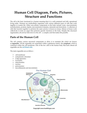
CHAPTER 1BY ALISA ZAPP MACHALEKAn Owner’s Guide to the CellWelcome! I hope the transformationBut from where we are, you can’t see nearly thatwasn’t too alarming. You have shrunkfar. Clogging your view is a rich stew of mole down to about 3 millionths of your normal size.cules, fibers, and various cell structures calledYou are now about 0.5 micrometers tallorganelles. Like the internal organs in your(a micrometer is 1/1000 of a millimeter). Butbody, organelles in the cell each have a uniquedon’t worry, you’ll return to your normal sizebiological role to play.before you finish this chapter.Now that your eyes have adjusted to theAt this scale, a medium-sized human celllooks as long, high, and wide as a football field.darkness, let’s explore, first-hand and up close,the amazing world inside a cell.MitochondriaNucleusGolgiRough ERSmooth ERLysosomesA typical animal cell, sliced open to reveal cross-sections of organelles.
Inside the Cell I An Owner’s Guide to the Cell 7Nucleus: The Cell’s BrainLook down. Notice the slight curve? You’re stand ing on a somewhat spherical structure about 50feet in diameter. It’s the nucleus — basically thecell’s brain.The nucleus is the most prominent organelleand can occupy up to 10 percent of the spaceinside a cell. It contains the equivalent of the cell’sgray matter — its genetic material, or DNA. Inthe form of genes, each with a host of helperis pockmarked with octagonal pits about an inchmolecules, DNA determines the cell’s identity,across (at this scale) and hemmed in by raisedmasterminds its activities, and is the officialsides. These nuclear pores allow chemicalcookbook for the body’s proteins.messages to exit and enter the nucleus. But we’veGo ahead—jump. It’s a bit springy, isn’t it?That’s because the nucleus is surrounded by twopliable membranes, together known as thenuclear envelope. Normally, the nuclear envelopecleared the nuclear pores off this area of thenucleus so you don’t sprain an ankle on one.If you exclude the nucleus, the rest of thecell’s innards are known as the cytoplasm.EUKARYOTIC CELLSPROKARYOTIC CELLSThe cells of “complex” organisms, including allplants and animals“Simple” organisms, includingbacteria and blue-green algaeContain a nucleus and many other organelles,each surrounded by a membrane (the nucleusand mitochondrion have two membranes)Lack a nucleus and othermembrane-encased organellesCan specialize for certain functions, such as absorbingnutrients from food or transmitting nerve impulses;groups of cells can form large, multicellular organsand organismsUsually exist as single, virtuallyidentical cellsMost animal cells are 10–30 micrometers across,and most plant cells are 10–100 micrometers acrossMost are 1–10 micrometers acrossVirtually all forms of life fall into one of two categories: eukaryotes or prokaryotes.
8National Institute of General Medical SciencesSugar ChainsEndoplasmic Reticulum: ProteinClothier and Lipid FactoryIf you peer over the side of the nucleus, you’llnotice groups of enormous, interconnected sacssnuggling close by. Each sac is only a few inchesacross but can extend to lengths of 100 feet orCholesterolmore. This network of sacs, the endoplasmicreticulum (ER), often makes up more thanLipidsProteins10 percent of a cell’s total volume.Take a closer look, and you’ll see that the sacsare covered with bumps about 2 inches wide.The membrane thatsurrounds a cell is madeup of proteins and lipids.Depending on the mem brane’s location and rolein the body, lipids canmake up anywhere from20 to 80 percent of themembrane, with theremainder being proteins.Cholesterol, which is notfound in plant cells, is atype of lipid that helpsstiffen the membrane.Cell Membrane: Specialist inContaining and CommunicatingYou may not remember it, but you crossed amembrane to get in here. Every cell is containedwithin a membrane punctuated with special gates,channels, and pumps. These gadgets let in —orforce out —selected molecules. Their purpose isto carefully protect the cell’s internal environment,a thick brew (called the cytosol) of salts, nutrients,Those bumps, called ribosomes, are sophisticatedmolecular machines made up of more than 70proteins and 4 strands of RNA, a chemical relativeof DNA. Ribosomes have a critical job: assemblingall the cell’s proteins. Without ribosomes, life aswe know it would cease to exist.To make a protein, ribosomes weld togetherchemical building blocks one by one. As naked,and proteins that accounts for about 50 percentinfant protein chains begin to curl out ofof the cell’s volume (organelles make up the rest).ribosomes, they thread directly into the ER.The cell’s outer membrane is made up of aThere, hard-working enzymes clothe themmix of proteins and lipids (fats). Lipids givewith specialized strands of sugars.membranes their flexibility. Proteins transmitNow, climb off the nucleus and out ontochemical messages into the cell, and they alsothe ER. As you venture farther from the nucleus,monitor and maintain the cell’s chemical climate.you’ll notice the ribosomes start to thin out. BeOn the outside of cell membranes, attached tocareful! Those ribosomes serve as nice hand- andsome of the proteins and lipids, are chains offootholds now. But as they become scarce orsugar molecules that help each cell type do its job.disappear, you could slide into the smooth ER,If you tried to bounce on the cell’s outer surface asunable to climb out.you did on the nuclear membrane, all these sugarIn addition to having few or no ribosomes,molecules and protruding proteins would make itthe smooth ER has a different shape and functionrather tricky (and sticky).than the ribosome-studded rough ER. A labyrinth
Inside the Cell I An Owner’s Guide to the Cell 9Rough ERSmooth ERof branched tubules, the smooth ER specializes insynthesizing lipids and also contains enzymes thatbreak down harmful substances. Most cell typesThe endoplasmic reticulum comes in two types:Rough ER is covered with ribosomes and preparesnewly made proteins; smooth ER specializes inmaking lipids and breaking down toxic molecules.have very little smooth ER, but some cells —likethose in the liver, which are responsible for neu tralizing toxins—contain lots of it.Next, look out into the cytosol. Do you seesome free-floating ribosomes? The proteins madeon those ribosomes stay in the cytosol. In contrast,proteins made on the rough ER’s ribosomes endup in other organelles or are sent out of the cellto function elsewhere in the body. A few examplesof proteins that leave the cell (called secretedproteins) are antibodies, insulin, digestiveSUSUMU ITOenzymes, and many hormones.Rough ERRx: Ribosome BlockersAll cellular organisms, including bacteria, have ribo somes. And all ribosomes are composed of proteinsand ribosomal RNA. But the precise shapes of thesebiological machines differ in several very specificways between humans and bacteria. That’s a goodthing for researchers trying to develop bacteria-killingmedicines called antibiotics because it means thatscientists may be able to devise therapies that knockout bacterial ribosomes (and the bacteria along withthem) without affecting the human hosts.Several antibiotic medicines currently on themarket work by inhibiting the ribosomes of bacteriathat cause infections. Because many microorganismshave developed resistance to these medicines, weurgently need new antibiotics to replace those thatare no longer effective in fighting disease.Using sophisticated imaging techniques like X-raycrystallography, researchers have snapped molecularpictures of antibiotics in the act of grabbing onto aIn a dramatic technical feat,scientists obtained the firststructural snapshot of an entireribosome in 1999. This more recent imagecaptures a bacterial ribosome in the act of makinga protein (the long, straight spiral in the lightest shadeof blue). It also shows that — unlike typical cellularmachines, which are clusters of proteins (shown hereas purple ribbons) — ribosomes are composedmostly of RNA (the large, light blue and grey loopyladders). Detailed studies of ribosomal structurescould lead to improved antibiotic medicines.IMAGE COURTESY OF HARRY NOLLERbacterial ribosome. Studying these three-dimensionalimages in detail gives scientists new ideas abouthow to custom design molecules that grip bacterialribosomes even more strongly. Such molecules maylead to the development of new and more effectiveantibiotic drugs. —Alison Davis
National Institute of General Medical SciencesGolgi: Finishing, Packaging,and Mailing CentersNow, let’s slog through the cytosola bit. Notice that stack of a halfdozen flattened balloons, each a fewinches across and about 2 feet long?That’s the Golgi complex, also called theGolgi apparatus or, simply, the Golgi. Likean upscale gift shop that monograms, wraps,and mails its merchandise, the GolgiLysosomes: Recycling Centersand Garbage Trucksreceives newly made proteins and lipidsSee that bubble about 10 feet across? That’sfrom the ER, puts the finishing touchesa lysosome. Let’s go —I think you’ll like this.on them, addresses them, and sends them toPerhaps even more than other organelles,their final destinations. One of the places theselysosomes can vary widely in size—from 5 inchesmolecules can end up is in lysosomes.to 30 feet across.Go ahead, put your ear next to it. Hear thesizzling and gurgling? That’s the sound of power ful enzymes and acids chewing to bits anythingthat ends up inside.But materials aren’t just melted into oblivionin the lysosome. Instead, they are precisely chippedinto their component parts, almost all of whichthe cell recycles as nutrients or building blocks.Lysosomes also act as cellular garbage trucks,hauling away unusable waste and dumping itTINA CARVALHO10outside the cell. From there, the body has variousways of getting rid of it.Golgi
Inside the Cell I An Owner’s Guide to the Cell 11Mitochondria:Cellular Power PlantsLike all other organelles, mitochondriaare encased in an outer membrane. But theyalso have an inner membrane.movements — as well as the many chemicalRemarkably, this inner membrane isreactions that take place inside organelles —four or five times larger than the outerrequire vast amounts of cellular energy. The mainmembrane. So, to fit inside the organelle, itenergy source in your body is a small moleculedoubles over in many places, extending long,called ATP, for adenosine triphosphate.fingerlike folds into the center of the organelle.ATP is made in organelles called mitochondria.Let’s see if we can find some. They look like blimpsabout as long as pickup trucks but somewhat nar These folds serve an important function: Theydramatically increase the surface area availableto the cell machinery that makes ATP. In otherwords, they vastly increase the ATP-productionrower. Oh, a few of them are over there. As we getcapacity of mitochondria.nearer, you may hear a low whirring or hummingsound, similar to that made by a power station.The mazelike space inside mitochondriais filled with a strong brew of hundreds ofIt’s no coincidence. Just as power plants convertenzymes, DNA (mitochondria are the onlyenergy from fossil fuels or hydroelectric dams intoorganelles to have their own genetic material),electricity, mitochondria convert energy from yourspecial mitochondrial ribosomes, and other mole food into ATP.cules necessary to turn on mitochondrial genes.ACTUAL SIZE (AVERAGE)PERCEIVED SIZE WHENMAGNIFIED 3 MILLION TIMESCell diameter30 micrometers*300 feetNucleus diameter5 micrometers50 feetMitochondrion lengthTypically 1–2 micrometers but canbe up to 7 micrometers long18 feetLysosome diameter50–3,000 nanometers*5 inches to 30 feetRibosome diameter20–30 nanometers2–3 inchesMicrotubule width25 nanometers3 inchesIntermediate filament width10 nanometers1.2 inchesActin filament width5–9 nanometers0.5–1 inch*A micrometer is one millionth (10-6) of a meter. A nanometer is one billionth (10-9) of a meter.D.S. FRIEND, BRIGHAM AND WOMEN'S HOSPITALBlink. Breathe. Wiggle your toes. These subtle
12National Institute of General Medical Sciences The three fibers of thecytoskeleton—microtubulesin blue, intermediate fila ments in red, and actin ingreen — play countlessroles in the cell.Cytoskeleton:The Cell’s Skeleton and MoreNow, about all those pipes, ropes, and rods you’vebeen bumping into. Together, they are called thecytoskeleton — the cell’s skeleton. Like the bonyskeletons that give us stability, the cytoskeletongives our cells shape, strength, and the ability tomove, but it does much more than that.Think about your own cells for a moment.Right now, some of your cells are splitting in half,newly released eggs from your ovaries into youruterus. And all that is thanks to the cytoskeleton.As you can see, the cytoskeleton is incrediblyversatile. It is made up of three types of fibers thatconstantly shrink and grow to meet the needs ofthe cell: microtubules, intermediate filaments,and actin filaments. Each type of fiber looks, feels,and functions differently.The 3-inch-wide flexible pipes you just bangedmoving, or changing shape. If you are a man, youryour head on are called microtubules. Made of thesperm use long tails called flagella to swim. If youstrong protein tubulin, microtubules are the heavyare a woman, hairlike fibers called cilia sweeplifters of the cytoskeleton. They do the toughKATHRYN HOWELLGolgi Spelunking: Exit Here, There, But Not AnywhereScientists use a variety of techniques tostudy organelles like the endoplasmicreticulum and Golgi, gaining ever moredetailed understanding of these minute butvery complicated structures. For example,Kathryn Howell of the University of ColoradoSchool of Medicine in Denver uses a special ized high-voltage electron microscope, rapidfreezing methods, and a computer modelingprogram to obtain a vivid three-dimensionalview of the Golgi and the pathways that proteinsuse to exit it.Howell begins by quick-freezing living cells,embedding them in plastic, and slicing the plasticcoated sample into thin sections. As she tilts themicroscope stage, she can capture many imagesof the same region of the sample. A computerassembles these images to form a threedimensional view, called a tomogram, of theGolgi and other organelles. Based on thetomogram, Howell’s research team can producea movie of a virtual journey through the cell.You can see one such movie at tras.Howell’s research shows that there are severalpathways for proteins and other molecules to exitthe Golgi. The findings are revealing, as earlierstudies using different methods had suggestedthat there was only one road out of this organelle.No doubt new chapters to this story will be writtenas biologists and computer scientists create evenmore sophisticated tools for imaging cells. —A.D.
Inside the Cell I An Owner’s Guide to the Cell 13TORSTEN WITTMANNIn these cells, actin filaments appear light purple, microtubules yellow, and nuclei greenish blue.This image, which has been digitally colored, won first place in the 2003 Nikon Small World Competition.physical labor of separating duplicate chromo they can determine the origin of —and possiblesomes when cells copy themselves and serve astreatments for—some kinds of cancer.sturdy railway tracks on which countless mole See that bundle of long rods near the edgecules and materials shuttle to and fro. They alsoof the cell? You can touch it, but don’t try to bendhold the ER and Golgi neatly in stacks and formthe rods. They shatter easily. These rods, slightlythe main component of flagella and cilia.thinner than intermediate filaments, are actinGrab one of those inch-thick ropes. Yeah,filaments. They are made up of two chains ofyou can swing on it —it won’t snap. These strands,the protein actin twisted together. Although actincalled intermediate filaments, are unusual becausefilaments are the most brittle of the cytoskeletalthey vary greatly according to their location andfibers, they are also the most versatile in termsfunction in the body. For example, some inter of the shapes they can take. They can gathermediate filaments form tough coverings, such astogether into bundles, weblike networks, or evenin nails, hair, and the outer layer of skin (not tothree-dimensional gels. They shorten or lengthenmention animal claws and scales). Others areto allow cells to move and change shape. Togetherfound in nerve cells, muscle cells, the heart,with a protein partner called myosin, actin fila and internal organs. In each of these tissues, thements make possible the muscle contractionsfilaments are made of different proteins. So ifnecessary for everything from your action on adoctors analyze intermediate filaments in tumors,sports field to the automatic beating of your heart.
14National Institute of General Medical SciencesThe Tour Ends HereCool Tools for Studying CellsYou’ve seen quite a bit of the cell in a short time.Cell biologists would love to do what you justHowever, this tour covered only the highlights;did—shrink down and actually see, touch, andthere are many other fascinating processes thathear the inner workings of cells. Because that’soccur within cells. Every day, cell biologists learnimpossible, they’ve developed an ever-growingmore, but much remains unexplained.collection of approaches to study cellular innardsYou will now regain your normal size.from the outside. Among them are biochemistry,There should be no lasting side effects of thephysical analysis, microscopy, computer analysis,miniaturization, except, I hope, a slight tinglingand molecular genetics. Using these techniques,sensation caused by new knowledge and a growingresearchers can exhaustively inventory theexcitement about what scientists know—and stillindividual molecular bits and pieces that makedon’t know—about cells.up cells, eavesdrop on cellular communication,and spy on cells as they adapt to changingenvironments. Together, the approaches providevivid details about how cells work together in thebody’s organs and tissues. We’ll start by discussingthe traditional tools of the trade—microscopes—then touch on the new frontiers of quantum dotsand computational biology.MICHAEL YAFFEMorphing MitochondriaIn this fruit fly cell,mitochondria (in red)form a web throughoutthe cell. Microtubulesare labeled in green.Scientists such as Michael P. Yaffeof the University of California, SanDiego, study what mitochondria looklike and how they change through out a cell’s life. To approach thisresearch problem, Yaffe uses simple organisms—such as yeast or fruit fly cells—which, like yourown cells, have membranes, a nucleus, andother organelles. This similarity makes theseorganisms important models for understandinghuman biology.Yaffe’s work helped change the textbook depictionof mitochondria as kidney bean-shaped organelles.Using advanced microscopy, Yaffe and others haveunveiled many different shapes for mitochondria,ranging from the classic beans to long snakesand weblike structures, all of which are thoughtto change on a constant basis. Researchers arediscovering that the different mitochondrialshapes accompany changes in cellular needs,such as when growing cells mature into specifictypes or when a cell responds to disease.Many scientists believe that mitochondria—which divide on their own, have their owngenome and protein-making machinery, andresemble prokaryotes in many ways—aredescendents of oxygen-loving microorganismsthat were taken in by primitive cells. This historicalevent set the stage for advanced life forms likeplants and animals. —A.D.
Inside the Cell I An Owner’s Guide to the Cell 15Light Microscopes:The First Windows Into CellsScientists first saw cells by using traditional lightmicroscopes. In fact, it was Robert Hooke(1635–1703), looking through a microscope ata thin slice of cork, who coined the word “cell.”He chose the word to describe the boxlike holes inthe plant cells because they reminded him of thecells of a monastery.CONLY RIEDERScientists gradually got better at grindingglass into lenses and at whipping up chemicals toselectively stain cellular parts so they could see thembetter. By the late 1800s, biologists already had iden tified some of the largest organelles (the nucleus,This fireworks explosion of color is a dividing newt lung cell seenunder a light microscope and colored using fluorescent dyes:chromosomes in blue, intermediate filaments in red, and spindlefibers (bundled microtubules assembled for cell division) in green.mitochondria, and Golgi
far. Clogging your view is a rich stew of mole cules, fibers, and various cell structures called organelles. Like the internal organs in your body, organelles in the cell each have a unique biological role to play. Now that your eyes have adjusted to the darkness, let’s explore,