Transcription
RESEARCHTwo New Rhabdoviruses(Rhabdoviridae) Isolated fromBirds During Surveillance forArboviral Encephalitis,Northeastern United StatesAmelia P.A. Travassos da Rosa,* Thomas N. Mather,† Tsutomu Takeda,†Chris A. Whitehouse,† Robert E. Shope,* Vsevolod L. Popov,* Hilda Guzman,*Lark Coffey,* Tais P. Araujo,* and Robert B. Tesh*Two novel rhabdoviruses were isolated from birds during surveillance for arboviral encephalitis in thenortheastern United States. The first, designated Farmington virus, is a tentative new member of theVesiculovirus genus. The second, designated Rhode Island virus, is unclassified antigenically, but its ultrastructure and size are more similar to those of some of the plant rhabdoviruses. Both viruses infect birdsand mice, as well as monkey kidney cells in culture, but their importance for human health is unknown.Since the appearance of West Nile virus (WNV) in NorthAmerica in 1999 (1), interest in surveillance of bird mortality has heightened among epidemiologists and other publichealth personnel (2). This interest is based on recent experience indicating that surveillance of bird deaths, especially ofcrows and other members of the family Corvidae, is a sensitive method for detecting WNV activity in a region (2–5).Consequently, many public health diagnostic laboratories inthe United States are now actively testing dead birds for WNV.We describe two new rhabdoviruses that were isolated frombirds during surveillance studies for WNV and Eastern equineencephalitis virus (EEEV) activity in the northeastern UnitedStates. This finding serves as a reminder that WNV and EEEVare not the only viruses that may be associated with bird deathsin this region.thawed, and a small portion was completely homogenized in3.0 mL of medium 199 (Sigma, St. Louis, MO). Homogenizedbrain tissue was centrifuged at 3,500 g for 20 minutes at 2oCin a refrigerated centrifuge; then 100 µL of the supernatantswas immediately added to 25-mL flasks containing Vero cellmonolayers. Tissue cultures were incubated at 37oC and 5%CO2 and examined for cytopathic effect (CPE) on days 3–7postinoculation.The third virus, designated CT-114, was originally isolatedfrom an unknown wild bird captured in central Connecticut in1969 by the late Robert B. Wallis, during surveillance forEEEV (6). The original isolation of CT-114 virus was made byintracerebral injection of newborn mice; no other informationis available about this isolate.Antigens and Immune ReagentsMethodsViruses StudiedWe examined three virus isolates from birds. Virus strainsRI-166 and RI-175 were both isolated from brain tissue ofdead pigeons (Columba livia) collected at two localities inRhode Island in summer 2000, as part of WNV surveillanceactivities. The two dead pigeons were collected on September15 and 16 in Barrington, Bristol County (#175), and East Providence, Providence County (#166), respectively. No trauma orobvious gross pathology was noticed in the brain of either birdat necropsy. Brain tissue, including nearly equal portions ofcerebrum and cerebellum, was collected and immediately frozen at –80oC until processed for culture. Frozen tissue was*University of Texas Medical Branch, Galveston, Texas, USA; and †University of Rhode Island, Kingston, Rhode Island, USA614Antigens for the three virus unknowns were prepared frominfected newborn mouse brain by the sucrose-acetone extraction method (7). Hyperimmune mouse ascitic fluids (HMAF)to RI-166 and CT-114 viruses were prepared in adult mice asdescribed (8). The adult mouse immunization schedule wasfour intraperitoneal injections per week of 10% crude suspensions of infected suckling mouse brain in phosphate-bufferedsaline mixed with Freund’s adjuvant. To induce ascites formation, sarcoma 180 cells were given intraperitoneally with thefinal injection.Of the other rhabdovirus antigens and immune reagentsused to characterize the three virus unknowns, some antigenswere sucrose-acetone–extracted infected mouse brain, whileothers were medium from infected Vero cell cultures. The latter viruses, antigens, and HMAF were from the Arbovirus Reference and Reagent Collection maintained at the University ofTexas Medical Branch (UTMB).Emerging Infectious Diseases Vol. 8, No. 6, June 2002
RESEARCHSerologic TestsComplement fixation (CF) tests were performed by amicrotechnique (7) with two full units of guinea pig complement. Titers were recorded as the highest dilutions giving 3 or 4 fixation of complement on a scale of 0 to 4 .Indirect immunofluorescent antibody (IFA) tests weredone on Vero and mosquito cells grown in eight-chamber LabTek tissue culture slides (Nunc, Inc., Naperville, IL). The mosquito cells tested were the C6/36 clone of Aedes albopictuscells (9) and a Culex quinquefasciatus cell line (10). Afteraddition of virus, the Vero and mosquito cells were incubatedwith appropriate media at 37oC and 28oC, respectively. Culture slides with Vero cells were fixed in cold acetone when thecells showed 2 to 3 viral CPE; the mosquito cells were fixedafter 6 days of incubation. The IFA tests were performed byusing HMAF at dilutions of 1:10 and 1:20 and a commercialfluorescein isothiocyanate-conjugated goat antimouse immunoglobulin G (Sigma, St. Louis, MO) (11).Transmission Electron MicroscopyImmediately after removal of the medium, Vero cell monolayers infected with RI-175 and CT-114 viruses were fixed in amixture of 1.25% formaldehyde and 2.5% glutaraldehyde in0.05 M cacodylate buffer at pH 7.3, to which 0.03% trinitrophenol and 0.03% CaCl2 were added, as described (12). Afterprimary fixation, monolayers were washed in cacodylatebuffer. Then the cells were scraped off the plastic, pelleted bylight centrifugation in buffer, and postfixed in 1% OsO4 in thesame buffer. They were stained en bloc with 1% uranyl acetatein 0.1 M maleate buffer at pH 5.0, dehydrated in ethanol, andembedded in Poly/Bed 812 (Polysciences, Warrington, PA).Ultrathin sections were cut on a Reichert/Leica Ultracut Sultramicrotome (Leica Microsystems, Inc., Bannockburn, IL),stained with 2% aqueous uranyl acetate and 0.4% lead citrate,and examined with Philips 201 or Philips CM-100 electronmicroscopes at 60 kV (Philips Electron Optics, Eindhoven, theNetherlands).ResultsBiological CharacteristicsViruses RI-166 and RI-175 were initially isolated in cultures of Vero cells at the Center for Vector-Borne Disease(CVBD), University of Rhode Island. Media from the positivecultures were tested by immunoassay for WNV, EEEV, Highlands J virus, Jamestown Canyon virus, La Crosse virus, SaintLouis encephalitis virus, and Flanders virus antigens, with specific monoclonal and polyclonal antibodies; results were negative. Both viruses RI-166 and RI-175 were subsequently sentto UTMB for further study and characterization. When addedto Vero cell cultures, both viruses produced extensive CPEwithin 48 hours. Newborn Institute for Cancer Research outbred mice that were injected intracerebrally with both RI-166and RI-175 viruses became sick and moribund within 96hours. RI-166 virus was also added to cultures of C6/36 andCx. quinquefasciatus cells; it did not produce CPE, and noviral antigen could be detected in the mosquito cells whenexamined by IFA 6 days later.Virus CT-114 was initially isolated by Robert Wallis at theDepartment of Epidemiology and Public Health, Yale University School of Medicine, following intracerebral injection of ahomogenate of bird tissue into newborn mice. The virus wassubsequently transferred to UTMB. Virus CT-114 producedillness and death in newborn mice 24–48 hours after intracerebral injection, as well as massive CPE in Vero cells within 48hours; however, it did not produce CPE in the mosquito cells.Specific viral antigen was detected by IFA in Cx. quinquefasciatus cells injected with CT-114 virus, but not in C6/36 cells.Ultrastructure of IsolatesVirions of isolate CT-114 were bullet shaped and werefound budding mostly into the intracytoplasmic vacuoles,either as single virions into a small vacuole, or as several virions budding into the same large vacuole (Figure, A and B).Virions of CT-114 were 55 nm–60 nm in diameter and 145nm–150 nm long, with a periodicity of striations of 10.5 nm(Figure, B).Virions of the isolate RI-175 were seen budding predominantly into the extracellular space from the plasmalemma ofthe Vero cells (Figure, C and D). The virions were bacilliform,measuring 90 nm–100 nm in diameter, up to 500 nm long, andwith a 20- to 25-nm periodicity of striations. In some crosssections, the spiral packaging of the nucleocapsid could beseen and had the appearance of tubules 9 nm in diameter (Figure, C). Large groups of virions could be observed outside thecells.Antigenic CharacteristicsOn the basis of their rhabdovirus-like morphology, RI-166,RI-175, and CT-114 antigens and HMAFs were examined byCF against 36 rhabdovirus antigens and HMAFs in our reference collection. The 36 agents included Carajas virus; Chandipura virus; Cocal virus; Isfahan virus; Maraba virus; Piryvirus; vesicular stomatitis virus, types Alagoas, Indiana, andNew Jersey; Vesiculovirus species Boteke, Jurona, Klamath,La Joya, Malpais Spring, Radi, and Yug Bogdanovac; Iririvirus, Flanders virus, Mosqueiro virus, Mossuril virus, KernCanyon virus, Nkolbisson virus, Le Dantec virus, Connecticutvirus, New Minto virus, sawgrass virus, Chaco virus, Timbovirus, Bangoran virus, Inhangapi virus, Joinjakaka virus, Kannamangalam virus, Kotonkan virus, Marco virus, Tibrogarganvirus, and Yata virus (13,14).In addition, RI-166 antigen was also tested against 26other rhabdovirus HMAFs: Calchaqui, Gray Lodge, Kwatta,Mount Elgon bat, Perinet, Porton, Duvenhage, Lagos bat,Mokola, Rabies, Bahia Grande, Hart Park, Kamese, Keuraliba,Almpiwar, Aruac, Bimbo, Charleville, Coastal Plains, Gossas,Kolongo, Navarro, Obodhiang, Parry Creek, Rio Grande, andSandjimba. In CF tests, RI-166 (selected as the prototype) andRI-175 viruses were indistinguishable (Table 1); but RI-166Emerging Infectious Diseases Vol. 8, No. 6, June 2002615
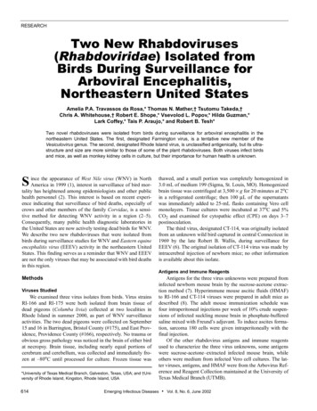
RESEARCHFigure. Ultrastructure of the new rhabdoviruses in infected Vero cells. A. Virions of isolate RI-175 budding from the surface of a Vero cell and fromcell surface projections (arrows). Arrowheads mark cross-sections of virions. The virion indicated with a large arrow is enlarged in B. B. A virion ofisolate RI-175 budding from host cell plasmalemma into an extracellular space. C. Details of the virion ultrastructure of isolate RI-175, showing spiral packaging of the nucleocapsid and its tubular structure in the cross-sections (arrows). D. A virion of the isolate CT-114 budding into an intracytoplasmic vacuole. Bar 100 nm.antigen and HMAF did not react with any of the other rhabdovirus antigens or HMAFs listed. Because of the geographicregion where they were isolated, we initially suspected thatRI-166 and CT-114 might be Connecticut or Flanders viruses.However, no antigenic relationship was shown by CF test(Table 1). The antigen RI 907-36 was prepared from a 1999isolate of Flanders virus from Rhode Island. Likewise, no relationship could be demonstrated between RI-166, CT-114, Connecticut, or Flanders viruses by IFA test (data not shown).Based on these findings, we conclude that RI-166 is probablya new, unassigned vertebrate rhabdovirus. The name RhodeIsland virus is proposed for this virus.In CF tests, CT-114 HMAF reacted with five vesicular stomatitis serogroup antigens: Chandipura, Isfahan, Maraba,Jurona, and La Joya (Table 2). Antigenically, CT-114 was mostclosely related to Jurona and La Joya viruses. Both Jurona andLa Joya viruses are tentative members of the genus Vesiculovirus (14–16). Based on the morphology and antigenic relationships of CT-114, we conclude that it is also a provisional616member of the Vesiculovirus genus. The name Farmington isproposed for this new virus.ConclusionThe isolation of these new rhabdoviruses from birds demonstrates the value of direct culture for detecting new andunexpected viral agents. Rhode Island virus was initially isolated in Vero cells; Farmington virus was detected by intracerebral inoculation of newborn mice. To save time and reducecosts, many arbovirus diagnostic laboratories in the UnitedStates have stopped culturing field specimens and instead areusing techniques such as antigen-capture enzyme-linkedimmunosorbent assay (17) or polymerase chain reaction (18–20) to detect viral antigens or nucleic acids in insect pools,blood, and tissue samples. While these newer techniques arerapid and quite sensitive, they detect only those viruses forwhich one has a capture antibody or a specific primer set. Furthermore, these techniques do not detect novel or unexpectedviral agents nor antigenic or virulence changes in knownEmerging Infectious Diseases Vol. 8, No. 6, June 2002
RESEARCHTable 1. Cross-reaction of CT-114 and RI-166 viruses with other selected rhabdoviruses by complement fixation testHyperimmune ascitic fluidAntigenConnecticutConnecticutNew MintoSawgrassFlandersCT-114RI-166256/ 640128/32000aNew Minto0256/ 640000Sawgrass16/3216/321,024/64000RI 907-36000 256/ 3200CT-1140000256/640b00000128/ 8RI-175 b00000128/ 8RI-166aReciprocalbof ascitic fluid titer/reciprocal of antigen titer.RI-166 and RI 175 antigens were fluids from infected cell cultures.viruses. A recent commentary (21) on the changing paradigmfor arbovirus identification discussed these limitations of themore rapid molecular methods and stressed the importance ofisolating viruses and obtaining phenotypic as well as genotypic information on them.The isolation of Rhode Island virus from dead pigeonssuggests that this virus may be an occasional avian pathogen.During the summer of 2000, a total of 335 birds, representing31 avian species, were tested for virus at the CVBD. RhodeIsland virus was isolated from 2 of 15 pigeons tested, suggesting that its host range may be restricted. Further experimentalstudies are needed to determine its pathogenesis and hostrange. In the northeastern United States, WNV, and to a lesserdegree, EEEV, are the arboviruses usually associated with birddeaths (3,22). However, as surveillance for WNV continuesand more dead birds are collected and cultured, other novelavian viral pathogens, such as Rhode Island virus, will probably be encountered.At present, little is known about the ecology of RhodeIsland or Farmington viruses. The ultrastructure and antigenicrelationships of Farmington virus suggest that it is a novelvesiculovirus. The ability of Farmington virus to infect the Cx.quinquefasciatus cell line is also compatible with a vesiculovirus, since most of the rhabdoviruses in this genus are arthro-pod associated (23,24). Jurona and La Joya viruses, thevesiculoviruses most closely related antigenically to Farmington virus, were both isolated from New World mosquitoes.Jurona virus has been isolated from Haemagogus sp. and froma human in northern Brazil (25); La Joya was isolated fromCx. dunni in Panama (13).Rhode Island virus is more intriguing. Its isolation fromdead birds and its ability to infect mice (both newborn andadult) as well as Vero cells, are strong evidence that it is a vertebrate rhabdovirus. Yet its ultrastructure and relatively largesize more closely resemble some of the plant rhabdoviruses(26). Further studies of this interesting new rhabdovirus andpotential avian pathogen are warranted.AcknowledgmentsThe authors thank Franklin D. Meglio for technical assistance,Violet C. Han for expert assistance in electron microscopy, and DoraSalinas for help in preparing the manuscript.This work was supported in part by National Institutes of Healthgrant AI-10984, the Rhode Island Department of EnvironmentalManagement, and the Island Fund of the New York CommunityTrusts. It is contribution no. 3891 of the Rhode Island AgricultureExperimental Station.Table 2. Cross-reaction of CT-114 and RI-166 viruses and selected vesicular stomatitis serogroup viruses by complement fixation testHyperimmune ascitic fluidAntigenChandipuraIsfahanMarabaJuronaLa JoyaCT-114RI-166256/ 32a00008/80Isfahan32/ 1664/ 328/8008/160Maraba8/ 80512/ 32008/160Jurona0001,024/ 32016/ 320La Joya0000512/ 3216/ 320CT-11400000256/ 160RI-166b000000128/ 8ChandipuraaReciprocalbof ascitic fluid titer/reciprocal of antigen titer.RI-166 antigen was fluid from an infected Vero cell culture.Emerging Infectious Diseases Vol. 8, No. 6, June 2002617
RESEARCHMs. Travassos da Rosa is a research associate at the Center forTropical Diseases, Department of Pathology, University of TexasMedical Branch in Galveston. She was formerly chief of the Arbovirus Department at the Evandro Chagas Institute in Belem, Para, Brazil. Her research interests focus on the identification and taxonomyof arthropodborne and other zoonotic viruses.References1. Centers for Disease Control and Prevention. Outbreak of West Nile-likeviral encephalitis—New York, 1999. MMWR Morb Mortal Wkly Rep1999;48:845–9.2. Centers for Disease Control and Prevention. Epidemic/epizootic WestNile virus in the United States: revised guidelines for surveillance, prevention and control. Fort Collins: US Department of Health and HumanServices; 2001. p. 7–14.3. Eidson M, Kramer L, Stone W, Hagiwara Y, Schmit K, and the New YorkState West Nile Virus Avian Surveillance Team. Dead bird surveillance asan early warning system for West Nile virus. Emerg Infect Dis2001;7:631–5.4. Eidson M, Komar N, Sarhage F, Nelson R, Talbut T, Mostashari F, et al.Crow deaths as a sentinel surveillance system for West Nile virus in thenortheastern United States, 1999. Emerg Infect Dis 2001;7:615–20.5. Steele KE, Linn MJ, Schoepp RJ, Komar N, Geisbert TW, Manduca RM,et al. Pathology of fatal West Nile virus infections in native and exoticbirds during the 1999 outbreak in New York City. J Vet Pathol2000;37:208–24.6. Wallis RC, Howard JJ, Main AJ, Frazier C, Hayes C. An increase inCuliseta melanura coinciding with an epizootic of eastern equine encephalitis in Connecticut. Mosquito News 1974;34:63–5.7. Beaty BJ, Calisher CH, Shope RE. Arboviruses. In: Lennette EH, Lennette DA, Lennette ET, editors. Diagnostic procedures for viral, rickettsial and chlamydial infections, 7th ed. Washington: American PublicHealth Association; 1995. p. 189–212.8. Travassos da Rosa APA, Tesh RB, Pinheiro FP, Travassos da Rosa JFS,Peterson NE. Characterization of eight new phlebotomus fever serogrouparboviruses (Bunyaviridae: Phlebovirus) from the Amazon Region ofBrazil. Am J Trop Med Hyg 1983;32:1164–71.9. Igarashi A. Isolation of a Singh’s Aedes albopictus cell clone sensitive todengue and chikungunya viruses. J Gen Virol 1978;40:531–44.10. Hsu SH. Growth of arboviruses in arthropod cell culture: comparativestudies. 1. Preliminary observation on growth of arboviruses in a newlyestablished line of mosquito cell (Culex quinquefasciatus Say). Curr TopMicrobiol Immunol 1971;55:140–8.11. Tesh RB. A method for the isolation of dengue viruses, using mosquitocell cultures. Am J Trop Med Hyg 1979;28:1053–9.12. Moncayo A, Hice CL, Watts DM, Travassos da Rosa APA, Guzman H,Russell KL, et al. Allpahuayo virus, a new arenavirus from the PeruvianAmazon. Virology 2001;284:277–86.13. Karabatsos N, editor. International catalogue of arboviruses includingcertain other viruses of vertebrates, 3rd ed. San Antonio: American Society of Tropical Medicine and Hygiene; 1985.61814. Walker PJ, Benmansour A, Dietz R, Fang R-X, Jackson AO, Kurath G, etal. Family Rhabdoviridae. In: van Regenmortel MHV, Fauquet CM,Bishop DHL, Carstens EB, Estes MK, Lemon SM, et al., editors. Virustaxonomy: classification and nomenclature of viruses. San Diego: Academic Press; 2000. p. 563–83.15. Tesh RB, Travassos da Rosa APA, Travassos da Rosa JS. Antigenic relationship among rhabdoviruses infecting terrestrial vertebrates. J GenVirol 1983;64:169–76.16. Calisher CH, Karabatsos N, Zeller H, Digoutte JP, Tesh RB, Shope RE, etal. Antigenic relationships among rhabdoviruses from vertebrates andhematophagous arthropods.
nm–150 nm long, with a periodicity of striations of 10.5 nm (Figure, B). Virions of the isolate RI-175 were seen budding predomi-nantly into the extracellular space from the plasmalemma of the Vero cells (Figure, C and D). The virions were bacilliform, measuring 90 nm–100 nm