Transcription
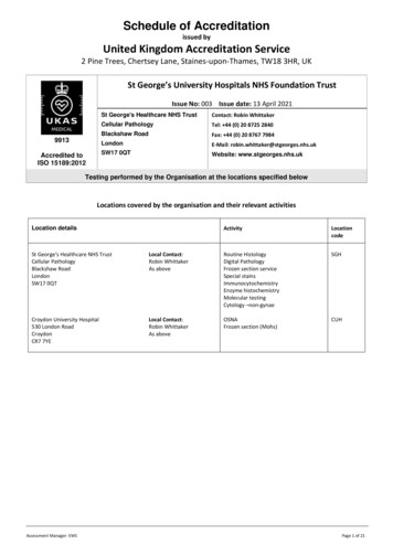
Schedule of Accreditationissued byUnited Kingdom Accreditation Service2 Pine Trees, Chertsey Lane, Staines-upon-Thames, TW18 3HR, UKSt George’s University Hospitals NHS Foundation TrustIssue No: 003 Issue date: 13 April 20219913Accredited toISO 15189:2012St George's Healthcare NHS TrustContact: Robin WhittakerCellular PathologyTel: 44 (0) 20 8725 2840Blackshaw RoadFax: 44 (0) 20 8767 7984LondonE-Mail: robin.whittaker@stgeorges.nhs.ukSW17 0QTWebsite: www.stgeorges.nhs.ukTesting performed by the Organisation at the locations specified belowLocations covered by the organisation and their relevant activitiesLocation detailsActivityLocationcodeSt George's Healthcare NHS TrustCellular PathologyBlackshaw RoadLondonSW17 0QTLocal Contact:Robin WhittakerAs aboveRoutine HistologyDigital PathologyFrozen section serviceSpecial stainsImmunocytochemistryEnzyme histochemistryMolecular testingCytology –non-gynaeSGHCroydon University Hospital530 London RoadCroydonCR7 7YELocal Contact:Robin WhittakerAs aboveOSNAFrozen section (Mohs)CUHAssessment Manager: EWSPage 1 of 21
S c he dul e of Ac c re di ta ti onissued byUnited Kingdom Accreditation Service2 Pine Trees, Chertsey Lane, Staines -upon-Thames, TW18 3HR, UKSt George’s University Hospitals NHS Foundation Trust9913Accredited to15189:2012ISOIssue No: 003Issue date: 13 April 2021Testing performed at main address onlySite activities performed away from the locations listed above:Location detailsHead and Neck FNA clinicsUltrasound Room St James Wing X-RayDepartmentLocal Contact:Robin WhittakerAs aboveActivityLocationcodeFNA material/slide preparationCellularity assessmentSGHUltrasound Lanesborough Wing ScanningDepartmentMelanoma clinic at The Rose CentreSt George’s Healthcare NHS trustBlackshaw RoadLondonEBUS – Endobronchial Ultrasound clinic,H&N Clinic, Interventional radiology suiteCUHCroydon University Hospital530 London RoadCroydonCR7 7YEAssessment Manager: EWSPage 2 of 21
S c he dul e of Ac c re di ta ti onissued byUnited Kingdom Accreditation Service2 Pine Trees, Chertsey Lane, Staines -upon-Thames, TW18 3HR, UKSt George’s University Hospitals NHS Foundation Trust9913Accredited to15189:2012ISOIssue No: 003Issue date: 13 April 2021Testing performed at main address onlyDETAIL OF ACCREDITATIONMaterials/ProductstestedHUMAN BODY TISSUE /FLUIDSFresh and formalin fixedtissue samplesType of test/Propertiesmeasured/Range ofmeasurementRoutine Histopathologyexaminations to assist inclinical investigations anddiagnosisStandard specifications/Equipment/Techniques usedLocationCodeMacroscopic and Microscopicexamination using in-houseprocedures (listed) in conjunction withmanufacturer’s instructions for thefollowing methods (where relevant)Macroanalysis and dissection using:SGHCEL-HIS-LAB-005 Specimen cut upCEL-MED-CUT-001 Medical protocolfor specimen cut upCEL-MED-CUT-006 Protocol for Cutup and Reporting of ThoracicSpecimensCEL-MED-CUT-007 Medical Protocolfor Cut-Up of Specimens from theCardiovascular SystemCEL-MED-CUT-010 Medical Protocolfor Cut-Up Of AppendicectomySpecimensCEL-MED-CUT-011 Medical Protocolfor Dealing with ColorectalPolypectomiesCEL-MED-CUT-012 Medical Protocolfor Cut-Up Colorectal CancerSpecimensCEL-MED-CUT-013 Medical Protocolfor Cut-Up Of Completion ProctectomySpecimensCEL-MED-CUT-014 Medical Protocolfor Cut-Up Of CholecystectomySpecimensCEL-MED-CUT-015 Medical protocolfor cut-up of urological specimensCEL-MED-CUT-016 Medical Protocolfor Cut-Up Endocrine (Adrenal andParathyroid) Specimens and TissuePathways for ReportingCEL-MED-CUT-017 GynaecologicalPathology Specimen DissectionCEL-MED-CUT-018 Medical Protocolfor Cut-Up of Colectomy Specimensfor Inflammatory Bowel DiseaseAssessment Manager: EWSPage 3 of 21
S c he dul e of Ac c re di ta ti onissued byUnited Kingdom Accreditation Service2 Pine Trees, Chertsey Lane, Staines -upon-Thames, TW18 3HR, UKSt George’s University Hospitals NHS Foundation Trust9913Accredited to15189:2012ISOIssue No: 003Issue date: 13 April 2021Testing performed at main address onlyMaterials/ProductstestedHUMAN BODY TISSUE /FLUIDS (cont’d)Fresh and formalin fixedtissue samplesType of test/Propertiesmeasured/Range ofmeasurementRoutine Histopathologyexaminations to assist inclinical investigations anddiagnosis (cont’d)Standard specifications/Equipment/Techniques usedLocationCodeMacroscopic and Microscopicexamination using in-houseprocedures (listed) in conjunction withmanufacturer’s instructions for thefollowing methods (where relevant)Macroanalysis and dissection (cont’d)using:CEL-MED-CUT-020 Medical Protocolfor Cut-Up of Paediatric BowelSpecimens in Hirschsprung’s DiseaseCEL-MED-CUT-021 Medical Protocolfor Paediatric Tumour Specimen CutUpSGHCEL-MED-CUT-022 Medical Protocolfor Cut-Up Of PlacentaeCEL-MED-CUT-023 Medical Protocolfor Cut-Up of Breast SpecimensCEL-MED-CUT-024 Medical Protocolfor Cut-Up of Lymph Node SpecimensCEL-MED-CUT-025 Medical Protocolfor Cut-Up of NeuropathologySpecimensCEL-HIS-APX-006 Routine Usage ofthe VANTAGE Workflow SolutionTracking SystemFormalin fixed tissuesincluding cell blocksTissue ProcessingAutomated method using:CEL-HIS-EQP-007 Tissue-Tek VIP6AICEL-HIS-LAB-024 Re-processingCEL-HIS-LAB-012 Fresh PaediatricTissuesCEL-HIS-LAB-013 Fresh SurgicalTissuesSGHFormalin fixed, processedtissues and cell blocksTissue EmbeddingManual method using:CEL-HIS-LAB-015Sakura Tissue Tek embedding centresCEL-HIS-EQP-008 Operation andMaintenance of the Tissue Tek Embedding CentresSGHAssessment Manager: EWSPage 4 of 21
S c he dul e of Ac c re di ta ti onissued byUnited Kingdom Accreditation Service2 Pine Trees, Chertsey Lane, Staines -upon-Thames, TW18 3HR, UKSt George’s University Hospitals NHS Foundation Trust9913Accredited to15189:2012Issue No: 003ISOIssue date: 13 April 2021Testing performed at main address onlyMaterials/ProductstestedHUMAN BODY TISSUE /FLUIDS (cont’d)Type of test/Propertiesmeasured/Range ofmeasurementRoutine Histopathologyexaminations to assist inclinical investigations anddiagnosis (cont’d)Formalin fixed, paraffinembedded (FFPE) tissueand cell blocksStandard specifications/Equipment/Techniques usedLocationCodeMacroscopic and Microscopicexamination using in-houseprocedures (listed) in conjunction withmanufacturer’s instructions for thefollowing methods (where relevant)SGHMicrotomyManual sectioning using:CEL-HIS-LAB-006Leica 2135 and Sakura SRM 200Accucut microtomesSGHFresh / frozen tissueIntra-operative diagnosis ofbasophilic and eosinophilictissue structureRapid frozen sectionCryotomy, automated and manualHaematoxylin & Eosin (H&E) stainingmethod using Leica CM1850 andLeica CM1860 UV CryostatsCEL-HIS-EQP-004 Operation andMaintenance of the Leica CM1850 &CM1860 UV CryostatCEL-HIS-LAB-030 CryotomyCEL-HIS-LAB-004 Freezing tissuesCEL-HIS-LAB-012 Paediatric tissueCEL-HIS-LAB-013 Surgical tissueCEL-HIS-LAB-017 Infective tissueCEL-HIS-LAB009 Handling andorientation of muscle biopsiesSGHFresh / frozen tissueDetection of:IgAIgMComplementManual ImmunofluorescenceCEL-HIS-LAB-026 Skin SpecimensRequiring ImmunofluorescenceCEL-ICC-GEN-008Fluorescence microscopyZeiss Axioplan 2SGHFresh / frozen tissue (skinand mucosa)Frozen Section Histologyexaminations to assist inclinical investigationsCEL-HIS-LAB-013 MohsLeica CM1850 cryostatShandon Linistain – Automated H&EstainingOlympus BX41 microscopeCEL-MOH-GEN-001 Mohs TechniqueCUHAssessment Manager: EWSPage 5 of 21
S c he dul e of Ac c re di ta ti onissued byUnited Kingdom Accreditation Service2 Pine Trees, Chertsey Lane, Staines -upon-Thames, TW18 3HR, UKSt George’s University Hospitals NHS Foundation Trust9913Accredited to15189:2012Issue No: 003ISOIssue date: 13 April 2021Testing performed at main address onlyMaterials/ProductstestedType of test/Propertiesmeasured/Range ofmeasurementHUMAN BODY TISSUE /FLUIDS (cont’d)Special staining ofHistopathology examinationsto assist in detection of clinicalabnormalitiesManual tinctorial and histochemicalmethods using:SGHSlides prepared fromsamples aboveIdentification of basophilic andeosinophilic tissue structuresand diastase resistantpolysaccharidesRoutine Haematoxylin & Eosin (H&E)and PAS /- D staining andcoverslippingAutomated method using:CEL-HIS-EQP-003Sakura Tissue Tek Prisma StainerShandon LinistainerSGHSections (slides) fromFFPE tissue / cell blocksAcid and neutral mucinsAlcian Blue PAS CEL-HIS-STA-003Acid mucopolysaccharideAlcian Blue CEL-HIS-STA-002AmyloidCongo Red CEL-HIS-STA-008Diastase resistantpolysaccharidesPAS /- Diastase CEL-HIS-STA-010GlycogenABPASD CEL-HIS-STA-004Connective tissueElastic Van Geison CEL-HIS-STA-011H. pylori and H. pylori likeorganismsGiemsa CEL-HIS-STA-013Bone canaliculaeGiemsa – Bone marrow CEL-HIS-STA012Gram Stain CEL-HIS-STA-014Gram positive and negativebacteriaAssessment Manager: EWSStandard specifications/Equipment/Techniques usedGram positive and negativebacteriaGram Twort CEL-HIS-STA-015Basement membrane, fungiGrocott CEL-HIS-STA-017Cellular ComponentsHaematoxylin and Eosin CEL-HISSTA-018LocationCodePage 6 of 21
S c he dul e of Ac c re di ta ti onissued byUnited Kingdom Accreditation Service2 Pine Trees, Chertsey Lane, Staines -upon-Thames, TW18 3HR, UKSt George’s University Hospitals NHS Foundation Trust9913Accredited to15189:2012Issue No: 003ISOIssue date: 13 April 2021Testing performed at main address onlyMaterials/ProductstestedHUMAN BODY TISSUE /FLUIDS (cont’d)Sections (slides) fromFFPE tissue / cell blocksAssessment Manager: EWSType of test/Propertiesmeasured/Range ofmeasurementStandard specifications/Equipment/Techniques usedCollagen, muscle and bilepigmentsSpecial staining ofHistopathology examinationsto assist in detection of clinicalabnormalities (cont’d)HVG CEL-HIS-STA-019Connective tissueMSB CEL-HIS-STA-023MelaninMasson Fontana CEL-HIS-STA-021Connective tissueHepatitis B surface antigenand copper associated proteinMasson Trichrome CEL-HIS-STA-022Orcein CEL-HIS-STA-027Fungi, glycogen, neutral mucinand basement membraneGlycogenPAS CEL-HIS-STA-028HaemosiderinPerls CEL-HIS-STA-0Reticulin fibresReticulin CEL-HIS-STA-034CopperRhodanine CEL-HIS-STA-033AmyloidThioflavin T CEL-HIS-STA-040Mast cell granules and acidmucinToluidine Blue CEL-HIS-STA-026Calcium depositsVon Kossa CEL-HIS-STA-036Leprosy bacilliWade Fite CEL-HIS-STA-037Spirochetes, HelicobacterWarthin-Starry CEL-HIS-STA-041MycobacteriaZiehl-Neelson CEL-HIS-STA-039Manual tinctorial and histochemicalmethods using:LocationCodeSGHPAS /- D – Muscle method CEL-HISSTA-051Page 7 of 21
S c he dul e of Ac c re di ta ti onissued byUnited Kingdom Accreditation Service2 Pine Trees, Chertsey Lane, Staines -upon-Thames, TW18 3HR, UKSt George’s University Hospitals NHS Foundation Trust9913Accredited to15189:2012Issue No: 003ISOIssue date: 13 April 2021Testing performed at main address onlyMaterials/ProductstestedType of test/Propertiesmeasured/Range ofmeasurementStandard specifications/Equipment/Techniques usedHUMAN BODY TISSUE /FLUIDS (cont’d)Enzyme HistochemistryPreparation and examinationto identify or excludemorphological & cytologicalabnormalitiesManual histochemical methods using:Fresh / frozen tissueAcid phosphataseAcid Phosphatase CEL-HIS-STA-043Fresh / frozen rectalbiopsy tissueAcetylcholinesterase – nervecellsAcetylcholinesterase CEL-HIS-STA005 for Diagnosing Hirschsprung’sMuscle fresh/frozensectionsCytochrome oxidase activityCOX-SDH CEL-HIS-STA-044Muscle fresh/frozensectionsType 1 and 2 muscle fibresMyoadenylate Deaminase CEL-HISSTA-052Muscle fresh/frozensectionsTetrazolium reductase activityNicotinamide adenosine CEL-HISSTA-049Muscle fresh/frozensectionsPhosphorylase activityPhosphorylase CEL-HIS-STA-048Muscle fresh/frozensectionsMuscle, mitochondria andcollagenGomori Trichrome CEL-HIS-STA-047Muscle fresh/frozensectionsLipidsOil Red O CEL-HIS-STA-025Assessment Manager: EWSLocationCodeSGHPage 8 of 21
S c he dul e of Ac c re di ta ti onissued byUnited Kingdom Accreditation Service2 Pine Trees, Chertsey Lane, Staines -upon-Thames, TW18 3HR, UKSt George’s University Hospitals NHS Foundation Trust9913Accredited to15189:2012Issue No: 003ISOIssue date: 13 April 2021Testing performed at main address onlyMaterials/ProductstestedType of test/Propertiesmeasured/Range ofmeasurementHUMAN BODY TISSUE /FLUIDS (cont’d)Immunocytochemistry ofHistopathology examinationsto assist in detection of clinicalabnormalitiesSections (slides) fromFFPE tissue / cell blocksAssessment Manager: EWSStandard specifications/Equipment/Techniques usedLocationCodeIn house documented procedures &manufacturer’s instructionsImmunohistochemistryAutomated methods using:Ventana Benchmark ULTRA and inhouse methodsCEL-ICC-MAN-020 BenchMarkULTRA Advanced Staining SystemOperator GuideCEL-ICC-EQP-009 Operation of theBenchMark ULTRACEL-ICC-GEN-003 Procedure forManagement of Control Material forImmunocytochemistryCEL-ICC-GEN-006 Formic Acid PreTreatment in Immunocytochemistry forNeurodegeneration AntibodiesCEL-ICC-GEN-020 Testing for MGMTMethylation StatusCEL-ICC-QMS-001 Internal QualityControl in Immunocytochemistry andthe following antibodiesCEL-ICC-FRM-004 List of antibodiesand controlsPituitary and adrenalACTHAnti-smooth muscleActinEpithelial tissue, tumours, andmetastatic carcinoma in lymphnodesAE1/AE3Anaplastic large celllymphomaALK-1Hepatocellular carcinoma andgerm cell tumoursAlpha feto proteinSGHPage 9 of 21
S c he dul e of Ac c re di ta ti onissued byUnited Kingdom Accreditation Service2 Pine Trees, Chertsey Lane, Staines -upon-Thames, TW18 3HR, UKSt George’s University Hospitals NHS Foundation Trust9913Accredited to15189:2012Issue No: 003ISOIssue date: 13 April 2021Testing performed at main address onlyMaterials/ProductstestedType of test/Propertiesmeasured/Range ofmeasurementHUMAN BODY TISSUE /FLUIDS (cont’d)Immunocytochemistry ofHistopathology examinationsto assist in detection of clinicalabnormalities (cont’d)Automated methods using:Ventana Benchmark ULTRA and inhouse methods (cont’d)Sections (slides) fromFFPE tissue / cell blocksGenetic disease inhepatocytes and for normalhistiocytes and hepatocytesAlpha-1-antitrypsinProstate carcinoma vs benignprostateAMACRApocrine, breast, Paget andsebaceous markerAndrogen receptorAnaplastic lymphoma andnon-small cell carcinomaAnti-ALK (D5F3)Mutations encoding cardiacactin, desmin, d-sarcoglycan,b-sarcoglycan, cardiactroponin T and tropomyosinA-SARCOGLYCANFollicular hyperplasia vsfollicular lymphoma. Detectsimmature enteric ganglion anddiffuse large cell lymphomaBCL2Germinal centre marker alsoCD10, follicular lymphomaBCL6Lung adenocarcinoma vsmesotheliomaMutations associated withmesenchymal tumours,colonic Ca, papillary thyroidCa and endometrioid CaBEREP4Germ cell and gestationaltrophoblastic tumoursBeta HCGOvarian tumoursCA 125Assessment Manager: EWSStandard specifications/Equipment/Techniques usedLocationCodeSGHBeta-CateninPage 10 of 21
S c he dul e of Ac c re di ta ti onissued byUnited Kingdom Accreditation Service2 Pine Trees, Chertsey Lane, Staines -upon-Thames, TW18 3HR, UKSt George’s University Hospitals NHS Foundation Trust9913Accredited to15189:2012Issue No: 003ISOIssue date: 13 April 2021Testing performed at main address onlyMaterials/ProductstestedType of test/Propertiesmeasured/Range ofmeasurementHUMAN BODY TISSUE /FLUIDS (cont’d)Immunocytochemistry ofHistopathology examinationsto assist in detection of clinicalabnormalities (cont’d)Automated methods using:Ventana Benchmark ULTRA and inhouse methods (cont’d)C cell hyperplasia andmedullary thyroid CaCalcitoninSmooth muscle cellsCaldesmonPulmonary subtypes, reactivemesothelial cells,Hirschsprung disease, ovarianSertoli-Leydig tumours,SchwannomasCalretininLymphoma markerCD10GIST, sarcoma and Hodgkinlymphoma markerCD117Acute myeloid leukaemiaCD13Keratoacanthoma, myelomaand plasmablastic lymphomaCD138Lymphoid tumours, Hodgkin’sdiseaseCD15Langerhan cell histiocytosisCD1AT cell markerCD2 & CD3B cell markerCD20Follicular dendritic andlymphoid cellsAnaplastic large cell andHodgkin’s lymphomasCD21/CD23Endothelial cells/ tumoursCD31/Factor 8Endothelial cells, dermalneoplasms, GIST andLeukaemia’sCD34 (QB10)Sections (slides) fromFFPE tissue / cell blocksAssessment Manager: EWSStandard specifications/Equipment/Techniques usedLocationCodeSGHCD30Page 11 of 21
S c he dul e of Ac c re di ta ti onissued byUnited Kingdom Accreditation Service2 Pine Trees, Chertsey Lane, Staines -upon-Thames, TW18 3HR, UKSt George’s University Hospitals NHS Foundation Trust9913Accredited to15189:2012Issue No: 003ISOIssue date: 13 April 2021Testing performed at main address onlyMaterials/ProductstestedType of test/Propertiesmeasured/Range ofmeasurementHUMAN BODY TISSUE /FLUIDS (cont’d)Immunocytochemistry ofHistopathology examinationsto assist in detection of clinicalabnormalities (cont’d)Automated methods using:Ventana Benchmark ULTRA and inhouse methods (cont’d)Myeloma and plasmablasticlymphomaCD38Lymphoma and T cell markerCD4AML-M7CD42BT and B cell lymphomasCD43Intestinal intraepitheliallymphocytes and inflammatorycellsCD45CLL and mantle celllymphomaCD5NK lymphomas andneuroendocrine disordersCD56Histiocytes/histiocytic tumoursand hepatocytesCD68T-ALLCD7B cell lymphoproliferativedisordersCD79aT cellsCD8Ewing sarcoma and T celllymphomaCD99 (MIC2)Acute myeloid leukaemiaCD123GI and selectedadenocarcinomaCDX2Adenocarcinomas (lung) andcolorectal carcinomasCEANeuroendocrine tumoursChromogranin ASections (slides) fromFFPE tissue / cell blocksAssessment Manager: EWSStandard specifications/Equipment/Techniques usedLocationCodeSGHPage 12 of 21
S c he dul e of Ac c re di ta ti onissued byUnited Kingdom Accreditation Service2 Pine Trees, Chertsey Lane, Staines -upon-Thames, TW18 3HR, UKSt George’s University Hospitals NHS Foundation Trust9913Accredited to15189:2012Issue No: 003ISOIssue date: 13 April 2021Testing performed at main address onlyMaterials/ProductstestedType of test/Propertiesmeasured/Range ofmeasurementHUMAN BODY TISSUE /FLUIDS (cont’d)Immunocytochemistry ofHistopathology examinationsto assist in detection of clinicalabnormalities (cont’d)Automated methods using:Ventana Benchmark ULTRA and inhouse methods (cont’d)Sections (slides) fromFFPE tissue / cell blocksMicroinvasion in VIN or CINCollagen IVMantle cell lymphoma andproliferation markerCyclin D1Basal subtypes of DCIS,breast papilloma andodontogenic neoplasmsCytokeratin 14Basal DCIS, epithelioidmesothelioma and spindlessquamous cell carcinomaCK5Bile duct tumoursCK7Epithelial tissueCK8/18CMV, hepatocytes, biliaryepithelium, endothelial cellsand Kupffer cellsCMVLymphatic endotheliumD2-40Skeletal/straiate myogenictissueDesminGIST, pancreatic andoesophageal neoplasmsDOG-1LCIS vs DCISE-CadherinMeningioma markerEMAMuscle diseaseEmerinBreast originERAssessment Manager: EWSStandard specifications/Equipment/Techniques usedLocationCodeSGHPage 13 of 21
S c he dul e of Ac c re di ta ti onissued byUnited Kingdom Accreditation Service2 Pine Trees, Chertsey Lane, Staines -upon-Thames, TW18 3HR, UKSt George’s University Hospitals NHS Foundation Trust9913Accredited to15189:2012Issue No: 003ISOIssue date: 13 April 2021Testing performed at main address onlyMaterials/ProductstestedType of test/Propertiesmeasured/Range ofmeasurementHUMAN BODY TISSUE /FLUIDS (cont’d)Immunocytochemistry ofHistopathology examinationsto assist in detection of clinicalabnormalities (cont’d)Automated methods using:Ventana Benchmark ULTRA and inhouse methods (cont’d)Sections (slides) fromFFPE tissue / cell blocksDermal dendrocytes and fibrohistiocytic proliferationsFactor 13APituitary tissueFSHThyroid tumours, liver, colonand parathyroid carcinomaGalectinBreast carcinomaGCDFP-15CNS tumours and colonicschwannomaGFAPIslet cell tumour,pheochromocytomaGHErythroid cells and acuteerythroid leukaemia (AML-M6)Glycophorin A (CD235a)T cellGranzyme BMesothelial cells and thyroidcarcinomasHBME1Helicobacter pylori markerHelicobacter PyloriHepatocytesHepar1Breast cancerHER-2Breast cancerHER-2 Dual ISH DNACEL-ICC-GEN-023Kaposi’s sarcomaHHV8AML, acute basophilicleukaem
United Kingdom Accreditation Service 2 Pine Trees, Chertsey Lane, Staines-upon-Thames, TW18 3HR, UK Blackshaw Road 9913 Accredited to ISO 15189:2012 St George’s University Hospitals NHS Foundation Trust Issue No: 003 Issue date: 13 April 2021 St George's Healthcare NHS Trust Cellula
