Transcription
CHAPTER 9MUSCLES
MUSCLE TISSUE TYPES
SPECIAL CHARACTERISTICS OFMUSCLE
FUNCTIONS OF MUSCLE
The ability of muscle to be stretched is ticity
SKELETAL MUSCLE:GROSS ANATOMY
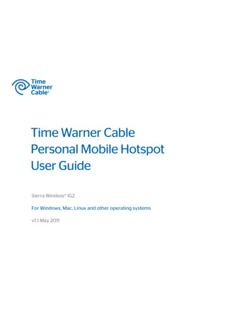
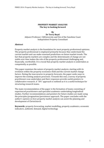
Figure 9.1 Connective tissue sheaths of skeletal muscle: epimysium, perimysium, and endomysium.EpimysiumBone EpimysiumPerimysiumEndomysiumTendon(b)Perimysium Fascicle(a)Copyright 2010 Pearson Education, Inc.Muscle fiberin middle ofa fascicleBlood vesselFascicle(wrapped by perimysium)Endomysium(between individualmuscle fibers)Muscle fiber
The connective tissue sheath around asingle muscle fiber is 1) Endomysium2) Epimysium3) Perimysium
SKELETAL MUSCLE:GROSS ANATOMY – ATTACHMENTS
SKELETAL MUSCLE:MICROSCOPIC ANATOMY
Figure 9.2a Microscopic anatomy of a skeletal muscle fiber.NucleiDark A bandLight I bandFiber(a) Photomicrograph of portions of two isolated musclefibers (700x). Notice the obvious striations (alternatingdark and light bands).Copyright 2010 Pearson Education, Inc.
Figure 9.2b Microscopic anatomy of a skeletal muscle fiber.SarcolemmaMitochondrionMyofibrilDark A band Light I band Nucleus(b) Diagram of part of a muscle fiber showing the myofibrils. Onemyofibril is extended afrom the cut end of the fiber.Copyright 2010 Pearson Education, Inc.
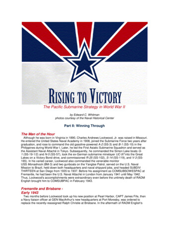
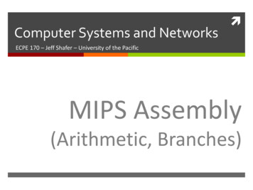
Figure 9.2c Microscopic anatomy of a skeletal muscle fiber.Thin (actin)filamentThick (myosin)filamentZ discI bandH zoneA bandSarcomereZ discI bandM line(c) Small part of one myofibril enlarged to show the myofilamentsresponsible for the banding pattern. Each sarcomere extends fromone Z disc to the next.Copyright 2010 Pearson Education, Inc.
The functional contractile unit of amuscle is the 1)2)3)4)FascicleMuscle fiberSarcomereEpimysium
Figure 9.2d Microscopic anatomy of a skeletal muscle fiber.SarcomereZ discM lineZ discThin (actin)filamentElastic (titin)filamentsThick(myosin)filament(d) Enlargement of one sarcomere (sectioned lengthwise). Notice themyosin heads on the thick filaments.Copyright 2010 Pearson Education, Inc.
Figure 9.2e Microscopic anatomy of a skeletal muscle fiber.MyosinfilamentActinfilamentI bandthinfilamentsonlyH zonethickfilamentsonlyM lineOuter edgeof A bandthick filamentslinked bythick and thinaccessoryfilaments overlapproteins(e) Cross-sectional view of a sarcomere cut through in different locations.Copyright 2010 Pearson Education, Inc.
Figure 9.3 Composition of thick and thin filaments.Longitudinal section of filamentswithin one sarcomere of a myofibrilThick filamentThin filamentIn the center of the sarcomere, the thickfilaments lack myosin heads. Myosin headsare present only in areas of myosin-actin overlap.Thick filamentThin filamentEach thick filament consists of manyA thin filament consists of two strandsmyosin molecules whose heads protrude of actin subunits twisted into a helixat opposite ends of the filament.plus two types of regulatory proteins(troponin and tropomyosin).Portion of a thick filamentPortion of a thin filamentMyosin headTropomyosinTroponinActinActin-binding sitesATPbindingsiteHeadsTailFlexible hinge regionMyosin moleculeCopyright 2010 Pearson Education, Inc.Active sitesfor myosinattachmentActinsubunitsActin subunits
Figure 9.4 Transmission electron micrograph of part of a sarcomere clearly showing the myosin heads forming crossbridges that generate the contractile force.Thin filament (actin)Copyright 2010 Pearson Education, Inc.Myosin headsThick filament (myosin)
SKELETAL MUSCLE:SLIDING FILAMENT MODEL OFMUSCLE CONTRACTION
Figure 9.6 Sliding filament model of contraction.ZZHAI1 Fully relaxed sarcomere of a muscle fiberZIIZAI2 Fully contracted sarcomere of a muscle fiberCopyright 2010 Pearson Education, Inc.
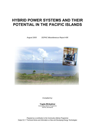
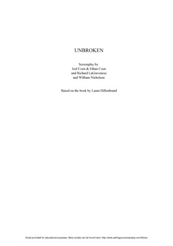
Figure 9.12 Cross Bridge CycleThin filamentActinMyosinheadCa2 ADPPiThickfilamentMyosin1 Cross bridge formation.ADPADPPiPiATPhydrolysis2 The power (working)stroke.4 Cocking of myosin head.ATPATP3 Cross bridgedetachment.Copyright 2010 Pearson Education, Inc.
Figure 9.5 Relationship of the sarcoplasmic reticulum and T tubules to myofibrils of skeletal muscle.Part of a skeletalmuscle fiber (cell)MyofibrilI bandA bandI bandZ discH zoneZ discM lineSarcolemmaSarcolemmaTriad: T tubule Terminalcisternaeof the SR (2)Tubules ofthe SRMyofibrilsMitochondriaCopyright 2010 Pearson Education, Inc.
Contraction of a sarcomere occurs becauseOf sliding of past .1)2)3)4)Actin / myosinMyosin / actinTropomyosin / myosinTropomyosin / actin
Before a cross-bridge cycle can occur, calciumions must bind to 1)2)3)4)actinMyosinTropomyosinTroponin
Once troponin binds calcium, it moves,exposing binding sites on .1)2)3)4)Actin / myosinMyosin / actinTropomyosin / actinTropomyosin / myosin
SKELETAL MUSCLE:PHYSIOLOGY OF MUSCLE FIBERSOr: How does the calcium get released?
Figure 9.8 Events at the Neuromuscular Junction (1 of 4)Axon terminal ofneuromuscularjunctionAction potentialSarcolemma(AP)of the musclefiberNucleusMyelinated axonof motor neuronCopyright 2010 Pearson Education, Inc.
Figure 9.8 Events at the Neuromuscular Junction (2 of 4)1 Action potentialarrives at axon terminalof motor neuron.2 Voltage-gated Ca2 channels open andCa2 enters the axonterminal.3 Ca2 entrycauses somesynaptic vesiclesto release theircontents(acetylcholine)by exocytosis.4 Acetylcholine, aneurotransmitter, diffusesacross the synaptic cleftand binds to receptors inthe sarcolemma.Copyright 2010 Pearson Education, Inc.Ca2 Ca2 Axon terminalof motor neuronSynaptic vesiclecontaining AChMitochondrionSynaptic cleftFusingsynapticvesiclesAChJunctionalfolds ofsarcolemmaSarcoplasm of muscle fiber
Figure 9.9 Summary of events in the generation and propagation of an action potential in a skeletal muscle fiber.Axon terminalOpen Na ChannelNa SynapticcleftClosed K ChannelACh–AChNa K K Na K Generation and propagation ofthe action potential (AP)2Closed Na Open K ChannelChannel NaLocal depolarization:generation of the endplate potential on thesarcolemma1Sarcoplasm of muscle fiberCopyright 2010 Pearson Education, Inc.3K Repolarization
The electrical signal from a neuron is carriedto a muscle by the neurotransmitter called 1)2)3)4)EpinephrineCalciumAcetylcholineAction potential
Figure 9.11 Excitation-Contraction Coupling (1 of 4)Setting the stageAxon terminalof motor neuronSynaptic cleftAChTerminal cisterna of SRMuscle fiber Ca2 Action potentialis generatedSarcolemmaTriadOne sarcomereCopyright 2010 Pearson Education, Inc.
Figure 9.11 Excitation-Contraction Coupling (3 of 4)1 Action potential isSteps inE-C Coupling:propagated along thesarcolemma and downthe T tubules.Voltage-sensitivetubule proteinSarcolemmaT tubuleCa2 releasechannelTerminalcisternaof SRCa2 Copyright 2010 Pearson Education, Inc.2 Calciumions arereleased.
Figure 9.11 Excitation-Contraction Coupling (4 of 4)ActinCa2 TroponinTropomyosinblocking active sitesMyosin3 Calcium binds totroponin and removesthe blocking action oftropomyosin.Active sites exposed andready for myosin binding4 Contraction beginsMyosincrossbridgeThe aftermathCopyright 2010 Pearson Education, Inc.
The depolarization of the membrane reachesthe sarcoplasmic reticulum via 1)2)3)4)T-tubulesVoltage-sensitive tubule proteinsSR calcium-release channelsAll of the above
SKELETAL MUSCLE:CONTRACTION
Figure 9.13a A motor unit consists of a motor neuron and all the muscle fibers it innervates.Spinal cordMotor Motorunit 1 unit 2Axon terminals atneuromuscular junctionsNerveMotor neuroncell bodyMotorMuscleneuronaxonMusclefibers(a) Axons of motor neurons extend from the spinal cord to themuscle. There each axon divides into a number of axonterminals that form neuromuscular junctions with musclefibers scattered throughout the muscle.Copyright 2010 Pearson Education, Inc.
Figure 9.13b A motor unit consists of a motor neuron and all the muscle fibers it innervates.Axon terminalsat neuromuscularjunctionsBranching axonto motor unitMusclefibers(b) Branching axon terminals formneuromuscular junctions, one permuscle fiber (photomicrograph 330x).Copyright 2010 Pearson Education, Inc.
Figure 9.14a The muscle twitch.Latent Period ofperiod contractionPeriod ofrelaxationSinglestimulus(a) Myogram showing the three phases of an isometric twitchCopyright 2010 Pearson Education, Inc.
Figure 9.14b The muscle twitch.Latent periodExtraocular muscle (lateral rectus)GastrocnemiusSoleusSinglestimulus(b) Comparison of the relative duration of twitch responses ofthree musclesCopyright 2010 Pearson Education, Inc.
During the period of relaxation, what ishappening in the muscle fiber on a molecularlevel?1)2)3)4)The membrane is depolarizingCa2 is being released from the SRCa2 is being taken up into the SRThe cross-bridge cycle is beginning
Figure 9.15a Muscle response to changes in stimulation frequency.Single stimulussingle twitchContractionRelaxationStimulus(a) A single stimulus is delivered. The musclecontracts and relaxesCopyright 2010 Pearson Education, Inc.
Figure 9.15b Muscle response to changes in stimulation frequency.Low stimulation frequencyunfused (incomplete) tetanusPartial relaxationStimuli(b) If another stimulus is applied before the musclerelaxes completely, then more tension results.This is temporal (or wave) summation and resultsin unfused (or incomplete) tetanus.Copyright 2010 Pearson Education, Inc.
Figure 9.15c Muscle response to changes in stimulation frequency.High stimulation frequencyfused (complete) tetanusStimuli(c) At higher stimulus frequencies, there is no relaxationat all between stimuli. This is fused (complete) tetanus.Copyright 2010 Pearson Education, Inc.
Figure 9.16 Relationship between stimulus intensity (graph at top) and muscle tension (tracing below).Stimulus strengthMaximalstimulusThresholdstimulusProportion of motor units excitedStrength of muscle contractionMaximal contractionCopyright 2010 Pearson Education, Inc.
Figure 9.17 The size principle of recruitment.Motorunit 1Recruited(smallfibers)Copyright 2010 Pearson Education, Inc.Motorunit 2recruited(mediumfibers)Motorunit 3recruited(largefibers)
Figure 9.18a Isotonic (concentric) and isometric contractions (1 of 2).(a) Concentric isotonic contractionOn stimulation, muscle develops enough tension (force) to liftthe load (weight). Once the resistance is overcome, the muscleshortens, and the tension remains constant for the rest of action)Tendon3 kgCopyright 2010 Pearson Education, Inc.3 kg
Figure 9.18b Isotonic (concentric) and isometric contractions (1 of 2).(b) Isometric contractionMuscle is attached to a weight that exceeds the muscle’s peaktension-developing capabilities. When stimulated, the tensionincreases to the muscle’s peak tension-developing capability,but the muscle does not shorten.Musclecontracts(isometriccontraction)6 kgCopyright 2010 Pearson Education, Inc.6 kg
When you perform a bicep curl with a bar bellweight, your biceps brachii is doing a(n) 1) Concentric isotonic contraction2) Isometric contraction3) All of the above
SKELETAL MUSCLE:METABOLISM
Figure 9.19a Pathways for regenerating ATP during muscle activity.(a)Direct phosphorylationCoupled reaction of creatinephosphate (CP) and ADPEnergy source: CPCPADPCreatinekinaseCreatineATPOxygen use: NoneProducts: 1 ATP per CP, creatineDuration of energy provision:15 secondsCopyright 2010 Pearson Education, Inc.
Figure 9.19b Pathways for regenerating ATP during muscle activity.(b)Anaerobic pathwayGlycolysis and lactic acid formationEnergy source: glucoseGlucose (fromglycogen breakdown ordelivered from blood)Glycolysisin cytosol2O2ATPnet gainReleasedto bloodPyruvic acidO2Lactic acidOxygen use: NoneProducts: 2 ATP per glucose, lactic acidDuration of energy provision:60 seconds, or slightly moreCopyright 2010 Pearson Education, Inc.
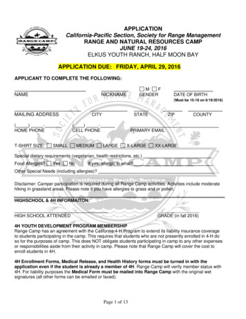
Figure 9.19c Pathways for regenerating ATP during muscle activity.(c)Aerobic pathwayAerobic cellular respirationEnergy source: glucose; pyruvic acid;free fatty acids from adipose tissue;amino acids from protein catabolismGlucose (fromglycogen breakdown ordelivered from blood)O2Pyruvic acidFattyacidsO2AerobicrespirationAerobic respirationin mitochondriamitochondriaAminoacids32CO2H2OATPnet gain perglucoseOxygen use: RequiredProducts: 32 ATP per glucose, CO2, H2ODuration of energy provision: HoursCopyright 2010 Pearson Education, Inc.
Figure 9.20 Comparison of energy sources used during short-duration exercise and prolonged-duration exercise.Short-duration exerciseATP stored inmuscles isused first.ATP is formedfrom creatinePhosphateand ADP.Copyright 2010 Pearson Education, Inc.Glycogen stored in muscles is brokendown to glucose, which is oxidized togenerate ATP.Prolonged-durationexerciseATP is generated bybreakdown of severalnutrient energy fuels byaerobic pathway. Thispathway uses oxygenreleased from myoglobinor delivered in the bloodby hemoglobin. When itends, the oxygen deficit ispaid back.
The end result of all three types of musclemetabolism is 1)2)3)4)Recharging creatine phosphateBurning fatATP hydrolysisATP synthesis
SKELETAL MUSCLE:FORCE OF CONTRACTION
Figure 9.21 Factors influencing force of skeletal muscle contraction.Largenumber ncy ofstimulationContractile forceCopyright 2010 Pearson Education, Inc.Muscle andsarcomerestretched toslightly over 100%of resting length
Figure 9.22 Length-tension relationships of sarcomeres in skeletal muscles.SarcomeresgreatlyshortenedSarcomeres atresting lengthSarcomeres excessivelystretched75%100%170%Optimal sarcomereoperating length(80%–120% ofresting length)Copyright 2010 Pearson Education, Inc.
SKELETAL MUSCLE:VELOCITY & DURATIONOF CONTRACTION
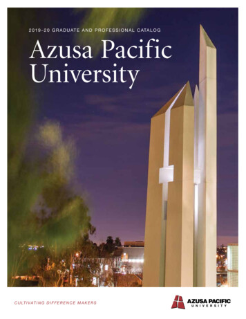
Figure 9.24 Cross section of the three types of fibers in skeletal muscle.FOSOFGCopyright 2010 Pearson Education, Inc.
Figure 9.23 Factors influencing velocity and duration of skeletal muscle contraction.Predominanceof fast glycolytic(fatigable) fibersContractilevelocityCopyright 2010 Pearson Education, Inc.Small loadPredominanceof slow ion
Figure 9.25 Influence of load on contraction duration and velocity.Light loadIntermediate loadHeavy loadStimulus(a) The greater the load, the less the muscleshortens and the shorter the duration ofcontractionCopyright 2010 Pearson Education, Inc.(b) The greater the load, theslower the contraction
Figure 9.25a Influence of load on contraction duration and velocity.Light loadIntermediate loadHeavy loadStimulus(a)The greater the load, the less the muscleshortens and the shorter the duration ofcontractionCopyright 2010 Pearson Education, Inc.
Figure 9.25b Influence of load on contraction duration and velocity.(b) The greater the load, theslower the contractionCopyright 2010 Pearson Education, Inc.
SKELETAL MUSCLE:EFFECT OF EXERCISE
SMOOTH MUSCLE:MICROSCOPIC STRUCTURE
Figure 9.26b Arrangement of smooth muscle in the walls of hollow organs.Longitudinal layer ofsmooth muscle (showssmooth muscle fibers incross section)Small intestineMucosa(b) Cross section of theintestine showing thesmooth muscle layers(one circular and the otherlongitudinal) running at rightangles to each other.Copyright 2010 Pearson Education, Inc.Circular layer of smoothmuscle (shows longitudinalviews of smooth muscle fibers)
Figure 9.27 Innervation of smooth muscle.VaricositiesAutonomicnerve fibersinnervatemost smoothmuscle fibers.SmoothmusclecellSynapticvesiclesCopyright 2010 Pearson Education, Inc.MitochondrionVaricosities releasetheir neurotransmittersinto a wide synapticcleft (a diffuse junction).
Figure 9.28 Intermediate filaments and dense bodies of smooth muscle fibers harness the pull generated by myosincross bridges.IntermediatefilamentCaveolaeNucleusGap junctionsDense bodies(a) Relaxed smooth muscle fiber (note that adjacent fibersare connected by gap junctions)NucleusDense bodies(b) Contracted smooth muscle fiberCopyright 2010 Pearson Education, Inc.
In smooth muscle, instead of neurotransmittersbeing released from axon terminals, they arereleased from 1)2)3)4)CaveolaeVaricositiesGap junctionsAll of the above
SMOOTH MUSCLE:CONTRACTION
Figure 9.29 Sequence of events in excitation-contraction coupling of smooth muscle.Extracellular fluid (ECF)Ca2 Plasma membraneCytoplasm1 Calcium ions (Ca2 )enter the cytosol fromthe ECF via voltagedependent or voltageindependent Ca2 channels, or fromthe scant SR.Ca2 Sarcoplasmicreticulum2 Ca2 binds to andactivates calmodulin.Ca2 Inactive calmodulinActivated calmodulin3 Activated calmodulinactivates the myosinlight chain kinaseenzymes.Inactive kinase4 The activated kinase enzymescatalyze transfer of phosphateto myosin, activating the myosinATPases.Activated kinaseATPADPPiPiInactivemyosin moleculeActivated (phosphorylated)myosin molecule5 Activated myosin forms crossbridges with actin of the thinfilaments and shortening begins.ThinfilamentThickfilamentCopyright 2010 Pearson Education, Inc.
True or false: in smooth muscle, calcium ionsbind to tropomyosin to allow myosin to bindto actin.1) True2) False
Figure 9.30 Formation of a multinucleate skeletal muscle fiber by fusion of myoblasts.Embryonicmesoderm cellsMyoblastsMyotube(immaturemultinucleatemuscle fiber)Satellite cell1 Embryonic2 Several3 Myotubemesoderm cellsundergo celldivision (toincrease number)and enlarge.myoblastsfuse togetherto form amyotube.matures intoskeletal musclefiber.Copyright 2010 Pearson Education, Inc.MatureSkeletalmusclefiber
A Closer Look 9.1 Athletes Looking Good and Doing Better with Anabolic Steroids?Copyright 2010 Pearson Education, Inc.
McPherron and Lee, Nature 2001
During the period of relaxation, what is happening in the muscle fiber on a molecular level? 1) The membrane is depolarizing 2) Ca2 is being released from the SR 3) Ca2 is being ta