Transcription
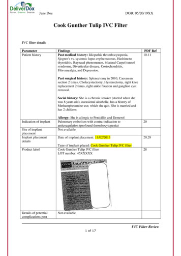
Jane DoeDOB: 05/20/19XXCook Gunther Tulip IVC FilterIVC filter detailsParameterPatient historyFindingsPast medical history: Idiopathic thrombocytopenia,Sjogren's vs. systemic lupus erythematosus, Hashimotothyroiditis, Raynaud phenomenon, bilateral Carpel tunnelsyndrome, Diverticular disease, Costochondritis,Fibromyalgia, and Depression.PDF Ref10-11Past surgical history: Splenectomy in 2010, Caesareansection 2 times, Cholecystectomy, Hysterectomy, right kneereplacement 2 times, right ankle fixation and ganglion cystremoval.Social history: She is a chronic smoker (started when shewas 8 years old), occasional alcoholic, has a history ofMethamphetamine use; which she quit. She is married andhas 2 children.Indication of implantSite of implantplacementImplant placementdetailsProduct labelDetails of potentialcomplications postAllergy: She is allergic to Penicillin and DemerolPulmonary embolism with contra-indication toanticoagulation (profound thrombocytopenia)Not available20Date of implant placement: 11/02/201320,28Type of implant placed: Cook Gunther Tulip IVC filterCook Gunther Tulip IVC filterLOT number: 45XXXXX28Not availableIVC Filter Review1 of 17
Jane DoeimplantManagement of thecomplicationsRadiology reportspertaining to the IVCfilterWas the deviceremovable/permanentfilterIf a removable filter,how long was the IVCfilter implanted for?Date of explantCurrent status of thepatient post IVC filterexplantDOB: 05/20/19XXNot availableAll the radiology images pertaining to the IVC filter has beenexplained belowCook Gunther Tulip IVC filter is a type of retrievable IVCfilterNot availableNot availableNot availableCase events:11/01/2013 - 11/11/2013: Patient is admitted for Thrombocytopenia and was discharged with adiagnosis of Idiopathic thrombocytopenic purpura, accessory spleen status post open splenectomy,acute pulmonary embolism and S/P IVC filter placement. (PDF page: 2-5)11/02/2013: IVC filter placement. (PDF page: 20)11/13/2013: Patient was admitted for persistent thrombocytopenia. (PDF page: 6-9)01/10/2014: Patient’s thrombocytopenia resolved after IVC filter placement, and she had resumed hertherapeutic anticoagulation. She has had 2 complications of anticoagulation. One was intra abdominalbleeding in the retroperitoneum and the second was a hemorrhagic stroke on 01/01/2014. She istaking intermittent doses of Lovenox and is not on Warfarin. As she was having recurrent bleedingcomplications, IVC filter removal was not recommended. (PDF page: 33-34)02/14/2014: On 12/31/2013, the patient had a sudden acute severe headache with neck pain,dysarthria, left upper and lower extremity weakness. CT revealed intraparenchymal hemorrhage in theleft fronto-temporal region with a 3mm shift in the midline. Changes were made in her medications.She was concluded to be contraindicated with anticoagulation. (PDF page: 54-61)09/06/2014: Cystoscopy, right retrograde ureteropyelogram, right ureteroscopy with laser lithotripsyand stone retrieval and right ureteral stent placement was performed for right mid ureteral stone. (PDFpage: 296-298)09/08/2014: Left sided video-assisted thoracoscopy with ligation of left atrial appendage wasperformed. (PDF page: 299-300)10/14/2014: Patient had bilateral leg pain and was diagnosed with a new left lower extremity DVT.(PDF page: 1291-1303)IVC Filter Review2 of 17
Jane DoeDOB: 05/20/19XX12/05/2014: Patient underwent pericardiocentesis, and 450cc of red fluid was removed. (PDF page:2036)Radiology Images:Image 1: IVC filter insertion on 11/02/2013(Note: Radiology images contain date in dd/mm/yyyy format. However, we have followed thestandard mm/dd/yyyy format in the report)Impression IVC filter placement procedureIVC Filter is seen in-situIVC filter is seen along the long axis of the IVCPost-cholecystectomy metallic clips are also seenIVC Filter Review3 of 17
Jane DoeDOB: 05/20/19XXImage 2: CT chest with contrast on 11/14/2013Impression: Incidental findings: Post splenectomy metallic clips are seen, and they are not to bemistaken for fractured fragments of IVC filterIVC Filter Review4 of 17
Jane DoeDOB: 05/20/19XXImage 3: CT abdomen, pelvis with contrast on 11/14/2013Impression: IVC filter is seen in-situ Tips of all the primary legs and secondary struts of the IVC filter are seenIVC Filter Review5 of 17
Jane DoeDOB: 05/20/19XXImage 4: CT abdomen, pelvis with contrast on 11/14/2013 Impression: Tips of the primary legs of the IVC filter are seen piercing the IVC luminal wallImage 5: CT abdomen, pelvis with contrast on 11/14/2013 Incidental findings: Post-cholecystectomy metallic clips are seen – not to be mistaken forfractured fragments of IVC filterIVC Filter Review6 of 17
Jane DoeDOB: 05/20/19XXImage 6: CT abdomen, pelvis with contrast –coronal on 11/14/2013 Impression: Incidental findings: Post-cholecystectomy metallic clips are seen – not to bemistaken for fractured fragments of IVC filterImage 7: CT abdomen, pelvis with contrast –coronal on 11/14/2013Impression: IVC filter is seen in-situ One of the tips of the primary leg on the right side is seen piercing the IVC luminal wallIVC Filter Review7 of 17
Jane DoeDOB: 05/20/19XXImage 8: CT abdomen, pelvis with contrast –coronal on 11/14/2013Impression: IVC filter is seen in-situ One of the tips of the primary leg on the right side is seen piercing the IVC luminal wall One of the tips of the primary leg on the left side is seen piercing the IVC luminal wallImage 9: Vascular, abdominal aortic angiogram on 12/02/2013 Impression: Incidental findings: Post-cholecystectomy metallic clips are seen – not to bemistaken for fractured fragments of IVC filterIVC Filter Review8 of 17
Jane DoeDOB: 05/20/19XXImage 10: Vascular, abdominal aortic angiogram on 12/02/2013Impression: Tips of the anterolateral & anteromedial primary legs are seen outside the IVC lumen The other 2 tips of the primary legs on the postero-lateral & posteromedial aspect are seenpiercing the IVC lumen Large left anterior abdominal wall intramuscular bleed seenImage 11: Vascular, abdominal aortic angiogram on 12/02/2013 Impression: Possible coil placement for left anterior abdominal wall intramuscular bleederIVC Filter Review9 of 17
Jane DoeDOB: 05/20/19XXImage 12: Abdomen stone study – axial on 09/06/2014Antero- lateralAntero-medialPostero-medialPostero-lateral Tips of all the primary legs are seen piercing the IVC luminal wall The tip of the primary leg in the posteromedial aspect is seen abutting the adjacent lumbarvertebral body The tip of the primary leg in the anteromedial aspect is seen abutting the adjacent aortic wall The tip of the primary leg in the anterolateral aspect is seen abutting possibly the gonadal/lumbarvein The tip of the primary leg in the posterolateral aspect is seen placed adjacent to right PsoasmuscleCoronal view Impression: Two tips of the primary legs of the IVC filter are seen outside the IVC andpiercing the adjacent structuresIVC Filter Review10 of 17
Jane DoeDOB: 05/20/19XXImage 13: Abdomen, Pelvis on 04/18/2017 All the tips of primary legs of the IVC filter are piercing the IVC luminal wall Tips of the primary legs of the IVC filter are seen outside the IVC and piercing the adjacentstructures Distortion the IVC filter is seenImage 14: Abdomen, Pelvis on 04/18/2017 All the tips of primary legs of the IVC filter are piercing the IVC luminal wall Tips of the primary legs of the IVC filter are seen outside the IVC and piercing the adjacentstructuresIVC Filter Review11 of 17
Jane DoeDOB: 05/20/19XXCoronal view IVC filter is seen in-situ One of the primary leg tips on the right side is seen outside the IVC An infrarenal segment of the Inferior vena cava appears significantly smaller and irregular incaliper with distortion of the IVC filter within Inferior venacavaImage 15: Abdomen, Pelvis on 04/18/2017 One of the primary leg tips is seen piercing and entering the lumen of the right common iliacartery No signs of bleeding or edema are seen around the right common iliac arteryIVC Filter Review12 of 17
Jane DoeDOB: 05/20/19XXImage 16: Abdomen, Pelvis on 04/18/2017 An infrarenal segment of the Inferior vena cava appears significantly smaller and irregular incaliper with distortion of the IVC filter within Inferior venacava One of the primary legs is seen piercing the L4 vertebral body and reaching L4-L5intervertebral disc space Secondary to chronic IVC occlusion, multiple prominent lymph nodes are noted in thepericaval, para-aortic and along the bilateral iliac groups of lymph nodes – secondary topossible underlying malignant pathologyComparison retation Tips of the primary legs of the IVCfilter are seen piercing the IVCluminal wall All the tips of primary legs of the IVCfilter are piercing the IVC luminal wall Tips of the primary legs of the IVCfilter are seen outside the IVC andpiercing the adjacent structures Distortion the IVC filter is seenIVC Filter Review13 of 17
Jane DoeDOB: 05/20/19XXFindingsWe note the following: The IVC filter placed in Jane Doe is Cook Gunther IVC filter Tips of the primary legs of the IVC filter are seen piercing the IVC wall (These changes arenoted 2 weeks after filter placement) An infrarenal segment of the Inferior vena cava appears significantly smaller and irregular incaliper with distortion of the IVC filter within Inferior venacava One of the primary legs is seen piercing the L4 vertebral body and reaching L4-L5intervertebral disc space In the latest dated available images (04/18/2017) IVC is seen chronically occluded withsignificant distortion of IVC filter and the primary legs are seen piercing the adjacent L4vertebral body/L4-L5 disc and right common iliac artery. However, No demonstratable fracture/migration of IVC filter seen in rest of theabdomen/chest images availableConclusion IVC filter inserted in Jane Doe is Cook Gunther IVC filter IVC perforation is the complication that occurred in Jane Doe Distortion of IVC filter and piercing of primary legs into the adjacent structures such asthe adjacent L4 vertebral body/L4-L5 disc and right common iliac artery. Chronic IVC occlusion might be secondary to her multiple underlying medicalconditions/long term IVC filter placementIVC Filter Review14 of 17
Jane DoeDOB: 05/20/19XXAnnexure 1: Details of Cook Gunther Tulip IVC filterName & ImageCook Gunther TulipIVC cal characteristics & FeaturesPhysical characteristics:The basic design of the filter is conical with fourlegs. The end of each leg is slightly hookedoutward. "Webbed" wires (like tulip petals)between the legs are bent strands of the samealloy which maintain the shape of the filter bypressing outward toward the vein walls. Thesewebs also increase the area into which theemboli can be trapped.Features: There are two types of Gunther TulipVena Cava Filter Sets a femoral setwhich is introduced through the femoralvein and a jugular set which isintroduced through the jugular veinSimple placement- Tulip’s hook enablesaccurate and simple jugular placementand superior retrievability.Specially designed anchors to achievestrong caval fixation.Indications:Used for the prevention of recurrent pulmonaryembolism via placement in the vena cava in thefollowing situations: Pulmonary thromboembolism whenanticoagulant therapy is contraindicated;Failure of anticoagulant therapy inthromboembolic diseases;Emergency treatment following massivepulmonary embolism where anticipatedIVC Filter Review15 of 17
Jane DoeDOB: 05/20/19XX benefits of conventional therapy arereduced; andChronic, recurrent pulmonary embolismwhere anticoagulant therapy has failedor is contraindicated.Contraindications: Unsuccessful retrieval attempts are morelikely to occur when IVCF position isangulated.Problems: The Günther Tulip is a retrievable IVCfilter, which means it is only intendedfor short-term protection againstpulmonary embolism. If it is left in apatient for more than 3-4 months, thereis a higher risk of complications likefilter fracture or migration. This canmake it very difficult to retrieve thefilter.Embolization occurs when brokenpieces of an IVC filter travel to theheart, where they are impossible toremove. This can lead to long-termcomplications, perforation of the heartmuscle, arrhythmia (abnormal heartrhythm), bleeding, sudden heart attack,and death.After analyzing data on 50 patients who wereimplanted with a Cook Celect or Günther Tulipfrom July 2007 to March 2009, researchersfound: All of the filters showed some degree ofvena caval perforation within 71 days.Filter tilt was also seen in 40% of thepatients.In 86% of patients, at least onecomponent of the filter completelyperforated the vena cava.IVC Filter Review16 of 17
Jane DoeDOB: a.gov/cdrh -8445-44a72334cb6d/IVC Filter Review17 of 17
Annexure 1: Details of Cook Gunther Tulip IVC filter Name & Image FDA Approval Date Terminated Date Physical characteristics & Features Cook Gunther Tulip IVC filter pulmonary embolism where anticipated 10/18/2000 Physical characteristics: The basic design of the filter is conical with f