Transcription
PhD Committee MeetingMarta Kersten
Previous ResearchMobile Emergency Triage (2002-2005)Dr. Wojtek Michalowski,Telfer School of Management; Faculty of Medicine, University of OttawaSz. Wilk, W. Michalowski, K. Farion, and M. Kersten, “Interaction Design for Mobile Clinical Decision Support Systems: the MET SystemSolutions.”, Foundations of Computing and Decision Sciences, 32(1):47–62, 2007.W. Michalowski, M. Kersten, S. Wilk, and R. Slowinksi, “Designing man-machine interactions for mobile clinical systems: MET triagesupport using palm handhelds.” European Journal of Operational Research, 177(3):1409–1417, March 2007.
Previous ResearchStereoscopic Volume Rendering of Medical Images (Master’s Thesis, 2005)Dr. Randy Ellis & Dr. James Stewart, School of Computing, Queen’s UniveristyM. Kersten, J. Stewart, N. Troje, and R. Ellis, “Depth Perception in Translucent Volumes”. IEEE Transactions on Visualization and ComputerGraphics, 12(6):1117–1123, September/October, 2006.J. Inoue, M. Kersten, B. Ma, J. Stewart, J. Rudan and R. Ellis, “Fast assessment of acetabular coverage using stereoscopic volumerendering.” Medicine Meets Virtual Reality (MMVR), January 2006.
Previous ResearchAngiography to CT/MRI Registration (2006-2007)Dr. Dirk Bartz, GRIS, Univeristy of Tübingen, GermanyM. Kersten, J. Hoffmann, D. Freudenstein, M. Tatagiba, U. Ernemann, D. Bartz. “Registration of 3D rotational angiography to CT (or MR)angiography.” Proceedings of the Deutschen Gesellshaft für Computer- und Roboterassisierte Chirugie (CURAC), Hannover,Oct. 12 14, 2007.
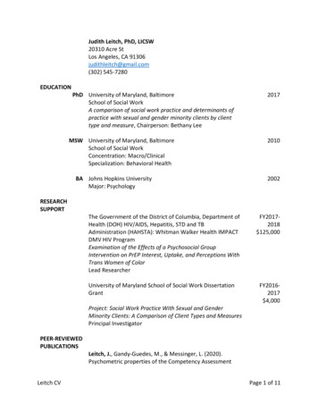
Current ResearchVisualization of Angiography DataDr. Louis Collins, BIC, McGill University(a) No depth Cues(b) Fog/Simulated Aerial Perspective(c) Edge Enhancement(d) Edge and Fog(e) Chromadepth(f) Wobble Stereo
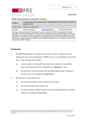
physical registration is performed by attaching imaging markers to the patient prior to scanning, andthese with physical localisation caps at surgery which can be localised using a tracked pointer.are particular issues involved in developing an AR system for surgical navigation. Since the mis moved relatively infrequently during surgery the latency of the system is not a problem. Of muchmportance is the accuracy of the overlaid structures with respect to their real equivalents. Surgeonsally use the system to avoid critical structures such as arteries or nerves, so a misalignment of even ae could have disastrous effects. For this reason accuracy has been a prime concern in developing thestem. A numericalsimulationwas producedwhichthe effectof differentmarker and LEDEnhancethe realview oftheenabledsurgeonwithpreoperativelyacquired data fromions to be analysed. The overlay accuracy of the system is now less than 1mm. Achieving good 3Dmultiple modalities.n of virtual structures beneath a physical surface in the magnified view of the surgical microscoped to be problematic, and the display paradigm of the virtual structures has been optimised to makeinformationdisplayedfieldble. FigurePreoperative1 shows an image acquiredduring a recentclinical withinevaluationtheof theoperativeMAGI system,in of view:An Image Overlay System for Medical Data Visualizationaccurate overlay ofOna partiallyvisible tumourisgiven.The surgeon is able to use the AR display totheocularsofthemicroscopeowards the surgical target. Texture mapping on the virtual tumour is used to help 3D perception.head-mounteddisplaymmary, MAGI is a Usingworking exampleof an AR systemin a domain which raises particular issues fore issues have been Ontoaddressedtheduringthe developmentof the system, and the resultsof this work willpatient(half silvered-mirror,projection)Displayrest to other researchers in the AR field.Augmented Reality 237Head TrackerStereo GlassesBeam Splitter3D OverlayworkspaceWork Tableb)a)Augmentedrealityin the E.,operatingmicroscope-Edwards,a) TheMAGIsysteminDetheoperatingtheatreP.J., King,A.P., Maurer,C.R., Jr.,Cunha,D.A.,Jannin, P., Fleig,O. J., Seigneuret,Grova, C., Morandi,X., andBlackwell,andM., Nikou, C., DiGioia, A., Kanade, T.: An Image OverlayHawkes, D.J., Hill, D.L.G., Gaston, R.P., Fenlon, M.R.,Scarabin, J. M. A data fusion environment for multimodal andsystem for medical visualization. Transactionslay ofmulti-informationalthe lesion. Jusczyzck, A., Strong,A.J., Chandler,C.L., (IAM),and Gleeson,whichM.J.neuronavigation.Computersurgeryto the internalon MedicalImage Analysis, 2000; 4: 67–72Designand evaluationof a thesystemfor microscope-assisted2000;5(1):1-10.with the section of the tumour thatprotrudesintoIAM.Fig.5.The new Image Overlay protoguided interventions (MAGI), IEEE Transactions on MedicalFig. 4. Configurationof1082-1093.original protoImaging, 2000;19(11):type in a pelvic screw fixation simulatype Image Overlay system.tion.
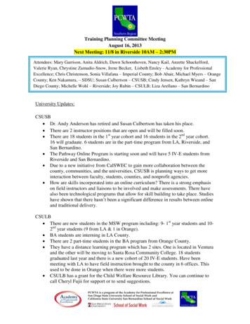
Augmented VirtualityAugment the “virtual world”, i.e., the 3D multi-modal preoperative datasets, withintraoperative views from microscope/camera.(a) Augmented Reality(b) Augmented VirtualityPaul, P,. Fleig. O., and Jannin, P. Augmented virtuality based on stereoscopic reconstruction in multimodal image-guided neurosurgery: methods and performance evaluation. IEEETransactions on Medical Imaging. 2005;24(11):1500-11.
Proposed Research: MotivationAugmented Reality/Virtuality for Multi-modal ImageGuided Neurosurgery Want to overcome the surgeons limited visual perception, restricted view ofthe region of intervention Little research on which methods and techniques are best and how they shouldbe incorporated into the surgical workflow Not enough evaluation of current systems from a workflow/human factors/interface perspective Need for evaluation of different visualization methods (e.g. Can we get similarperceptual results using other perspective cues than stereo?)
Proposed ResearchAugmented Reality/Virtuality for Multi-modal ImageGuided Neurosurgery Implementation and evaluation of an augmented reality and augmented virtualityprotocol for intraoperative guidance of neurosurgeries Evaluation of non-photorealistic rendering (NPR) techniques for IntraoperativeGuidance Systems (IGSs) Incorporation of psychophysical knowledge (e.g. perception) to drivevisualization Incorporation of intraoperative (i.e. ultrasound) data into visualizations How to best overlay preoperative images into the surgical field of view withoutdisrupting work flow and comprehension of the real world Evaluation of these different methods
Courses TakenMcGill: PHGY 314 – Integrative Neuroscience BMDE 650 – Advanced Medical ImagingCOMP 764 – Computer Graphics & AnimationECSE 626 – Statistical Computer VisionECSE 618 – HapticsECSE 683 – Topics in Vision and Robotics (Audited)Queen’s: CISC 850 – Topics in Computer Application & Algorithms CISC 857 – Image Processing CISC 868 – Computational Geometry CISC 874 – Foundations of Neural Networks CISC 875 – Bioinformatics (Audited)
Dr. Wojtek Michalowski, Telfer School of Management; Faculty of Medicine, University of Ottawa Sz. Wilk, W. Michalowski, K. Farion, and M. Kersten, “Interaction Design for Mobile Clinical Decision Support Systems: the MET System