Transcription
HindawiBioMed Research InternationalVolume 2019, Article ID 5636423, 12 pageshttps://doi.org/10.1155/2019/5636423Research ArticleA Deep Learning Segmentation Approach in Free-BreathingReal-Time Cardiac Magnetic Resonance ImagingFan Yang ,1,2 Yan Zhang,3 Pinggui Lei ,3 Lihui WangHong Xie,3 and Zhu Zeng1,4 Yuehong Miao,1,21Key Laboratory of Biology and Medical Engineering, Guizhou Medical University, Guiyang 550025, ChinaSchool of Biology & Engineering, Guizhou Medical University, Guiyang 550025, China3Department of Radiology, The Affiliated Hospital of Guizhou Medical University, Guiyang 550004, China4Key Laboratory of Intelligent Medical Image Analysis and Precise Diagnosis of Guizhou Province,School of Computer Science and Technology, Guizhou University, Guiyang 550025, China2Correspondence should be addressed to Pinggui Lei; 570465670@qq.com and Lihui Wang; wlh1984@gmail.comReceived 20 April 2019; Accepted 17 July 2019; Published 30 July 2019Academic Editor: Kwang Gi KimCopyright 2019 Fan Yang et al. This is an open access article distributed under the Creative Commons Attribution License, whichpermits unrestricted use, distribution, and reproduction in any medium, provided the original work is properly cited.Objectives. The purpose of this study was to segment the left ventricle (LV) blood pool, LV myocardium, and right ventricle (RV)blood pool of end-diastole and end-systole frames in free-breathing cardiac magnetic resonance (CMR) imaging. Automatic andaccurate segmentation of cardiac structures could reduce the postprocessing time of cardiac function analysis. Method. We proposeda novel deep learning network using a residual block for the segmentation of the heart and a random data augmentation strategyto reduce the training time and the problem of overfitting. Automated cardiac diagnosis challenge (ACDC) data were used fortraining, and the free-breathing CMR data were used for validation and testing. Results. The average Dice was 0.919 (LV), 0.806(myocardium), and 0.818 (RV). The average IoU was 0.860 (LV), 0.699 (myocardium), and 0.761 (RV). Conclusions. The proposedmethod may aid in the segmentation of cardiac images and improves the postprocessing efficiency of cardiac function analysis.1. IntroductionFree-breathing cardiac magnetic resonance (CMR) cineimaging techniques have been developed for the evaluationof cardiac function [1–5]. It is an accurate and reproducibletechnique for chamber volume, myocardial mass, and strokevolume measurements [5]. Compared with breath-hold CMRcine imaging, it has a short acquisition time and eliminates the unnecessary breath-hold stage. It is beneficialfor children and patients who are unable to hold theirbreath during data acquisition. However, the postprocessingof free-breathing CMR cine imaging is time-consumingand laborious. Although some commercial software couldautomatically segment the left ventricle (LV) blood pool, LVmyocardium, and right ventricle (RV) blood pool contoursof end-diastole (ED) and end-systole (ES) frames, manual adjustment of the segmented contour is still requiredby an expert. However, the procedure could introduceintraobserver and interobserver variability [6]. Hence, a fullyautomatic segmentation method of ED and ES frames isnecessary for improving the postprocessing efficiency of thefree-breathing CMR cine imaging.The segmentation process of cardiac imaging was previously divided into two stages, i.e., localization and segmentation. For the localization task, some studies used varianceimage [7], the Fourier Transform [8], and the circular Houghtransform [9] to locate the heart. Due to the diaphragmmotion in free breathing CMR cine imaging, these methodscannot be applied directly for the heart localization, especiallyfor the apex slice. For the segmentation task, the levelset was previously widely used for the LV, myocardium,and RV [10–13]. Other methods include threshold, pixelclassification, cardiac atlas, shaped registration, and activeshape model, among others [6, 14–19]. However, most algorithms require prior information and manual operation.An alternative automatic method to locate and segmentthe heart from the cardiac image is to use deep learningtechniques.
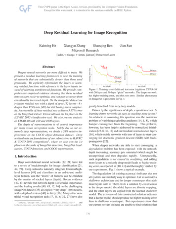
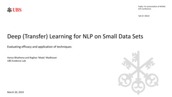
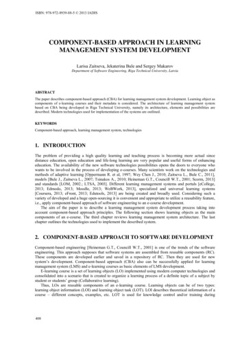
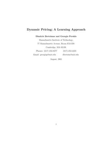
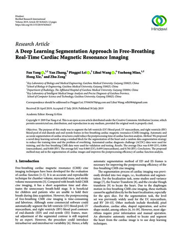
2Recently, deep learning approaches have been widely usedin medical image segmentation, especially for CMR images[20–35]. Bernard et al. [20] summarized all the types ofsegmentation methods using deep learning. Avendi et al. [21]presented a combined deformable model and deep learningmethod for LV segmentation. Ngo et al. [22] proposed adeep learning method combined with the level set for LVsegmentation. Tan et al. [23] designed a regression networkon the segmentation of short-axis LV. Isensee et al. [24]used an ensemble of modified 2D and 3D U-Net to tacklethe segmentation. Baumgartner et al. [25] tested variousconvolution neural networks with hyperparameters. Zottiet al. [26] proposed an extension of the U-Net for thecardiac segmentation and an automated analysis methodfor CMR images [27]. Vigneault et al. [28] presented anovel network for localization and semantic segmentation ofcardiac images. Zheng et al. [29] applied a novel variant of UNet for the cardiac segmentation on short axis MRI image.Khened et al. [30] utilized multiscale residual DenseNetsfor cardiac segmentation. Zhang et al. [31] proposed a noveldeep network for the segmentation of myocardial infarction.Bai et al. [32] used a fully convolutional network for CMRimage analysis. Qin et al. [33] proposed a motion estimationand segmentation method for cardiac images. Oktay et al.[34] developed neural networks to cardiac image enhancement and segmentation. Romaguera et al. [35] studied themyocardial segmentation with deep learning approach. Thesemethods were successful for cardiac segmentation. However,the image quality of free-breathing CMR imaging is lowerthan the breath-hold CMR imaging (see Figure 1) [36]. Thedeep learning techniques for the accurate segmentation offree-breath CMR data remain a challenge.As mentioned above, the development of a fully automaticmethod for the segmentation of free-breathing CMR imagescould assist experts in analyzing cardiac function. In thispaper, we propose a one-stage deep learning network basedU-Net [37] for the segmentation of the heart. The LV, LVmyocardium, and RV were directly segmented by usingpresented deep learning model. The ACDC data were usedfor training, and free-breathing CMR data were used forvalidation and testing. We also used a random augmentationdata strategy during training. The data were only augmentedduring the training process and were not stored, making itfaster than data augmentation before training. Furthermore,we proposed an improved loss function to yield highersegmentation accuracy. The proposed method is validatedand tested on free-breathing CMR data. The experimentalresults validate its accuracy for cardiac segmentation. Thelayout of the paper is as follows: The datasets are introducedin detail in Section 2; the methods are presented in Section 3;Section 4 demonstrates the experimental results; in Section 5,we discuss and analyze the results; and the conclusions aregiven in Section 6.2. Materials2.1. Free-Breathing CMR Data. The study was approved bythe institutional Review Board. Twelve subjects (7 males; 5BioMed Research Internationalfemales; age 25 4), with informed consent, were recruitedfor the study. The heart function assessments were carriedout using a 3.0T MR scanner (Siemens, Germany). The heartrate was monitored using ECG. Ten short axis slices coveringthe whole heart from apex to base were imaged using a freebreathing 2D real-time SSFP, to which the Karhunen-Loevetransform filter was applied along the temporal direction toincrease the signal-to-noise ratio. The imaging parameterswere as follows: slice thickness 8 mm with a 2 mm gap,field of view (FOV) 340 287 mm2 , pixel spacing 2.25mm/pixel, repetition time/echo time (TR/TE) 2.5/1.1 ms,matrix size 160 128, TPAT 4, bandwidth 1488 Hz/pixel,temporal resolution 59.5 ms, and cine duration of 5 s foreach slice, containing 84 frames covering end-expiration andend-inspiration. The ED and ES frames in end-expiratorystage of each slice of twelve subjects were used to validate thefinal network model. The segmentation contours for the LV,LV myocardium, and RV of each frame were provided by theradiologist. The ED and ES frames in other respiratory stagesof each slice were used for testing.2.2. 2017 ACDC Data. The data was obtained from the automated cardiac diagnosis challenge (ACDC) data [20], whichwas initiated at the 2017 MICCAI Segmentation Challenge inthe STACOM workshop. It consists of 150 subjects with normal, previous myocardial infarction, dilated cardiomyopathy,hypertrophic cardiomyopathy, and abnormal right ventricle.The data is divided into the training and testing sets, with100 and 50 cases, respectively. As the training set contains theLV, LV myocardium, and RV contours, we used the trainingset to determine the parameters for segmentation in ourstudy.2.3. Data Processing. The ACDC data varied in size from 154 224 to 428 512. We resized all images to 160 128 by bilinear interpolation without any image cropping operations.Since the data acquisition from different imaging acquisitionsequences can introduce inconsistencies in image intensityand pixel intensity, the 16-bit images were normalized to 8bit images. Thereafter, contrast-limited adaptive histogramequalization (CLAHE) [38] was used to enhance the contrastof the grayscale image. Finally, 1902 images (100 subjects)from ACDC data were used for training, and 80 images (4subjects) and 160 images (8 subjects) from free-breathingCMR data were used for validation and testing, respectively.For the ground truth (labeled image) in the training stage, LVblood pool, LV myocardium, RV blood pool, and backgroundare labeled as 4, 3, 2, and 1, respectively.3. Methods3.1. Outline of the Method. The block diagram of the proposed method is shown in Figure 2. In our method, theproposed segmentation network can be divided into twostages: encoder and decoder. The encoder stage was used forCMR image representation and pixel-level classification, anda decoder stage was used to restore the original spatial resolution. To better display the segmentation result, red, green,
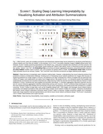
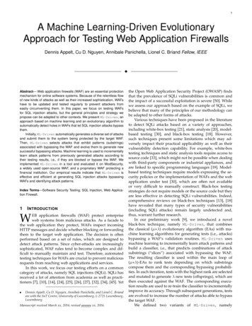
BioMed Research International3(a)(b)(c)(d)Figure 1: Comparison of breath-hold CMR data (a, c) and free-breath CMR data (b, d), where (a, b) are ED frames and (c, d) are ES frames.Segmentation NetworkEncoderDecoderLVLV myocardiumRVFigure 2: The block diagram of the proposed method for segmentation.and blue colors indicate the region of LV, LV myocardium,and RV in Figure 2.3.1.1. Heart Segmentation. For LV, LV myocardium, and RVsegmentation, it was always necessary to initially locate theheart region when using older methods [7–9, 30], whichis highly time-consuming especially for the deep learningmethods. We proposed a deep learning network based onU-Net and ResNet [39] to directly locate and segment theheart region. Figure 3 demonstrates the architecture of thenetwork for heart segmentation. The architecture consistsof a down-sample path (encoder) followed by an up-samplepath (decoder) to restore the size of the input image. In thedown-sample path (left section in Figure 3), the input image
4BioMed Research International2448 2424160 12848160 12896 4811929640 3240 32384192Residual block3 33 3Conv, bn Conv, bn10 810 82 2 max pooling38420 1619220 163 3 conv, bn, relu160 12880 6480 64160 1289632 41 1Conv, bn1 1 convconcatenation2 2 transposed convrelusumFigure 3: The proposed network architecture for heart segmentation. The label value of LV, LV myocardium, RV, and background is 4, 3, 2,and 1, respectively.is 160 128 in size. The residual block includes two 3 3convolution layers and one 1 1 convolution layer whichare appended by batch normalization (BN) and subsequentlyby ReLU activation (see Figure 3). The max pooling of size2 2 is used to down-sample the convolved maps. Thedropout layer is used in the bottom of the convolution layer toprevent overfitting. The dropout ratio was set to 0.5. In the upsample path (right section in Figure 3), the 2 2 transposedconvolution was used to up-sample the convolved maps.Several skip connections were used to concatenate featuremaps between down-sample and up-sample paths, whichcould provide more feature information for localization andsegmentation. A 1 1 convolution layer maps the 24 featurechannels to 4 classes. The pixel value in the output image of4 to 1 indicates the LV, LV myocardium, RV, and background,respectively. Thereafter, the loss function layer is used tocalculate the loss value from the output of the SoftMax layer.3.1.2. Loss Function. Previous methods used Dice loss [40]to solve the problem of class imbalance between heartstructure and background in cardiac image segmentation.Recently, Sudre et al. presented an improved Dice loss, calledgeneralized Dice loss [41, 42], which is a robust and accurateloss function for unbalanced tasks and is formulated as𝐿𝑜𝑠𝑠 1 𝑤𝑘 𝑀2 𝐾𝑘 1 𝑤𝑘 𝑚 1 𝑌𝑘𝑚 𝑇𝑘𝑚𝑀22 𝐾𝑘 1 𝑤𝑘 𝑚 1 (𝑌𝑘𝑚 𝑇𝑘𝑚 )12( 𝑀𝑚 1 𝑇𝑘𝑚 )(1)(2)where 𝑌 and 𝑇 are predicted label image and ground truth,respectively, 𝐾 is the number of classes, 𝑀 is numberelements along the first two dimensions of 𝑌 or 𝑇, and 𝑤𝑘is a weighting factor for each class. In our study, some imagesin the ACDC data did not have full labels, as illustrated inFigure 4. In such cases, 𝑇𝑘𝑚 0 and 𝑤𝑘 infinite. To avoid thiskind of problem, the loss and 𝑤𝑘 are revised and given by𝐿𝑜𝑠𝑠 1 𝑤𝑘 𝑤𝑘 𝑀2 𝐾𝑘 1 𝑤𝑘 𝑚 1 𝑌𝑘𝑚 𝑇𝑘𝑚 𝜀𝑀22 𝐾𝑘 1 𝑤𝑘 𝑚 1 (𝑌𝑘𝑚 𝑇𝑘𝑚 ) 𝜀1 𝑀𝑚 1 𝑇𝑘𝑚1 𝑀𝑚 1 𝑌𝑘𝑚,(3)𝑇𝑘𝑚 ̸ 0(4),𝑇𝑘𝑚 0where 𝜀 10 8 is used to avoid the numerical issue of dividingby 0. When 𝑇𝑘𝑚 0, the weight of 𝑘 class is determined by the𝑌𝑘𝑚 ; it could improve the performance of segmentation.3.2. Implementation Details. The heart segmentation network was implemented in Matlab
A Deep Learning Segmentation Approach in Free-Breathing Real-Time Cardiac Magnetic Resonance Imaging FanYang , 1,2 YanZhang, 3 PingguiLei , 3 LihuiWang , 4 YuehongMiao, 1,2