Transcription
3.4 BIOLOGY OF MEMORYKaplan & Sadock’s Comprehensive Textbook of PsychiatryCHAPTER 3. CONTRIBUTIONS OF THE PSYCHOLOGICALSCIENCES3.4 BIOLOGY OF MEMORYLARRY R. SQUIRE, PH.D., AND KEN A. PALLER, PH.D.Memory as Synaptic ChangeCortical Organization of MemoryInsights from AmnesiaMemory SystemsImplicationsAssessment of Memory FunctionsThe topic of memory is fundamental to the discipline of psychiatry. Memory provides the essentialsubstrate for the cognitive activities that define human experience, it allows one to connect the presentmoment to what came before, and it is the basis of cultural evolution.An individual's personality reflects habits and dispositions that have developed from experience.Adaptive and maladaptive coping strategies, anxieties, and phobias are largely products of learning.Neurotic or psychotic symptoms can be the consequences of specific experiences or repeated patterns ofexperience. Psychotherapy is a process by which new behaviors are acquired through the accumulationof new experiences. Thus, memory is at the heart of psychiatry's concern with the effects of earlyexperience, the development of the individual, and the possibility of change.Disorders of memory and complaints about memory lapses are pervasive in both neurology andpsychiatry. Memory problems are also of special concern as side effects of psychopharmacologicaltreatments and electroconvulsive therapy. Accordingly, the effective clinician needs to understandmemory, its psychological and neurological foundations, the varieties of memory dysfunction, and howmemory can be evaluated. The biological perspective on memory developed here rests on a growingbody of neuroscientific evidence that relates mental events to the functioning of the brain.MEMORY AS SYNAPTIC CHANGEMemory is a special case of the more general phenomenon of neural plasticity. Neurons can show
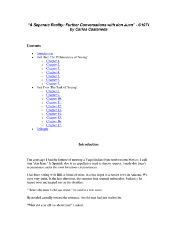
history-dependent behavior by responding differently as a function of recent input, and this plasticity ofnerve cells and synapses is the basis of memory. In the last decade of the nineteenth century, researchersproposed that the persistence of memory could be accounted for by nerve cell growth. Others haverestated this idea, developing the hypothesis that the synapse is the critical site of change. In principle,there are many possible ways for such structural change to be realized, including alterations in thenumber of synaptic contacts or in the strength of existing contacts.Plasticity Neurobiological evidence from animal studies supports two basic conclusions about thebiology of memory. First, specific synaptic events, including an increase in neurotransmitter release, areresponsible for short-lasting plasticity, which may last for seconds or minutes. Second, long-lastingmemory depends on new protein synthesis, physical growth of neural processes, and an increase in thenumber of synaptic connections.A major source of information has been the extended study of the marine mollusc Aplysia californica. Asufficient number of individual neurons and connections between neurons have been identified to allowthe wiring of some simple behaviors to be diagrammed. Aplysia is capable of both associative learning(including classical conditioning and operant conditioning) and nonassociative learning (habituation andsensitization). Figure 3.4-1 shows the circuitry responsible for the gill-withdrawal reflex, a defensivereaction whereby tactile stimulation causes the gill and siphon to retract. When tactile stimulation ispreceded by stimulation to the head, gill withdrawal is facilitated. The cellular mechanisms underlyingthis sensitization are based on an enhanced release of neurotransmitter by the facilitatory neuron (labeled“Int” in Fig. 3.4-1) and accompanied by covalent modifications of preexisting proteins. Under sometraining conditions, sensitization can persist for weeks, and these longer-lasting changes can also beproduced by repeated applications of serotonin, distributed over a period of 1½ hours. Although bothshort- and long-lasting plasticity are based on enhanced transmitter release, the long-lasting changeuniquely requires the expression of genes and the synthesis of proteins. In addition, the long-termchange, but not the short-term change, is accompanied by the growth of neural processes of neuronswithin the reflex circuit.FIGURE 3.4-1 A schematic diagram of the neuronal circuitunderlying behavioral habituation and sensitization of the gillwithdrawal reflect in Aplysia. The relative simplicity of thenervous system of Aplysia makes it a valuable organism forstudying cellular and synaptic mechanisms of memory. Thesynapse between the sensory neuron (SN) and the motor neuron(MN) is an important site of habituation. Sensitization resultsfrom activation of the interneuron (Int) pathway. (Reprinted withpermission from Kandel ER: Cellular Basis of Behavior. Freeman, San Francisco, 1976.)
In vertebrates, behavioral manipulations can also result in measurable changes in the brain's architecture.For example, rats reared in enriched environments show an increase in the number of synapses endingon individual neurons in neocortex. These changes are accompanied by small increases in corticalthickness, in the diameter of neuronal cell bodies, and in the number and length of dendritic branches.New synapses may be formed directly or synapses may be selectively preserved from a population thatis continuously being replaced. Behavioral experience thus exerts powerful effects on the wiring of thebrain.Many of these same structural changes have been found in adult rats exposed to an enrichedenvironment, and some have been found in adult rats given extensive maze training. In this case opaquecontact-lens occluders were used to restrict vision to one eye, and the corpus callosum was transected toprevent information received by one cerebral hemisphere from reaching the other hemisphere. In thesemonocularly trained animals, increases in the size of dendritic fields of pyramidal neurons of occipitalcortex were found only in the trained hemisphere. This finding rules out a number of nonspecificinfluences including motor activity, indirect effects of hormones, and overall level of arousal. Thereforeit seems likely that long-term memory in vertebrates is generally based on specific changes within theneurons that lie along specific pathways.Long-Term Potentiation The phenomenon of long-term potentiation (LTP) is a form of neuralplasticity likely to be important for memory in vertebrates. LTP is observed when a postsynaptic neuronis persistently depolarized following a brief burst of high-frequency stimulation. LTP has a number ofproperties that make it a promising candidate as a physiological substrate of memory. First, it isestablished quickly and then lasts for a long time. Second, it is associative in that it depends on the cooccurrence of presynaptic activity and postsynaptic depolarization. Third, it occurs only at thepotentiated synapses, not at all the synapses terminating on the postsynaptic cell. Finally, LTP occursprominently in the hippocampus, a structure with important memory functions. The induction of LTP isknown to be mediated postsynaptically and to involve activation of the N-methyl-D-aspartate (NMDA)receptor, which permits influx of calcium into the postsynaptic cell. The mechanism whereby LTP ismaintained is not clearly established, but evidence has been presented in favor of a presynaptic locus ofchange (increased transmitter release). Rapidly developing structural changes in the dendritic spines ofthe postsynaptic neuron have also been described in association with LTP.A new method for studying molecular mechanisms of memory relies on introducing specific mutationsinto the genome. For example, by altering a single cloned gene, a mutant strain of mice can be producedwith specific receptors or cell-signaling molecules inactivated or altered. This knock-out technique canprovide greater specificity than pharmacological blocking methods. Recently, it has been possible tostudy mice with a selective deletion of one type of NMDA receptor in the CA1 field of thehippocampus. Although many aspects of CA1 physiology remain intact, the CA1 cells do not exhibitLTP. In addition, an impairment is observed on a learning task. If reversible gene knock-outs can beachieved, it will be possible to induce specific molecular changes in a developmentally normal adult.
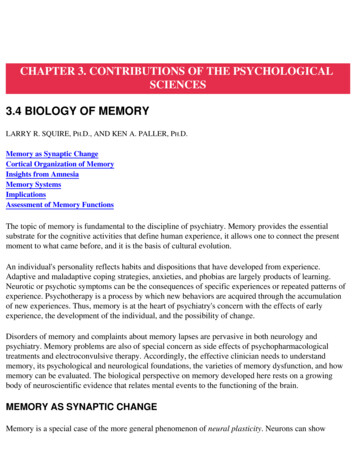
Associative Learning Additional insights into memory have been gleaned from the study of the neuralcircuitry underlying classical conditioning of the eyeblink-nictitating membrane response in rabbits.Repeated pairings of a tone (conditioned stimulus) and an airpuff to the eye (unconditioned stimulus)lead to a conditioned eyeblink in response to the tone. Reversible lesions of the deep nuclei of thecerebellum eliminate the conditioned response without affecting the unconditioned response, whichindicates that the cerebellum contains part of the essential circuitry for learned association, theconditioned stimulis–unconditioned stimulus link. Reversible lesions of the deep nuclei also preventlearning from occurring, and the rabbits begin learning from the naive state when the lesion is reversed.This finding does not mean that all the changes occurring in the animal during conditioning involve thecerebellum; it means only that essential neural changes responsible for the conditioned stimulus–unconditioned stimulus link depend on this circuitry. The relevant plasticity appears to be distributedbetween the cerebellar cortex and the deep nuclei (Fig. 3.4-2). An analogous pattern of plasticity isthought to underlie motor learning in the vestibulo-ocular reflex, and perhaps associative learning ofmotor responses in general. Based on the idea that learned motor responses depend on coordinatedcontrol of changes in both timing and strength of response, it has been suggested that synaptic change inthe cerebellar cortex is crucial for learned timing, whereas synaptic change in the deep nuclei is crucialfor learned changes in the strength of the response.FIGURE 3.4-2 A schematic diagram of the circuitry of themammalian cerebellum (top). In the classically conditionedblink response, input from the air-puff unconditioned stimulusand input from the auditory conditioned stimulus comes inthrough parallel pathways to the cerebellar cortex and to thedeep cerebellar nucleus, and plasticity occurs in both pathways(bottom). (Reprinted with permission from Raymond JL,Lisberger SG, Mauk MD: The cerebellum: A neuronal learningmachine? Science 272:1126, 1996. 1996 American Association for the Advancement of Science.)Understanding the biology of memory requires more than just an understanding of the synaptic eventsthat store memory. It is also essential to understand how and where synaptic events are organized in thebrain. Many levels of analysis can be identified between synaptic change and behavioral memory, andmany important questions about memory address levels of biological analysis that are intermediate tosynapses and behavior.CORTICAL ORGANIZATION OF MEMORYThe question of where memories are stored in the brain has long been a major research issue. In the1920s Karl Lashley carried out a series of experiments that were directed at this problem. Lashley
recorded the number of trials that rats needed to relearn a preoperatively learned maze problem afterremoval of different amounts of cerebral cortex. The deficit was proportional to the amount of cortexremoved and, further, it seemed to be qualitatively similar, regardless of what region of cortex wasremoved. Lashley concluded that memory for the maze habit was not localized in any one part of thebrain, but instead was distributed equally over the entire cortex. Subsequent work has led to a revision ofthis idea. Maze learning in rats depends on many forms of information, including visual, tactual, spatial,and olfactory information. These various forms of information are processed and stored in differentareas. Thus, the correlation between retention score and lesion size that Lashley observed reflects theprogressive encroachment on specialized cortical areas serving the many components of cognitionimportant to maze learning.The specialized cortical areas responsible for processing and storing visual information have beenstudied most extensively in nonhuman primates. Nearly half of the primate neocortex is specialized forvisual functions. Cortical pathways for visual information processing (Fig. 3.4-3) begin in primaryvisual cortex (V1) and proceed from there along parallel pathways or streams. One stream projectsventrally to the inferotemporal cortex (area TE in the monkey) and processes information about thequality of visual percepts. Another stream projects dorsally to the parietal cortex and processesinformation about spatial location. Electrophysiological studies in the monkey show that neurons in areaTE register specific and complex features of visual stimuli, like shape, and may even respond selectivelyto patterns and objects. These specific visual processing areas, along with connections to correspondingregions in dorsolateral prefrontal cortex, are involved in the immediate experience of perceptualprocessing, and in what has been called immediate memory or working memory. These areas also serveas the ultimate repositories of the memories that result from their activity. Accordingly, lesions in theseareas lead to impairments in visual perception as well as in visual learning and memory, althoughelementary visual functions such as acuity remain intact. Inferotemporal cortex can thus be thought ofboth as a higher-order visual processing system and a storehouse of the visual memories that result fromthat processing. These stored visual memories can be used and manipulated according to currentprocessing demands, and they can also be quite long-lasting.FIGURE 3.4-3 Summary of cortical visual areas and some oftheir connections. There are two major pathways from striatecortex (V1). The processing stream for object vision follows aventral route into the temporal lobe via V4 (dark gray boxes) andthe processing stream for spatial vision follows a dorsal routeinto the parietal lobe via MT (light gray boxes). Solid linesindicate connections arising from both central and peripheralvisual field representations; dotted lines indicate connectionrestricted to peripheral field representations. Shaded region on the lateral view of the brain represents theextent of the cortex included in the diagram. Abbreviations: DP, dorsal prelunate area; FST, fundus ofsuperior temporal area; HIPP, hippocampus; LIP, lateral intraparietal area; MSTc, medial superior
temporal area, central visual field representation; MSTp, medial superior temporal area, peripheralvisual field representation; MT, middle temporal area, MTp, middle temporal area, peripheral visualfield representation; PO, parieto-occipital area; PP, posterior parietal sulcal zone; STP, superior temporalpolysensory area; VIP, ventral intraparietal area; STS, rostral superior temporal sulcus; and VTF, visualresponsive portion of area TF. (Reprinted with permission from Ungerleider LG: Functional brainimaging studies of cortical mechanisms for memory. Science 270:769, 1995. 1995 AmericanAssociation for the Advancement of Science.)Many parts of the nervous system participate in storing representations of an event in memory. Duringan event, visual information is stored in inferotemporal cortex so that the same visual material can laterbe recognized as familiar. Concurrently, other components of the event—including spatial, temporal,tactile, olfactory, emotional, and other sorts of information—are processed and stored separately.Memory storage in the cerebral cortex thus depends on a fractionation of experience as follows. First,any particular event or learning task is composed of a number of components. Second, each componentengages a particular processing site or set of sites. Third, each processing site stores information as anoutcome of the processing that is done.Thus, memory is both distributed and localized in the nervous system. It is distributed in the sense that,as Lashley concluded, there is no unitary cortical center dedicated solely to the storage of memories.Yet, memory is localized in the sense that different aspects or dimensions of events are stored at specificcortical sites—the same regions specialized to analyze and process those particular aspects ordimensions of information.INSIGHTS FROM AMNESIAThe idea that the functional specialization of cortical regions governs both information processing andinformation storage is important, but it does not provide a complete account of the organization ofmemory in the brain. If it did, then particular cortical injuries would disrupt only particular domains oflearning and memory (i.e., visual memory or spatial memory) and no global disruption of memorywould occur. Brain injury would always produce a difficulty in learning a restricted type of newinformation along with a loss of previously learned information of that same type. Yet commonneurological syndromes of memory impairment conflict with these expectations.The hallmark of neurological memory impairment is a profound anterograde amnesia, or loss of newlearning ability, that extends across all sensory modalities. Typically, this occurs together withretrograde amnesia, a memory loss for information acquired prior to the onset of amnesia. Theretrograde deficit often has a temporal gradient, such that recall for recent events is impaired, but recallfor remote events is intact. Other cognitive functions are preserved, including attention, immediatememory, personality, and social skills.
The selectivity of the memory deficit in amnesia implies that the brain has isolated intellectual andperceptual functions from the ability to lay down a record of information processing. The cognitivedysfunction experienced by amnesic patients affects memory storage but does not affect a wide range ofother intellectual capabilities. The fact that memory storage is affected for all sensory modalities withouta parallel disruption of perception implies that the memory function is superimposed on normal corticalprocessing. The fact that anterograde amnesia often occurs together with intact remote memory impliesthat viable retrieval mechanisms are intact, and also that the brain structures damaged in amnesia are notthe ultimate repositories of memory. Detailed studies of amnesic patients and models of amnesia innonhuman animals have illuminated these issues considerably.Specialized Memory Function Amnesia results from damage to either of two brain regions: the medialtemporal lobe or the midline diencephalon. Early studies of a severely amnesic patient known as HMmarkedly stimulated investigation of the role of the medial temporal lobe.H.M. became amnesic in 1953, when he sustained a bilateral resection of the medial temporal lobeto relieve severe epilepsy. The removal included approximately half of the hippocampus, most ofthe amygdala, and the neighboring entorhinal and perirhinal cortices. Following the surgery, H.M.'s seizure condition was much improved. Moreover, he retained normal language, normalintellectual functions, and normal immediate memory (e.g., as tested with a digit span test).However, he exhibited profound forgetfulness, and this deficit has persisted for more than 40years.Extensive investigations of other amnesic patients have also been used to explore the memoryfunctions of the medial temporal lobe. For example, patient R.B. became amnesic following anepisode of global ischemia. He suffered from a moderately severe anterograde amnesia withminimal retrograde amnesia. After his death 5 years later, extensive histological study of his brainrevealed a circumscribed bilateral lesion of hippocampal field CA1, whereas the minor additionalpathology that was found could not reasonably explain the memory impairment. Similarpathological findings in the hippocampus have also been observed in other amnesic patients (Fig.3.4-4). Magnetic resonance imaging (MRI) with high-resolution protocols can reveal pathology inthe hippocampal region of amnesic patients in vivo. Two conclusions about the anatomicalcorrelates of amnesia follow. First, damage limited to the hippocampus itself can result inclinically significant memory impairment; second, medial temporal regions in addition tohippocampal field CA1 also make a critical contribution to memory.FIGURE 3.4-4 Coronal sections through the hippocampal region stained with thionin in a normalsubject (A) and three amnesic patients with bilateral damage to the hippocampal formation (B-D). Thehippocampus proper can be divided into three distinct fields, designated CA1, CA2, and CA3. The CA1field extends to the subiculum (S). Other structures include the dentate gyrus (DG), presubiculum (PrS),parasubiculum (PaS), and entorhinal cortex (EC). In patient GD (B), damage included CA1; in patientLM (C), damage included CA1, CA2, CA3, DG, and EC; in patient WH (D), damage included CA1,
CA2, CA3, DG, S, and EC. For additional details, see RempelClower N, Zola SM, Squire LR, Amaral DG: Three cases ofenduring memory impairment following bilateral damagelimited to the hippocampal formation. J Neurosci 16:5233, 1996.(Reprinted with permission from Squire LR, Zola SM: Memory,memory impairment, and the medial temporal lobe. In ColdSpring Harbor Symposia on Quantitative Biology, vol 61. ColdSpring Harbor Laboratory Press, Plainview, New York, 1996.)The findings from human amnesia inspired the development of models of amnesia in experimentalanimals. Early animal studies yielded contradictory findings that could not be easily related to memoryimpairment. In part, the difficulty was that human amnesia itself was poorly understood. Memory is nowknown to be a collection of different abilities and not a unitary mental faculty. Human amnesia does notaffect all kinds of memory. Until researchers understood this, selecting memory tasks for makingcomparisons across species was problematic. Indeed, obtaining memory performance measures thatreflect parallel memory functions in humans and experimental animals requires a high degree of controlover the cognitive strategies used.Nonetheless, a model of human amnesia in the nonhuman primate became available in the early 1980s,and subsequent investigations identified the crucial structures and connections. In the medial temporallobe, these include the hippocampus proper (CA fields, dentate gyrus, and subiculum) and adjacentregions of entorhinal, perirhinal, and parahippocampal cortex. Monkeys with surgical damage to specificstructures were trained to perform tasks analogous to tasks sensitive to memory impairment in humans.Large medial temporal lesions intended to approximate the damage that occurred in patient H.M. causedmonkeys to exhibit many features of human amnesia. For example, the impairment occurred in morethan one sensory modality, short-term memory was intact, the deficit was enduring, skill learning waspreserved, and retrograde amnesia was temporally graded.Within the medial temporal lobe separate contributions can be identified for memory and emotion. Theparticipation of the medial temporal lobe region in emotional expression was first studied systematicallyin 1937 by Heinrich Kluver and Paul Bucy, who found that monkeys with bilateral temporal lobectomybecame tame, approached animals and objects without reluctance, examined objects by mouth instead ofby hand, and exhibited abnormal sexual behavior. Subsequent studies have indicated that emotionalbehavior is related not to the hippocampus but to the adjacent set of nuclei known collectively as theamygdala. In addition, other work has shown that the amygdala is part of a set of structures essential forfear conditioning.Amnesia can also result from circumscribed damage to structures of the medial diencephalon, includingthe mammillary nuclei, the dorsomedial nucleus of the thalamus, the anterior nucleus, the internalmedullary lamina, and the mammillothalamic tract. Korsakoff's syndrome is the best studied example ofdiencephalic amnesia. Patients with alcoholic Korsakoff's syndrome typically have frontal lobe
pathology in addition to diencephalic damage. Frontal lobe pathology produces a pattern of cognitiveimpairment that is dissociable from amnesia itself. In the case of the patient with Korsakoff's syndrome,frontal lobe pathology is superimposed on severe memory impairment (Table 3.4-1).Table 3.4-1 Associated and Dissociated Deficits in AmnesiaOne limitation of conventional methods for assessing neuropathology is that remote functional damagemay be overlooked. For example, standard MRI scans may show structural damage limited to aparticular region, but this damage may lead indirectly to disrupted functioning in other regions.Accordingly, functional neuroimaging may be useful for characterizing more fully the neuraldysfunction responsible for amnesia. In Korsakoff's syndrome, results from positron emissiontomography (PET) have revealed functional damage in widespread cortical regions (Fig. 3.4-5).Accordingly, diencephalic amnesia may often reflect a disruption of thalamocortical connections that arecritical for memory storage.FIGURE 3.4-5 PET and MRI scans in a patient with Korsakoff'ssyndrome. Neural dysfunction was evident as reduced glucoseutilization in multiple cortical regions in the frontal and parietallobes, and in the cingulate. Functional neuroimaging can revealbrain dysfunction that might otherwise not be evident if limitedto structural neuroimaging results. In Korsakoff's syndrome, thememory impairment probably reflects a disruption ofthalamocortical circuitry. (Reprinted with permission from PallerKA, Acharya A, Richardson BC, Plaisant O, Shimamura AP, Reed BR, Jagust WJ: Functionalneuroimaging of cortical dysfunction in alcoholic Korsakoff's syndrome. J Cogn Neurosci 9:277, 1997.)Although amnesia can result from damage to either the medial temporal lobe or to the diencephalon, thedistinctive functions of these two regions have been difficult to elucidate. It may be reasonable to expectthe medial temporal lobe and diencephalic brain regions to make different contributions to normal
memory, but there is currently no compelling evidence for a corresponding qualitative difference inmemory impairment. This could be because the two regions function together as one system thatfacilitates the formation of links between neocortical storage sites, or because the two regions functionseparately but each makes an essential contribution to linking neocortical storage sites. In any event,memory clearly relies on an elaborate complex of neural circuits extending across multiple brain areas;Figure 3.4-6 shows the chief components of this circuitry. Ongoing research continues to improve ourunderstanding of the neuroanatomy of amnesia and the normal functions of this neural circuitry.FIGURE 3.4-6 A schematic view of some of the chief brainregions critical for declarative memory. The entorhinal cortex isthe major source of projections to the hippocampus, and nearlytwo thirds of the cortical input to the entorhinal cortex originatesin the perirhinal and parahippocampal cortex. Entorhinal cortexalso receives direct connections from cingulate, insula,orbitofrontal, and superior temporal cortex.Retrograde Amnesia Memory loss in amnesia typically affects recent memories more than remotememories (Fig. 3.4-7). Temporally graded amnesia has been demonstrated retrospectively in studies ofamnesic patients and prospectively in studies of monkeys, rats, mice, and rabbits. These findings haveimportant implications for understanding the nature of the memory storage process. Memories aredynamic, not static. Apparently, memory storage can become more robust over time. As time passesafter learning, some memories are forgotten while others become stronger because of a process ofconsolidation that depends on cortical, limbic, and diencephalic structures. The limbic-diencephaliccontribution diminishes over time such that the neocortical component of the memory eventuallybecomes self-sufficient. In other words, the limbic-diencephalic structures are needed at the time oflearning and during this gradual process. After sufficient time has elapsed, long-term memories can beretrieved whether or not limbic-diencephalic structures are intact. Thus, the permanent repositories ofmemory are the distributed neocortical regions, not diencephalic or hippocampal regions.FIGURE 3.4-7 Remote memory performance of amnesicpatients with Korsakoff's syndrome (KOR), alcoholic controlsubjects (ALC), amnesic patients with confirmed or suspecteddamage to the hippocampal formation (AMN), healthy controlsubjects (CON), and patients with transient global amnesia(TGA). The left column shows recall scores for past publicevents that had occurred in one of the four decades from 1950 to
1985. The right column shows performance on a multiple-choicetest (four alternatives) involving the same public events. (Reprinted with permission from KritchevskyM, Squire LR: Transient global ischemia: Evidence for extensive, temporally graded retrograde amnesia.Neurology 39:213, 1989 and Squire LR, Haist F, Shimamura AP: The neurology of memory:Quantitative assessment of retrograde amnesia in two groups of amnesic patients. J Neurosci 9:828,1989.)Atypical patterns of amnesic impairment have also been reported. Patients have been described withsubstantial retrograde impairments together with little or no impairment in new learning ability, a patterntermed focal retrograde amnesia. Evidence that focal retrograde amnesia can result from damage toportions of the anterior temporal lobes or possibly other neocortical areas has important implications forspecifying the neuroanatomical substrates of memory. A likely explanation
Kaplan & Sadock's Comprehensive Textbook of Psychiatry CHAPTER 3. CONTRIBUTIONS OF THE PSYCHOLOGICAL SCIENCES 3.4 BIOLOGY OF MEMORY LARRY R. SQUIRE, PH.D., AND KEN A. PALLER, PH.D. Memory as Synaptic Change Cortical Organization of Memory Insights from Amnesia Memory Systems Implications Assessment of Memory Functions