Transcription
www.pathfinderacademy.in
Fundamentals and Techniques ofBiophysics andMolecular BiologyPranav KumarFormer faculty,Department of BiotechnologyJamia Millia Islamia, New Delhi, IndiaNew Delhi, India Pathfinder PublicationNew Delhi, Indiawww.pathfinderacademy.in
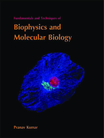
Pranav KumarFormer faculty,Department of BiotechnologyJamia Millia Islamia, New Delhi, IndiaNew Delhi, IndiaFundamentals and Techniques of Biophysics and Molecular BiologyISBN: 978-93-80473-15-4 (paperback)This book is printed on acid-free paper.Copyright 2014 by Pathfinder Publication, all rights reserved.About the cover:Visualizing intermediate filaments and the Golgi complex in bovine pulmonaryartery endothelial cells with immunofluorescence.This book contains information obtained from authentic and highlyregarded sources. Reasonable efforts have been made to publish reliable dataand information, but the author and the publisher cannot assume responsibilityfor the validity of all materials or for the consequences of their use.No part of this book may be reproduced by any mechanical, photographic, orelectronic process, or in the form of a phonographic recording, nor it may bestored in a retrieval system, transmitted, or otherwise copied for public orprivate use, without written permission from the publisher.Publisher : Pathfinder PublicationProduction editor : Ajay KumarCopy editor : Jomesh JosephIllustration and layout : Pradeep VermaCover design : Pradeep VermaMarketing director : Arun KumarProduction coordinator : Murari Kumar SinghPrinter : Ronit Enterprises, New Delhi, IndiaPathfinder PublicationA unit of Pathfinder Academy Private Limited, New Delhi, racademy.in
PrefaceThe field of biophysical and molecular biology continues to be one of the most exciting and dynamic areas of science. Over the past few decades, the spectacular progress in this field has occurreddue to the conceptual synthesis of ideas from biology, physics, chemistry, mathematics, statisticsand computer science.This textbook has the primary goal to teach students about theoretical principles and applicationsof the key biophysical and molecular methods used in biochemistry and molecular biology. I havetried to present the subject from a conceptual perspective. A substantial theoretical basis hasbeen covered to understand key experimental techniques so that students can make appropriatechoices and efficient use of techniques. There have been a number of major advances in molecularbiology in the past few years. But I have covered selected topics that provide the basic principlesfor understanding the structure and functional relationships of molecular biology.The most significant feature of this book is its clear, up-to-date and accurate explanations ofmechanisms, rather than the mere description of facts and events. The question of what to includeand what to omit is, for today’s authors, crucially important. With such a broad array of potentialtopics and techniques available, it is difficult to select those that students should experience andmaster. However, there are techniques and concepts that most of us would agree form a ‘core’ inbiophysics and molecular biology. I have tried to resist the temptation to describe more and moretechniques, adding detail but not increasing understanding of the basic concepts. I hope that thistext book will prove useful both to teachers and students. Finally, I have provided a concise list ofselected references (research papers, reviews and books) so that curious readers can trace fundamentals and ideas to their roots. These are arranged in alphabetical order.Although the chapters of this book can be read independently of one another, they are arranged ina logical sequence. Each page is carefully laid out to place related text, figures and tables near oneanother, minimizing the need for page turning while reading a topic. I have given equal importanceto text and illustrations as well.AcknowledgementsThere is an old proverb that says that you never really learn a subject until you teach it. We nowknow that you learn a subject even better when you write about it. Preparing this text has providedme with a wonderful opportunity to share our knowledge with students. Thanks go first and foremost to our students. In preparing this book, I have relied heavily on and benefited greatly fromthe advice and constructive criticism of numerous colleagues. I am particularly grateful to AjayKumar for his enthusiastic editing of the complete manuscript. I would also like to thank PrakashVardhan for invaluable contribution. This book is a team effort, and producing it would be impossible without the outstanding people of Pathfinder Publication. It was a pleasure to work with manyother dedicated and creative people of Pathfinder Publication during the production of this book,especially Pradeep Verma.Pranav Kumarwww.pathfinderacademy.inv
www.pathfinderacademy.inThis page intentionally left blank.
ReviewersThe followings, in alphabetical order, have reviewed one or more chapters, correcting errors of factor interpretation and helping to ensure they have the appropriate balance and emphasis. Theirthoughtful comments, suggestions, and encouragement have been of immense help to me in writingthis book.Ankur Roy (Associate Research scientist, Columbia University Medical Center, United States)Anup K. Biswas (Associate Research Scientist, Columbia University Medical Center, United States)Anand Yadav (Research fellow, JNU, New Delhi, India)Arvind Panday (Research fellow, Louisiana State University, United States)Deepak Singh (DST Young scientist, JNU, New Delhi, India)Lalit Kumar (Assistant professor, Department of Physics, Hindu College, Delhi University, India)Lekha Nath (Research fellow, Research Center Borstel, Germany)Maryam Imam (Post Doctoral Fellow, ICGEB, New Delhi, India)Sarad Mishra (Associate professor, Department of Biotechnology, D.D.U. Gorakhpur University, India)Usha Mina (Senior scientist, IARI, New Delhi, India)www.pathfinderacademy.invi
ContentsBiophysicsChapter 01Chromatography1Chapter 02Electrophoresis13Chapter 03Spectroscopy25Chapter 04Mass spectrometry45Chapter 05Centrifugation51Chapter 06Microscopy59Chapter 07Flow cytometry75Chapter 08X-ray crystallography85Chapter 09Patch clamp techniques90Chapter 10Immunotechniques95Chapter 11FRET and FRAP110Molecular BiologyChapter 12Molecular Biology Techniques12.1Polymerase chain reaction11812.2Nucleic acid hybridization13012.3Labeling of nucleic acids13212.4Blotting14012.5Phage display14212.6Yeast two-hybrid assay14512.7Transcript analysis14612.8DNA microarray14812.9Electrophoretic mobility shift assay15112.10Footprinting assay15212.11Site-directed mutagenesis15512.12DNA sequencing16212.13Chromatin immunoprecipitation169Self TestIndexwww.pathfinderacademy.invii
AbbreviationsμmmicrometerÅangstromA260absorbance at 260 nmAbantibodyADactivation domainADAadenosine deaminaseADPadenosine 5’-diphosphateAFLPamplified fragment length polymorphismAFMatomic force microscopyAgantigenAMPadenosine 5’-monophosphateATPadenosine 5’-triphosphatebisbisacrylamide N,N’-methylenebisacrylamidebpbase pairBrdU5-bromo-2-deoxyuridinecccDNAcovalently closed circular DNACDcircular dichroismcDNAcomplementary DNACHEFcontour-clamped homogeneous electric fieldCMcarboxymethylCNBrcyanogen bromidecpmcounts per minuteDadaltondATPdeoxyadenosine triphosphateDAPI4’,6-diamidino-2-phenylindole dihydrochlorideDBDDNA binding e triphosphateDEAEdiethylaminoethylDMSdimethyl sulfatedNTPdeoxynucleoside triphosphateELISAenzyme-linked immunosorbent assayEMSAelectrophoretic mobility shift assayERelectromagnetic radiationwww.pathfinderacademy.inix
EtBrethidium bromideFACSfluorescence activated cell sorterFISHfluorescence in situ hybridizationFITCfluorescein isothiocyanateFLIPfluorescence loss in photobleachingFRAPfluorescence recovery after photobleachingFRETfluorescence (Förster) resonance energy transferFSCforward scatterGCgas chromatographyGFPgreen fluorescent proteinGLCgas-liquid chromatographyGSCgas-solid eHGPRThypoxanthine-guanine phosphoribosyl transferaseHPLChigh performance liquid chromatographyIEFisoelectric focusingIFEimmunofixation ekcalkilocalorieKdpartition or distribution coefficientkDakilodaltonLCliquid chromatographymAbmonoclonal antibodyMALDImatrix-assisted laser desorption/ionizationMbmegabase pairMRImagnetic resonance imagingMSmass spectrometryNAnumerical aperturenmnanometerNMRnuclear magnetic resonanceORDoptical rotatory dispersionPAGEpolyacrylamide gel electrophoresisPCRpolymerase chain reactionPEphycoerythrinPFGEpulsed-field gel electrophoresiswww.pathfinderacademy.inx
PIpropidium iodidePMTphotomultiplier tubeRACErapid amplification of cDNA endsRAPDrandom amplification of polymorphic DNARCFrelative centrifugal fieldRfrelative frontRFLPrestriction fragment length polymorphismRIAradioimmunoassayRPMrevolution per minuteRT-PCRreverse transcription polymerase chain reactionSDSsodium dodecylsulfateSEMscanning electron microscopeSPsulfopropylSPRsurface plasmon resonanceSSCside scatterSTMscanning tunneling electron microscopyTaqThermus aquaticusTdTterminal deoxynucleotidyl transferaseTEMtransmission electron microscopeTEMEDN, N, N’, N’-tetramethylethylenediamineTLCthin layer chromatographyTmmelting isiblewww.pathfinderacademy.inxi
Units, Conversion factors and Physical constantsBase unitsLengthmeter (m)Masskilogram (kg)Timesecond (s)Electric currentampere (A)Temperaturekelvin (K)Amount of substancemole (mol)Luminous intensitycandela (cd)Conversion factorsMultiplication ofMillimeters (mm)Centimeters (cm)Meters (m)Kilometers 0000010000010001Length unitswww.pathfinderacademy.inxii
Volume unitsCubic centimeter (cm3)Cubic meter (m3)Liter n units1 revolution 2π radians 360 degrees1 degree 60 minutesπ radians 3.1416 radians 180 degrees1 Hz 1 cycle/sec.1 rpm (revolutions per minute) 60 rps (revolutions per second) 60Hz1 Hz (Hertz) 1 s–11 N (Newton) 1 kgm/s2 100,000 dyne1 dyne 10–5 Newton1 Pascal (Pa) 1 N/m2 6.895 kPa1 atm (metric atmosphere) 760 mm Hg at 0 C 1.0132 105 N/m21 microbar 0.1 N/m21 angstrom (Å) 10–10 mPhysical constantsIdeal gas law constant (R) 1.987 cal/mole KBoltzmann’s constant (K) 1.3 10–16 erg/K 1.3 10–23 J/KPlanck’s constant (h) 6.62 10–27 erg-sec 6.62 10–34 J.secAvogadro’s number 6.02 1023 mol–1Density of water 1 g/cm3Electron charge 1.60 10–19 coulombsElectron rest mass 9.11 10–31 kgProton rest mass 1.67 10–27 kgSpeed of light (c) 3.00 108 m/secGravitational constant (G) 6.67 10–11 Nm2/kg2Acceleration due to gravity (g) 9.8 m/s2www.pathfinderacademy.inxiii
www.pathfinderacademy.inThis page intentionally left blank.
Chapter 1ChromatographyChromatography is a physical method for separation of compounds. Tswet, Russian botanist(referred as the father of chromatography) is credited for the development of chromatography. He employed the technique to separate various plant pigments such as chlorophyllsand xanthophylls by passing solutions of these compounds through a glass column packedwith finely divided calcium carbonate. The separated species appeared as coloured bands onthe column, which accounts for the name he chose for the method (Greek chroma meaning‘colour’ and graphein meaning ‘writing’).Chromatography is based on the fact that sample distributes or partitions itself to differentextents in two different, immiscible phases, which is described by a partition or distributionThe International Unionof Pure and AppliedChemistry (IUPAC) hasdefined chromatographyas:A method, used primarilyfor separation of the components of a sample, inwhich the components arecoefficient, Kd. If we consider two immiscible phases A and B,Kd Concentration of sample in phase AConcentration of sample in phase BThe two immiscible phases could be a solid and a liquid, or a gas and a liquid or a liquid andanother liquid. One of the two phases is a stationary phase (a solid or a liquid supported ona solid) and does not move and the other is a mobile phase and moves with respect to first.The mobile phase may be a liquid (liquid chromatography) or a gas (gas chromatography).distributed between twoAll chromatographic methods involve passing a mobile phase through a stationary (immobile)phases, one of which isphase. The two phases are chosen so that the components of the sample distribute them-stationary while the otherselves between the mobile and stationary phases to varying degrees.moves. The stationaryphase may be a solid, or aClassification of chromatographic methodsliquid supported on a solid,Chromatographic methods can be classified in two fundamental ways. The first classificationor a gel. The stationaryis based on the physical means by which the stationary and mobile phases are brought intophase may be packed in acolumn, spread as a layer,contact. On this basis, chromatography is classified into column and planar chromatography.or distributed as a film,In planar chromatography, the stationary phase is present as or on a plane. The plane canetc.; in these definitionsbe a paper, serving as such or impregnated by a substrate as the stationary bed (paper chro-chromatographic bed ismatography, PC) or a layer of solid particles spread on a support e.g. a glass plate (thin layerused as a general term tochromatography, TLC). Planar chromatography is also termed open-bed chromatography. Indenote any of the differentforms in which the stationary phase may be used.thin layer chromatography, the stationary phase is a thin layer of silica gel or alumina on aglass, metal or plastic plate. Most commonly, silica gel is used as a stationary phase. In silicaThe mobile phase may begel, the silicon atoms are joined via oxygen atoms in a giant covalent structure. The othergas or liquid.commonly used stationary phase is alumina (aluminium oxide).www.pathfinderacademy.in1
www.pathfinderacademy.inPages 2 to 3 are not shown in this preview.
Rf Distance traveled by substanceDistance traveled by solvent frontNaturally the Rf can be calculated only in those instances when the solvent is not allowed toleave the end of the paper sheet.Solvent frontBROriginQAPFor B, Rf Q/RFor A, Rf P/RPaper chromatograms can be developed either by ascending or descending solvent flow.There is little difference in the quality of the chromatograms and the choice is usually a matter of personal preference. Descending chromatography has two advantages: 1. it is fasterbecause gravity aids the flow and 2. for quantitative separations of materials with very smallRf values, which therefore require long runs, the solvent can run off the paper.1.2Size exclusion chromatographySize exclusion chromatography or molecular sieve chromatography separates molecules onthe basis of size and shape. A column matrix filled with porous gel beads, made up of aninsoluble and hydrated polymer such as polyacrylamide (Sephacryl or BioGel P) or dextran(Sephadex) or agarose (Sepharose) acts as a stationary phase. Size exclusion chromatographyincludes: gel permeation chromatography and gel filtration chromatography. Gel permeationchromatography uses organic mobile solvent while gel filtration chromatography uses aqueous mobile solvent to separate and characterize molecules.MobilephaseFigure 1.2When thesample passes through theporous gel beads, smallsample molecules canenter the pores, causing them to flow slowerthrough the column. Largemolecules which cannot enter the pores, passthrough the column at afaster rate than the smaller ones. Correct pore sizesand solvents are crucial fora good separation.www.pathfinderacademy.in4Chromatography
www.pathfinderacademy.inThis page intentionally left blank.
3.The volume occupied by the packing material itself i.e. bed volume, Vg.Therefore, VT V0 Vi VgThe values of the V0 and Vi are determined experimentally by measuring the elution volumesof, respectively, a large solute that is totally excluded from the interior of the porous beadand a small solute that has access to all pores of the gel bead. The elution volume of a givensolute, Ve, is the volume of solvent required to elute the solute from the column after it hasfirst contacted the gel. The elution volume, Ve, of a solute that is partially included in thepores of the gel bead can be related to the void and internal volumes of a column by the following equation:Ve V0 σViwhere, σ is the partition coefficient of the solute.It is the partition coefficient, σ, which describes how much of the internal volume is available for the solute (0 σ 1). When σ is compared with the values measured for solutes ofknown size, it provides information about the molecular size of an unknown solute. If a seriesof solutes of known size is subjected to size exclusion chromatography, a linear relationshipbetween partition coefficient and size is observed.1.3Ion exchange chromatographyIon exchange chromatography is applicable for the separation of charged molecules. In thischromatographic technique, the stationary solid phase commonly consists of an insoluble matrix with covalently attached anions or cations (called ion exchanger). Solute ions of the opposite charge in the mobile liquid phase are attracted to the ion exchanger by electrostatic forces.Ion exchangerIon exchangers are made up of two parts – an insoluble matrix and chemically bondedcharged groups within and on the surface of the matrix. An ion exchanger is classified ascationic or anionic depending on whether it exchanges cations or anions.Cation exchanger (also called acidic ion exchanger) : It is used for cation separation.Anion exchanger (also called basic ion exchanger) : It is used for anion separation.Each type of exchanger is also classified as strong or weak according to the ionizing strengthof the functional group. An exchanger with a quaternary amino group is, therefore, a stronglybasic anion exchanger, whereas primary or secondary aromatic or aliphatic amino groupswould lead to a weakly basic anion exchanger. A strongly acidic cation exchanger containsthe sulfonic acid group.Table 1.2 Commonly used ion exchangersNameTypeFunctional groupDEAE-celluloseWeakly basicDiethylaminoethyl (DEAE)QAE-SephadexStrongly basicQuaternary aminoethyl (QAE)Q-SepharoseStrongly basicQuaternary ammonium (Q)CM-celluloseWeakly acidicCarboxymethyl (CM)SP-SepharoseStrongly acidicSulfopropyl (SP)SOURCE SStrongly acidicMethylsulphate (S)Anion exchangerCation exchangerwww.pathfinderacademy.in6Chromatography
www.pathfinderacademy.inThis page intentionally left blank.
The choice of ion exchanger for the purification of a protein largely depends on the isoelectricpoint, pI, of the protein. At a pH value above the pI of a protein, it will have a net negative chargeand adsorb to an anion exchanger. Below the pI, the protein will adsorb to a cation exchanger.pH of bufferpH pI, net negative charge, binds to anion exchangerpH pI, no net charge, no binding to ion exchanger Figure 1.5pH pI, net positive charge, binds to cation exchangerThe net charge on a protein is influenced by the pH of its solvent. At pH pI, the proteinhas zero net charge and, therefore, will not bind to a cation exchanger or an anion exchanger stationaryphase. Adjusting the pH above or below the pI of the protein will lead to a net charge and protein bindingto either an anion exchanger (pH pI) or a cation exchanger (pH pI) stationary phase.For example, if the pI is 4 then, in most cases, it is advisable to choose an ion exchangerwhich binds to the protein at a pH 4. Since at pH 4 this protein is negatively charged, theion exchanger has to be an anion exchanger, e.g. DEAE. One could also use a pH 4 and acation exchanger, but many proteins are not stable or aggregate under these conditions. If, incontrast, the protein we want to purify has a pI 10, it is positively charged at a pH around 7.Thus, in general for this protein type we have to choose a cation exchanger, which is negatively charged at neutral pH.Isoelectricpoint24681012pH rangeof stability–Figure 1.6Binds toanion exchanger}Ion exchange chromatography can also beused to separate DNAfrom a cell extract. It isbased on the interactionbetween anion exchanger(DEAE) and negativelycharged phosphates ofthe DNA backbone. Theanion-exchange resinconsists of silica beadswith a high charge density. When the cell extractpasses through the column, all the negativelycharged molecules bindto the resin and retainedin the column. If the saltsolution of gradually increasing concentration ispassed through the column, the different typesof molecule will elute inthe sequence protein,RNA and finally DNA.Net charge on protein Binds tocation exchangerDiagram shows how the net charge of a hypothetical protein changes as a function of pH.Below the isoelectric point, the molecule has a net positive charge and would be bound to a cation exchanger. Above the isoelectric point, the net charge is negative, and the protein would bind to an anionexchanger. Superimposed on this graph is the pH range of stability for the hypothetical protein. The rangeof stability refers to the pH range in which the biomolecule is not denatured. Because it is stable in therange of pH 7.0-9.0, the ion exchanger of choice is an anionic exchanger. In most cases, the isoelectricpoint of the protein is not known. The type of ion exchanger must be chosen by trial and error.1.4Affinity chromatographyAffinity chromatography is a technique enabling purification of a biomolecule with respect tobiological function or individual chemical structure. The substance to be purified is y
www.pathfinderacademy.inPages 9 to 12 are not shown in this preview.
Chapter 2ElectrophoresisElectrophoresis (Electro refers to the energy of electricity and Phoresis, from the Greek verbphoros, means to carry across) is a technique for separating or resolving charged molecules(such as amino acids, peptides, proteins, nucleotides, and nucleic acids) in a mixture underthe influence of an applied electric field. Charged molecules in an electric field move or migrate, at a speed determined by their charge to mass ratio. According to the laws of electrostatics, an ion with charge ‘Q’ in an electric field of strength ‘E’ will experience an electricforce, FelectricalFelectrical Q.EThe resulting migration of the charged molecule through the solution is opposed by a frictional forceFfrictional V.fwhere, V is the rate of migration of charged molecule and f is its frictional coefficient.Frictional coefficient depends on the size and shape of the migrating molecule and the viscosityof the medium. In constant electric field, the force on charged molecule balances each other;QE Vfso that each charged molecule moves with a constant characteristic velocity. The migrationof the charged molecule in the electric field is generally expressed in term of electrophoreticmobility (μ), which is the ratio of the migration rate of an ion to the applied electric field:μ V Q EfSo according to the equation, if two molecules have the same mass and shape, the one withthe greater net charge will move faster towards an electrode.Electrophoresis is of two types – moving boundary electrophoresis and zone electrophoresis.Moving boundary or free boundary electrophoresis is the electrophoresis in a free solution. Itwas developed by Tiselius in 1937. To separate the different charged molecules present in amixture, the sample (dissolved in a buffer solution that serves as electrolyte and maintainsthe desired pH) is placed in glass tube connected to electrodes. When an electrical potentialis applied across the tube, the charged molecules migrate toward one or the other electrode.Because different charged molecules migrate at different rates, a number of interfaces orboundaries are formed between the leading edge of each charged molecules and the remainingwww.pathfinderacademy.in13
www.pathfinderacademy.inPages 14 to 18 are not shown in this preview.
Isoelectric focusing is anelectrophoretic methodin which proteins areseparated on the basisof their pIs. It makesuse of the property ofproteins that their netcharges are determinedby the pH of their localenvironments. Proteinscarry positive, negative orzero net electrical charge,depending on the pH oftheir surroundings.The net charge of anyparticular protein is thesum of all of its positiveand negative charges.These are determined bythe ionizable acidic andbasic side chains of theconstituent amino acidsand prosthetic groupsof the protein. If thenumber of acidic groupsin a protein exceeds thenumber of basic groups,the pI of that protein willbe at a low pH value andthe protein is classifiedas being acidic. When thebasic groups outnumberthe acidic groups in aprotein, the pI will be highwith the protein classifiedas basic. Proteins showconsiderable variationIsoelectric focusing is carried out in an acrylamide gel. The gel contains ampholytes (forforming the pH gradient) together with 8M urea and a non-ionic detergent, both of whichdenature and maintain the solubility of the proteins being analysed. The denatured proteins,therefore, separate in this gel according to their isoelectric points.SDS electrophoresisIsoelectric focusing (IEF)Addition of proteinmixture in gel strip withimmobilized pH gradient410pH gradientSeparation in firstdimension (by charge)High massLoading ofgel strip onsecond gelLow massSeparation in seconddimension (by size or mass)Figure 2.7Two-dimensional gel electrophoresis. The first dimension in a 2-D gel electrophoresis experi-ment involves the separation of proteins according to their isoelectric point (pI) by isoelectric focusing(IEF). IEF works by applying an electric field to protein within a pH gradient. The proteins separate asthey migrate through the pH gradient in response to the applied voltage. When a protein reaches a pHvalue that matches its pI, its net electrical charge becomes neutral and stops migrating. In this way, eachprotein in a sample becomes focused according to its pI. The resulting gel strip is applied to an SDSpolyacrylamide gel and the proteins are separated into bands on the basis of their masses (size).Proteins that have been separated on an IEF gel can then be separated in a second dimensionbased on their size or mass. To accomplish this, the IEF gel is extruded from the tube andplaced lengthwise on a second polyacrylamide gel, this time formed as a slab saturated withSDS. When an electric field is imposed, the proteins will migrate from the IEF gel into the SDSslab gel and then separate according to their mass. The sequential resolution of proteins bytheir charge and mass can achieve excellent separation of cellular proteins.Native PAGEin isoelectric points, butpI values usually fall inthe range of pH 3-12with mostly having pIsbetween pH 4 to 7.SDS-PAGE is not used if a particular protein (e.g. an enzyme) has to be separated on theProteins are positivelycharged in solutions at pHvalues below their pI andnegatively charged abovetheir isoelectric points.Thus, at pH values belowthe pI of a particularprotein, it will migratetoward the cathode duringelectrophoresis. At pHvalues above its pI, aprotein will move towardthe anode. A protein at itsisoelectric point will notmove in an electric field.pH of the gel, proteins can be separated according to their different electrophoretic mobilities.basis of its biological activity, as the protein is denatured by the SDS-PAGE. In native gels,non-denaturing conditions are used. SDS is not used and the proteins are not denatured priorto loading. Since all the proteins in the sample being analyzed carry their native charge at the2.1ImmunoblottingSeparation of a mixture of proteins by electrophoretic techniques usually results in a complexpattern of protein bands or zones. Specific proteins can often be identified using an immunoblotting technique (also known as Western blotting). This technique requires an antibodyagainst the test protein. After the initial separation by electrophoretic technique in a gel, theproteins are transferred (or blotted) from the gel to a membrane, usually nitrocellulose orpolyvinylidene difluoride. During the transfer, the gel is at the negative electrode (cathode)side and the membrane at the positive electrode (anode) side. Proteins that are coated withnegatively charged SDS will move from the negative side, the gel, to the positive side, horesis19
www.pathfinderacademy.inPages 20 to 24 are not shown in this preview.
Chapter 3SpectroscopySpectroscopy is the study of the interaction between electromagnetic radiation and matter.The matter can be atoms, molecules or ions. The nature of the interaction between radiationand matter may include – absorption, emission or scattering. It is the absorption, emissionor scattering of radiation by matter that is used to quantitatively or qualitatively study thematter or a physical process. A study of the radiation absorbed or emitted by an atom or amolecule will give information about its identity and this technique is known as qualitativespectroscopy. Measurement of the total amount of radiation will give information about thenumber of absorbing or emitting atoms or molecules and is called quantitative spectroscopy.3.1Electromagnetic radiationElectromagnetic radiation is a form of energy and
Molecular Biology Chapter 12 Molecular Biology Techniques 12.1 Polymerase chain reaction 118 12.2 Nucleic acid hybridization 130 12.3 Labeling of nucleic acids 132 12.4 Blotting 140 12.5 Phage display 142 12.6 Yeast two-hybrid assay 145 12.7 Transcript analysis 146 12.8 DNA microarray 148 12.9 Electrophoretic mobility shift assay 151