Transcription
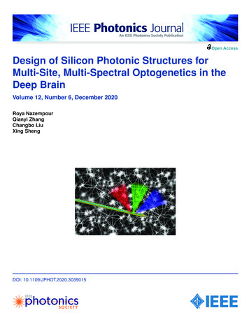
Open AccessDesign of Silicon Photonic Structures forMulti-Site, Multi-Spectral Optogenetics in theDeep BrainVolume 12, Number 6, December 2020Roya NazempourQianyi ZhangChangbo LiuXing ShengDOI: 10.1109/JPHOT.2020.3039015
IEEE Photonics JournalDesign Of Silicon Photonic StructuresDesign of Silicon Photonic Structures forMulti-Site, Multi-Spectral Optogenetics inthe Deep BrainRoya Nazempour,1 Qianyi Zhang,1 Changbo Liu,2and Xing Sheng 11 Department of Electronic Engineering, Beijing National Research Center for InformationScience and Technology, Center for Flexible Electronics Technology, Tsinghua University,Beijing 100084, China2 School of Materials Science and Engineering and Hangzhou Innovation Institute, BeihangUniversity, Beijing 100191, ChinaDOI:10.1109/JPHOT.2020.3039015This work is licensed under a Creative Commons Attribution 4.0 License. For more information, uscript received November 5, 2020; accepted November 15, 2020. Date of publication November18, 2020; date of current version December 9, 2020. This work was supported in part by the NationalNatural Science Foundation of China under Grant 61874064, in part by Beijing Municipal NaturalScience Foundation under Grant 4202032, and in part by Beijing Innovation Center for Future Chips.(R. Nazempour and Q. Zhang contributed equally to this work.) Corresponding author: Xing Sheng(e-mail: xingsheng@tsinghua.edu.cn).Abstract: Micro- and nanoscale photonic structures and devices play important roles inthe development of advanced biophotonic systems, in particular, implantable light sourcesfor optogenetic stimulations. In this paper, we numerically investigate silicon (Si) photonicsbased microprobes that can achieve multi-site, multi-spectral optical excitation in the deepanimal brain. On Si substrates, silicon nitride (Si3 N4 ) based planar waveguides can delivervisible light in the deep tissue with low losses, and couple to grating emitters diffractinglight in targeted brain regions. In our model, we combine near-field wave optic and far-fieldray tracing simulations, showing that the designed photonic structures spectrally split blue,green and red photons into different locations in the tissue. Furthermore, by introducingdual grating components, photons at different wavelengths can be spatially separated atdifferent depths. Therefore, these photonic probes can be used to selectively activate orinhibit specific neurons and nuclei, when expressing various corresponding light sensitiveopsins. We anticipate that such device strategies can find wide applications in the design ofadvanced implantable photonic systems for neuroscience and neuroengineering.Index Terms: Implantable device, silicon photonics, optogenetics.1. IntroductionOptogenetics plays an indispensable role in the field of neuroscience and neuroengineering fordecoding neural signals and modulating neural activities, which could enrich understandings inbrain functions and help discover novel strategies for neurological disease treatments [1], [2]. Suchoptically based neural interrogation methods employ various light-sensitive microbial opsins thatexhibit different excitation spectra and control different ion channels, providing precise controlover specific neural activities with light modulations at certain wavelengths [3], [4]. As excitationspectra of most opsins typically lie within the visible range, where light penetration is severelylimited by the tissue scattering and absorption, advanced waveguides and optoelectronic devicesVol. 12, No. 6, December 20204200107
IEEE Photonics JournalDesign Of Silicon Photonic Structureshave been exploited for effective light delivery into the deep tissue [5], [6]. Most commonly usedlight-guiding method for optogenetics is based on optical fibers made of silica [7], [8] owing to its lowcost, high optical transparency and structural stability in biological environments. However, simplesilica fibers cannot provide versatile functions like spatially and spectrally resolved light deliveryin the tissue, and the high mechanical stiffness of silica often causes unwanted tissue damagesand inflammation. Alternative strategies are also proposed, based on polymer waveguides, whichexhibit better biocompatibility and even dissolvability within the body [9]–[11]. Recently developedthin-film, microscale optoelectronic devices that can be directly implanted into the animal brainprovide alternative promising solutions to advanced optical neural interfaces. By integrating microscale light-emitting diodes (LEDs), photodetectors, and various electronic and chemical sensorsonto flexible substrates, as well as wirelessly operated circult modules, untethered, multifunctionalneural probes can be realized [12]–[15]. Nevertheless, challenges associated with electrical heatingand device’s long-term stability within the tissue remain daunting for these directly implanted activeemitters and sensors [16]. Apart from above solutions, planar waveguides based on silicon (Si)and related materials (silicon oxide SiO2 , silicon nitride Si3 N4 , etc.) offer an emerging method forimplantable light sources within the animal body. Taking advantage of mature micro- and nanofabrication techniques recently developed for silicon photonics in the past decade, sophisticatedoptical components like waveguides, splitters, resonators and couplers, can be fine-tuned to realizeminiaturized, compact structures used as implantable optical sources for various functionalities.Remarkable examples include the developments of Si photonics based waveguides, cavities andswitchable grating couplers for sensing biomarkers [17], intracranial pressure and temperature [18],and multi-site optogenetic stimulations [19], [20].With further explorations of the complex neural activities associated to different cell types andbrain regions [21], multi-site, multi-spectral light-guiding techniques become crucial in optogeneticstudies. By expressing different types of opsins in specific neural cells and brain regions, andemploying stimulations by photons with corresponding wavelengths, simultaneous, bi-directionalmodulations of neural activities for different cells and brain regions are becoming possible. Torealize this vision, the design of light emitters for multi-site and multi-spectral stimulation is highlydemanded. Recent works have achieved such multi-site stimulation functionalities using multipointemitting optical fibers [22], fiber bundles [23], micro-LEDs [24] or nanophotonic waveguides [19],[25], and yet only few research focuses on light-guiding systems combining both multi-site andmulti-spectral stimulation [26].In this paper, we propose implantable waveguide structures with grating emitters that can realizemulti-site and multi-spectral optical stimulations in the deep brain. Enabled by silicon photonicstechnology, Si3 N4 based thin-film waveguides with low losses and diffractive gratings with highcoupling efficiencies can be numerically designed and applied to spectrally split emitted photonsinto different locations when implanted into the brain tissue. Modeling results reveal that outcoupledlight with different colors (blue, green and red) can be directed at different emissive angles, anddistributed into different tissue locations in the far field accordingly. Furthermore, we also proposea photonic structure that is able to spatially separate blue and red photons and deliver theminto different regions via the waveguide. The designed structures provide guidelines for advancedoptical neural interfaces, and offer new opportunities to optogenetic interrogations of complex brainfunctions.2. Results and DiscussionsFig. 1 illustrates the conceivable design of the implantable photonic probe that can deliver photonsof different wavelengths into the animal brain. Based on a silicon substrate, the designed Si3 N4waveguide provides high transparency in the visible spectral range, with a grating emitter thatdiffracts photons at different wavelengths at various angles into the brain tissue.Fig. 2(a) displays the schematic structures of the waveguide and the grating coupler on the probetip. The Si3 N4 waveguide has a width of 20 μm and a thickness of 0.2 μm, embedded in SiO2 . Thethicknesses of the top and the bottom SiO2 layers are 0.66 μm and 5 μm, respectively. The gratingVol. 12, No. 6, December 20204200107
IEEE Photonics JournalDesign Of Silicon Photonic StructuresFig. 1. Conceptual sketch of an implanted waveguide with grating based emitters for multi-site, multicolor optogenetic stimulations in the mouse brain.Fig. 2. Optical simulations of light outcoupling for the designed grating emitter, based on the finitedifference time-domain (FDTD) method. (a) Schematic illustration of the waveguide and gratingstructures, showing that the outcoupled light is spectral split towards different emissive angles. (b)Normalized, near-field electric field distributions around the grating emitter at wavelengths of 450 nm(top), 550 nm (middle) and 620 nm (bottom), respectively. Arrows indicate the propagation directionsof the diffracted beams. (c) Far-field emission intensities at various wavelengths and emissive angles.(d) Far-field, angular dependent emission profiles for 3 different wavelengths: 450 nm (blue), 550 nm(green) and 620 nm (red).emitter has a period 0.26 μm, with a 50:50 duty cycle (fill factor F 0.5) and 20 periods.The entire structure is based on a silicon substrate with a thickness less than 50 μm, and can beformed using standard microfabrication processes, including chemical vapor deposition, reactiveion etching, photolithography, wafer thinning, etc. Finite-difference time-domain (FDTD) simulations[27] are utilized to analyze the wave propagation and beam diffraction properties. Optical propertiesof Si, SiO2 and Si3 N4 are quoted from literatures [28], [29]. SiO2 and Si3 N4 have refractive indicesnSiO2 1.46 1.47 and nSi3N4 2.04 2.08, respectively, and both are transparent across thevisible spectrum. The surrounding environment is assumed to be a biological tissue with a refractiveindex of 1.36. Since the grating width is much larger than the grating period, a two-dimensionalFDTD model is applied to reduce the running time. A source of plane wave from 400 nm to 700 nmin transverse magnetic (TM) mode is coupled to the waveguide. The waveguide loss is calculatedto be less than 1 dB/cm. When the wave reaches the grating, light beams are scattered into thetissue, with the first-order diffractive angle θ . Fig. 2(b) presents the profiles of electric field withinsuch a photonic structure at specific wavelengths λ 450 nm, 550 nm and 620 nm. Arrows in thegraphs indicate the diffractive directions upward into the tissue. The first-order diffractive angle θcan also be calculated analyticallynSiO2 · sin θ neff Vol. 12, No. 6, December 2020λ (1)4200107
IEEE Photonics JournalDesign Of Silicon Photonic StructuresFig. 3. Optical simulations of light emission from the designed grating emitter to the brain tissue, basedon the FDTD results in Fig. 2(d) and the ray tracing method, at various wavelengths: (a) 450 nm; (b) 550nm; (c) 620 nm, and (d) merged results for the 3 wavelengths, illustrating the spectral splitting effectsin the tissue. Iso-intensity lines show 10% and 1% of the maximum power.where the effective index of the grating neff is defined asneff F · nSi3N4 (1 F ) · nSiO2(2)For blue (450 nm), green (550 nm) and red (620 nm) photons, diffractive angles of this grating aredetermined to be 3.4 , 21.4 and 35.3 , respectively. The corresponding diffraction efficienciesare extracted from the FDTD models, which are 26.3%, 40.7% and 43.5%. It should be noted thatalmost half of the diffractive waves go downwards and absorbed by the Si substrate, and the resultscan be further optimized, for example, by applying a metal mirror or a Bragg reflector betweenthe Si substrate and the SiO2 cladding layer. Angular-dependent, far-field diffraction intensitydistributions can be further determined, as plotted in Fig. 2(c). Fig. 2(d) presents the results for thethree representative wavelengths (450 nm, 550 nm and 620 nm). In agreement with calculationsbased on Eq. (1), the first-order diffractive angle increases with the wavelength from 450 nm to700 nm, and diffraction beams are highly collimated ( 1.6 for 450 nm, 2.2 for 550 nm, and 2.6 for 620 nm). Such a design presents much smaller divergence compared with conventionallight-guiding methods based on implantable fibers ( 10 ) and microscale LEDs ( 120 ); therefore,it allows light beams to be more efficiently focused on targeted nuclei or regions.To predict the performance of the aforementioned waveguides and grating emitters within thebrain tissue, models based on the Monte-Carlo ray tracing method [30] are established, with resultspresented in Fig. 3. The area of the grating emitter is set to be 20 μm 20 μm, within a tissueassumed to be the grey matter of animal brains (refractive index 1.36). Results at the same threewavelengths (450 nm, 550 nm and 620 nm) as those used in Fig. 2 are modeled. Scattering andabsorption coefficients at these wavelengths are (13.13, 8.7 and 7.2 /mm), and (0.38, 0.28, and0.10 /mm), respectively [28], and the anisotropy of scattering is assumed to be 0.85 (highly forwardscattering). Angular-dependent emission profiles are collected from the simulated far-field resultsshown in Fig. 2(d), of which the emission peaks at diffractive angles of 3.4 , 21.4 and 35.3 ,respectively. A total of 105 rays are employed to simulate each wavelength. Figs. 3(a)–3(c) illustrateoptical propagations at the three distinct wavelengths, and one can observe the light attenuationassociated with the scattering and absorption within the tissue. Penetration depths (at which theoptical intensity falls to 1/e 0.368 of the original value) are estimated to be 126 μm, 155 μmand 182 μm, for blue (450 nm), green (550 nm) and red (620 nm) photons, respectively. Fig. 3(d)further depicts optical emissions from the three distinct colors, which clearly reveals that photonsat different wavelengths can be split into different parts of the brain tissue. The emission of eachcolored beam can cover a specifically targeted nucleus, which typically range from tens to hundredsof micrometers in the mammalian brain. By expressing light sensitive opsins (for example, ChR2 forblue, C1V1 for green and Chrimson for red [31]) in specific neurons at different locations, we couldrealize optical excitation and inhibition of different cell types, thereby modulating neural activitieswith more freedom compared to previous devices based on monochromatic stimulation.Vol. 12, No. 6, December 20204200107
IEEE Photonics JournalDesign Of Silicon Photonic StructuresFig. 4. Design of a grating emitter that delivers blue and red light at different locations. (a) Schematicillustration of the double grating coupler. (b) Normalized, near-field electric field distributions around thegrating coupler at wavelengths of 450 nm (top) and 620 nm (bottom), respectively. Arrows indicate thepropagation directions of the diffracted beams. (c) Far-field, angular dependent emission profiles forthe two wavelengths: 450 nm (blue) and 620 nm (red). (d) Ray tracing results showing spectrally splitemissions from the double grating coupler to the brain tissue. The distance between the two gratings isassumed to be around 100 μm. Iso-intensity lines show 10% and 1% of the maximum power.By leveraging the spectral-dependent diffraction properties of the grating emitter, we can furthercreate a waveguide structure that delivers multispectral photons at different depths into the braintissue. As shown in Fig. 4(a), the photonic structure consists of two gratings emitting blue (450 nm)and red (620 nm) light, respectively, which are connected by a planar waveguide and based on thesame material system as in Fig. 2(a). The first grating (on the left) is designed to have a periodof 163 nm (20 periods) and a duty cycle of 60:40 (Si3 N4 :SiO2 ). Based on Eq. (1), its first-orderdiffractive angle for blue photons (450 nm) can be obtained to be 50.3 with beam divergence of4.0 and its diffraction efficiency is 42.0%, while red photons (620 nm) can transmit through it withminimum scattering. The transmission efficiencies for 405 nm and 620 nm are 16.0% and 95.0%.The second grating (on the right) has a period of 406 nm (10 periods) and a duty cycle of 50:50,which outcouples red photons (620 nm) at a diffractive angle of 3.3 with beam divergence of 2.7 and its diffraction efficiency is 43.9%. Fig. 4(b) shows simulated electric field distributions of sucha double grating structure at 450 nm and 620 nm, based on the FDTD method. To reduce thesimulation time, the planar waveguide between the two gratings has a length of 4.8 μm. Far-field,angular-dependent emission profiles are plotted in Fig. 4(c). We further evaluate the light emissionproperties of such a double grating coupler within the brain tissue based on the Monte-Carlo raytracing method (Fig. 4(d)). Different from the FDTD simulations, the distance between the twogratings is increased to 100 μm. In practice, the grating separation can be tuned based on thelocations of targeted brain regions, which will not affect the results, since the waveguide loss isnegligible within a few millimeters. The spatially separated photon emissions clearly demonstratethat brain regions at different positions can be targeted, by expressing corresponding opsins in thespecific cells and nuclei.3. ConclusionTo summarize, here we present the numerical design of silicon photonic structures that can forman implantable optical neural interface and realize multi-site, multi-spectral stimulation of neural activities. Moving forward, the fabrication of this nanophotonic device is compatible with the standardcomplementary metal oxide semiconductor (CMOS) process. The application of such Si basednanophotonic technologies permits the molding of light flow with high spatial-temporal resolutions,low divergence and versatile adjustability for optical neural modulations. Another unique feature ofsuch thin-film Si based implants stems from the biocompatibility and aqueous degradation of Sirelated materials [17] showing the promise of physical transience in a controlled manner in vivo[32]. Other explorable applications include periscleral crosslinking [33] and phototherapy [34], [35],as demonstrated previously using polymer fibers. Additionally, reconfigurable photonic structuresVol. 12, No. 6, December 20204200107
IEEE Photonics JournalDesign Of Silicon Photonic Structuresbased on electronically, thermally, and/or mechanically activated devices could be incorporated toobtain tunable emission wavelengths or angles. The system platform also allows the integration withother electronic and photonic components, such as recording electrodes [24], microscale LED anddetectors [36], and even microfluidic channels [37] and electrochemical sensors [14]. The resultspresented here show promising paths to utilize the-state-of-the-art nanophotonic technologies foradvanced neural stimulators and medical sensors in general.References[1] K. M. Tye and K. Deisseroth, “Optogenetic investigation of neural circuits underlying brain disease in animal models,”Nat. Rev. Neurosci., vol. 13, no. 4, pp. 251–266, Apr. 2012.[2] H. Zhang and A. E. Cohen, “Optogenetic approaches to drug discovery in neuroscience and beyond,” TrendsBiotechnol., vol. 35, no. 7, pp. 625–639, Jul. 2017.[3] K. Deisseroth, “Optogenetics: 10 years of microbial opsins in neuroscience,” Nat. Neurosci., vol. 18, no. 9,pp. 1213–1225, Sep. 2015.[4] N. C. Klapoetke et al., “Independent optical excitation of distinct neural populations,” Nat. Methods, vol. 11, no. 3,pp. 338–346, Mar. 2014.[5] S. B. Goncalves, J. F. Ribeiro, A. F. Silva, R. M. Costa, and J. H. Correia, “Design and manufacturing challenges ofoptogenetic neural interfaces: A review,” J. Neural Eng., vol. 14, no. 4, Aug. 2017, Art. no. 041001.[6] H. Xu, L. Yin, C. Liu, X. Sheng, and N. Zhao, “Recent advances in biointegrated optoelectronic devices,” Adv. Mater.,vol. 30, no. 33, Aug. 2018, Art. no. 1800156.[7] L. A. Gunaydin et al., “Natural neural projection dynamics underlying social behavior,” Cell, vol. 157, no. 7,pp. 1535–1551, Jun. 2014.[8] A. G. Mignani and F. Baldini, “In-vivo biomedical monitoring by fiber-optic systems,” J. Lightw. Technol., vol. 13, no. 7,pp. 1396–1406, Jul. 1995.[9] A. Gierej et al., “On the characterization of novel step-index biocompatible and biodegradable poly(D,L-lactic acid)based optical fiber,” J. Lightw. Technol., vol. 38, no. 7, pp. 1905–1914, Apr. 2020.[10] R. Fu et al., “Implantable and biodegradable Poly(l-lactic acid) fibers for optical neural interfaces,” Adv. Opt. Mater.,vol. 6, no. 3, Feb. 2018, Art. no. 1700941.[11] R. Nazempour, Q. Zhang, R. Fu, and X. Sheng, “Biocompatible and implantable optical fibers and waveguides forbiomedicine,” Materials, vol. 11, no. 8, Aug. 2018, Art. no. 1283.[12] L. Li et al., “Heterogeneous integration of microscale GaN light-emitting diodes and their electrical, optical, and thermalcharacteristics on flexible substrates,” Adv. Mater. Technol., vol. 3, no. 1, Jan. 2018, Art. no. 1700239.[13] Y. Zhao et al., “Wirelessly operated, implantable optoelectronic probes for optogenetics in freely moving animals,” IEEETrans. Electron Devices, vol. 66, no. 1, pp. 785–792, Dec. 2018.[14] C. Liu et al., “A wireless optoelectrochemical probe for optogenetics stimulation and dopamine detection,” Microsyst.Nanoeng., vol. 6, 2020, Art. no. 64.[15] T. Kim et al., “Injectable, cellular-scale optoelectronics with applications for wireless optogenetics,” Science, vol. 340,no. 6129, pp. 211–216, Apr. 2013.[16] P. S. Yarmolenko et al., “Thresholds for thermal damage to normal tissues: An update,” Int. J. Hyperthermia, vol. 27,no. 4, pp. 320–343, Jun. 2011.[17] W. Bai et al., “Bioresorbable photonic devices for the spectroscopic characterization of physiological status and neuralactivity,” Nat. Biomed. Eng., vol. 3, no. 8, pp. 644–654, Aug. 2019.[18] J. Shin et al., “Bioresorbable optical sensor for monitoring of interacranial pressure and temperature,” Sci. Adv., vol. 5,no. 7, Jul. 2019, Paper eaaw1899.[19] A. Mohanty et al., “Reconfigurable nanophotonic silicon probes for sub-millisecond deep-brain optical stimulation,” Nat.Biomed. Eng., vol. 4, no. 2, pp. 223–231, Feb. 2020.[20] B. Li, K. Lee, S. C. Masmanidis, and M. Li, “A nanofabricated optoelectronic probe for manipulating and recordingneural dynamics,” J. Neural Eng., vol. 15, no. 4, pp. 046008.1–046008.8, May 2018.[21] G. Buzsáki et al., “Tools for probing local circuits: High-density silicon probes combined with optogenetics,” Neuron,vol. 86, no. 1, pp. 92–105, Apr. 2015.[22] F. Pisanello et al., “Multipoint-emitting optical fibers for spatially addressable in vivo optogenetics,” Neuron, vol. 82,no. 6, pp. 1245–1254, Jun. 2014.[23] Y. Sych, M. Chernysheva, L. T. Sumanovski, and F. Helmchen, “High-density multi-fiber photometry for studying largescale brain circuit dynamics,” Nat. Methods, vol. 16, pp. 553–560, Jun. 2019.[24] F. Wu, E. Stark, P. C. Ku, K. D. Wise, G. Buzsáki, and E. Yoon, “Monolithically integrated μLEDs on silicon neuralprobes for high-resolution optogenetic studies in behaving animals,” Neuron, vol. 88, no. 6, pp. 1136–1148, Dec. 2015.[25] Y. Son et al., “In vivo optical modulation of neural signals using monolithically integrated two-dimensional neural probearrays,” Sci. Rep., vol. 5, no. 1, Oct. 2015, Art. no. 15466.[26] K. Kampasi et al., “Dual color optogenetic control of neural populations using low-noise, multishank optoelectrodes,”Microsyst. Nanoeng., vol. 4, no. 1, Jun. 2018, Art. no. 10.[27] A. Taflove and S. C. Hagness, Computationai Electrodynamics. Norwood, MA, USA: Artech House, Jun. 2000.[28] E. S. Gebhart, W. Lin, and A. Mahadevan-Jansen, “In vitro determination of normal and neoplastic human brain tissueoptical properties using inverse adding-doubling,” Phys. Med. Biol., vol. 51, no. 8, Mar. 2006, Art. no. 2011.[29] K. Luke, Y. Okawachi, M. R. Lamont, A. L. Gaeta, and M. Lipson, “Broadband mid-infrared frequency comb generationin a Si3 N4 microresonator,” Opt. Lett., vol. 40, no. 21, pp. 4823–4826, Nov. 2015.Vol. 12, No. 6, December 20204200107
IEEE Photonics JournalDesign Of Silicon Photonic Structures[30] S. L. Jacques and L. Wang, Monte Carlo Modeling of Light Transport in Tissues. Berlin, Germany: Springer U.S., 1995.[31] H. Yawo, H. Kandori, and A. Koizumi, Optogenetics: Light-sensing proteins and Their Applications. Berlin, Germany:Springer, Jun. 2015.[32] S. W. Hwang et al., “A physically transient form of silicon electronics,” Science, vol. 337, no. 6102, pp. 1640–1644,Sep. 2012.[33] S. J. J. Kwok et al., “Flexible optical waveguides for uniform periscleral cross-linking,” Invest. Ophthalmol. Vis. Sci.,vol. 58, pp. 2596–2602, 2017.[34] S. Nizamoglu et al., “Bioabsorbable polymer optical waveguides for deep-tissue photomedicine,” Nat. Commun., vol. 7,Jan. 2016, Art. no. 10374.[35] Y. Jiang et al., “Green light-based photobiomodulation with an implantable and biodegradable fiber for bone regeneration,” Small Methods, vol. 4, no. 7, Jul. 2020, Art. no. 1900879.[36] L. Lu et al., “Wireless optoelectronic photometers for monitoring neuronal dynamics in the deep brain,” Proc. Nat.Acad. Sci., vol. 115, no. 7, pp. E1374–E1383, Feb. 2018.[37] J. W. Jeong et al., “Wireless optofluidic systems for programmable in vivo pharmacology and Optogenetics,” Cell,vol. 162, no. 3, pp. 662–674, 2015.Vol. 12, No. 6, December 20204200107
IEEE Photonics Journal Design Of Silicon Photonic Structures Fig. 1. Conceptual sketch of an implanted waveguide with grating based emitters for multi-site, multi-color optogenetic stimulations in the mouse brain. Fig. 2. Optical simulations of light outcoupling for the designed grating emitter, based on the finite-