Transcription
Gene. '59 (1987) 19-28Elsevier19 .GEN 02133Molecular cloning of the genome of human spumaretrovirus(Foamy virus; reverse transcriptase; recombinant DNA; AIDS; HIV; long terminal repeat; embryonie lung)A. Rethwilma, G. Daraib, A. Rösenb, B. Maurer 8 and R.M. Flügel 8InstituteofVirus Research, German Cancer Research Center, 6900 Heidelberg(F.R.G.)and 6 Institutfür Medizinische Virologieder Universität, 6900 Heide/berg (F.R. G.) Tel. (6221)56-3008aReceived 25 July 1987Accepted 25 August 1987SUMMARYDNA ofhuman spumaretrovirus (HSRV) was cloned from both cDNA and from viral DNA into phage A.and bacterial plasmid vectors. The recombinant plasm.ids harboring viral DNA were characterized by Southemblot hybridization and restriction mapping. Physical maps were constructed from cDNA and found to becolinear with the restriction maps obtained from viral DNA. The recombinant clones isolated contained viralDNA inserts which rangein size from 2.2 kb to 15.4 kb. The recombinant clones allowed to construct a physicalmap of the complete HSRV provirus of 12.2 kb.INTRODUCTIONSpumaretroviruses (foamy viruses) have beenknown for many years. They have been isolated frommany species including man (Hooks and Hooks,1981). It was particularly D.C. Gajdusek and bisgroup who succeeded in isolating many simianfoamy viruses during their classical experiments onCo"espondence to: Dr. R.M. Flügel, Institut für Virusforschung,Deutsches Krebsforschungszentrum, Im Neuenheimer Feld 280,6900 Heidelberg (F.R.G.) Tel. (6221)484-611.Abbreviations: bp, base pair(s) cDNA, DNA complementary toviral RNA; dNTP, deoxynucleoside triphosphate; EtdBr,ethidium bromide; HEL, human embryonie lung; HIV, humanimmunodeticiency virus; HSRV, human spumaretrovirus; kb,kilobase(s) or 1000 bp; LTR, long terminal repeat; nt, nucleotide(s); SOS, sodium dodecyl sulfate; SSC, 0.15 M NaCI, 0.015M Na 3 citrate, pH 7.0; TKM, 0.05 M Tris · HCI, 0.025 M KCI,0.0025 M MgC1 2 , pH 7.4.Kuru and slow virus infections (Rogers et al., 1967;Gajdusek, 1977). Epidemiological data indicate anatural prevalence of foamy virus infection amongmen in East Africa (Muller et al., 1980). Foamyviruses have been found in patients with variousdiseases (Weiss et al., 1982). In cell culture, foamyviruses replicate only poorly, but cause a cytopathogenic effect with multinucleated giant-cell fonnationconcomitant with typical vacuolization in a widerange of cells (Hooks and Hooks, 1981 ). They havebeen classified as a third subfamily of retroviridaeand termed spumaviruses. Since the molecularbiology of these viruses is virtually unknown, wemolecularly cloned spumaretroviral DNA. As starting material for cloning, either DNA complementaryto the HSRV genome or viral DNA from the genomicDNA ofHSRV-infected human cells was prepared.The molecular clones of HSRV DNA will facilitatefurther studies on the structure and function ofHSRV genome and its gene products.0378-1119/87/ 03.50 1987 Elsevier Science Publishers B.V. (Biomedical Division)
20MATERIALS AND METHODS(a) Cells and virusesHEL cells were grown as described (Flügel et al.,1987) and HSRV.was kindly provided by Dr. P. Loh(Loh and Ang, 1981 ). Virus was propagated byrnixing infected HEL cells with uninfected growingHEL cells at a ratio of 1: 4 (Neumann-Haefelinet al., 1983). For in vitro labeling ofviral RNA, 16.5J.LCi [ 3 H]uridine (spec. act. 38 Cijmmol) per ml ofculture medium was added. HSRV virions wereclarified from cell supernatants by ultracentrifugation on days 2-5 after infection when the cytopathiceffect was almost complete. Virus was resuspendedin TKM buffer (Obara et al., 1971) and stored at-20 o C until further processing.(b) Virus purification and synthesis ofHSRV cDNAHSRV was purified by centrifuging twice througha 20-60% sucrose gradient in TKM butTer for 4 hat 100000 x gin a SW41 rotor (Beckmann). Densityfractions of 1.155-1.175 g/ml shown to have maximum reverse transcriptase activity and in addition,to contain 3 H-labeled RNA, were used for synthesisof viral cDNA.For frrst-strand synthesis the method of Rotbenberg and Baltimore (1976) was modified. Thereaction volume of500 J.Ll contained 50 mM Tris (pH8.3), 8 mM MgC1 2 , 0.4 mM MnC12 , 100 mM KCI,1 mM dithiothreitol, 1 mM of each dNTP, 0.01%NP40, 75 J.Lg/ml oligo(dT) 12 primer (BoehringerMannheim), and 30 J.Lg/ml oligodeoxynucleotideprimers (provided by Dr. John Taylor) and anappropriate amount of virus. The reaction wascarried out at 37 C for 20 h. Aftertreatment withSDS and proteinase K the primary product wasextracted with phenol-chloroform, and precipitatedwith ethanol. For synthesis of the second strand theprocedure of Gubler and Hoffmann (1983) wasfollowed. The fmal product was labeled according toRigby et al. {1977), using 100 J.LCi each of the four( cx- 32 P]dNTPs, and used as probe for molecularhybridization.(c) Molecular cloning of riral cDNAStock-frozen viruswas used directly for synthesisof double-stranded HSRV cDNA. In two separatesets of experiments, cD NA was cut either withEcoRI or with Hindlll, Iigated to the correspondingsites of the phage vector A.NM 1149 (Murray, 1983)and packaged (Hohn, 1979). Recombinant phageswere selected by plating on Escherichia coli POP13b(Murray, 1983). HSRV-positive clones were identified by filter hybridization using [ 32 P]cDNA madefrom sucrose-gradient-purified virions as describedabove.Out of 5000 recombinant clones, 180 hybridized to32[P]cDNA when using EcoRI-cut cDNA. Cloningefficiency using Hindlll-digested cDNA was 10%{five out of 50 recombinant clones hybridized).DNAs from clones A.HSRV-EcoRI 1 to 30 thatgavestrong hybridization signals, and DNA from clonesA.HSRV-Hindlll 1 to 5 that hybridized positivelywere prepared according to standard protocols(Maniatis et al., 1982). Insert DNAs from clonesA.HSRV-EcoRI 1 to 30 were purified by 0.8% agarose gel electrophoresis and recloned into the Eco RIsites of the plasmid vector pACYC184 (Chang andCohen, 1978). Selection, amplification, and purification of the recombinants were carried out as described previously (Maniatis et al., 1982). HSRVpositive clones were identified by hybridization usingseparately synthesized [3 2 P]cDNA as probe. Theplasmid clone pyHSRV-E-02 was characterized indetail. DNA inserts from clones A.HSRV-Hindlll 1to 5 were inserted into the Hindill sites ofthe vectorpAT153 {Twigg and Sheratt, 1980), [ 32P]cDNA wasused to identify HSRV-positive clones, and clonepHSRV-H-C55 was further characterized.(d) Molecular cloning of viral DNABamHI-digested high-Mr DNA (0.5 J.Lg) fromHSRV-infected HEL cells was Iigated with BamHIdigested DNA from vector A.EMBL4 (Frischaufet al., 1983). DNA was packaged and recombinantphages were plated on E. coli NM539 (Frischaufet al., 1983 ). Out of 3 x 10⁴ recombinant clones thatwere screened, two recombinant clones were identified to bear HSRV-specific sequences by filterhybridization with nick-translated DNA frompyHSRV-E-02. The two recombinant phage cloneswere plaque-purified and re-screened. Insert DNA(15.4 kb) was extracted from low-melting-pointagarose and recloned into the BamHI sites of theplasmid vector pSP65 (Melton et al., 1984) and the
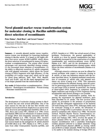
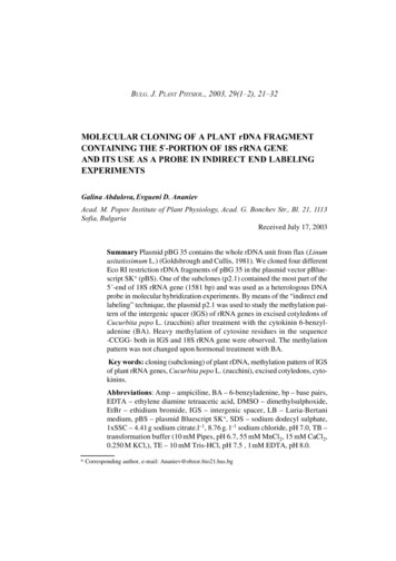
21clone designated pSPHSRV-B-C11 was characterized in detail.(e) Analysis of recombinant DNAONAs were cleaved with different restrictionendonucleases and were separated by agarose gelelectrophoresis. Fragments were transferred to nitrocellulose according to Southem (1975). IndividualONAs (0.5 Jlg) were labeled as described by Rigbyet al. (1977) using 1.6 mCi/ml of [cx- 32 P]dCTP and[ cx- 32P]dATP (6000 Cijmmol from Amersham).Hybridization was carried out in 50% formamideand 5 x SSC at 42 C for 24-48 h. Filters werewashed 3 x 30 min in,2 x SSC and 0.5% SOS at68 C.RESULTS AND DISCUSSION(a) Molecular cloning of cDNA complementary toHSRVRNAS pumaretrovirus particles were prepared fromHEL cell supematants, purified by sucrose gradientcentrifugations, and directly used for synthesis ofviral cONA as described in MATERIALS ANDMETHODS, section b. To clone HSRV cDNA, it wasdigested with Eco RI and the fragments were Iigatedto EcoRI-cleaved A.NM1149 DNA (Murray et al.,1983), packaged into phage envelopes and plated onE. coli. Plaques were screened by fdter hybridizationusing separately synthesized HSRV [ 32 P]c0NAfrom sucrose-gradient-purified virions as hybridization probe. Out of 5000 recombinant clones, 180clones were identified as positive. Several of thepositive recombinant clones were cleaved withEcoRI, and the insert DNA was recloned into theEcoRI sites of the plasmid vector pACYC184 andfound to hybridize to [l 2 P]cDNA (Fig. 1). Theresulting recombinant plasmid clones were termedpyHSRV-E-02, 03,04 and 06. One ofthem, 02,was characterized in detail by restriction enzyme andSouthem blot analysis (clone 2 in Fig. 3).Another series of recombinant clones wereobtained in an analogous way by using the Hindillinstead ofthe EcoRI enzyme to cleave viral cDNAsynthesized in vitro. Mter ligation and packaging,Fig. 1. Southern blot hybridization of different recombinantplasmids to 32 P-labeled cDNA of the HSRV genome. DNAsfrom recombinant plasmids constructed from viral DNA (lanes1 and 2) and from cDNA (lanes 3 to 9) were cleaved withrestriction endonucleases BamHI (lane I, pHSRV-B-34), C/ai(lane 2, pHHRV-B-52), EcoRI (lane 3, pyHSRV-E-C5),EcoRI Hindill (lane 4, pyHSRV-E-C8), and EcoRI (lanes 5to 9, pyHSRV-E-02, D3, D4, D6, and F8, respectively), andseparated electrophoretically on 0.8% agarose gels. Phage A.DNA (0.25 J.L8 unlabeled and 0.02 J.Lg 32P-Iabeled) cleaved withEcoRI served as marker (lanes M). (A) EtdBr staining; (8)autoradiogram ofthe same gel after hybridization to 32P-labeledcDNA. Sizes are given in kb on the margins. The C/ai cleavagepattern of B-52 DNA (lane 2) is the result of a partial digestion,with fragment sizes of 5.5 kb (thick arrow) and 2.3 kb (thinarrow).
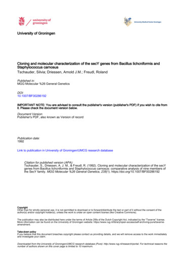
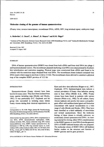
22five positive plaques were identified from a total of50 recombinant ,t phage clones that were screened.The DNA of one clone was cleaved with Hindlll,inserted into tbe Hindill site of the plasmid vectorpAT153, termed pHSRV-H-CSS, and was characterized in detail (clone 55 in Fig. 3).To confrrm the viral origin of the recombinantclones obtained, all clones were hybridized to DNAFig. 2. Southern blot hybridization of recombinant plasmid [32P)pyHSRV-E-D2 to human genomic DNAs from uninfected andHSRV infected HEL cells. Deproteinized DNAs (5 JL8 per lane including RNA), obtained from uninfected cells (lanes 1 to 6) and fromHSRV-infected cells (lanes 7 to 12), were cleaved with restriction endonucleases AlP718 (lanes 2 and 8), BamHI (lanes 3 and 9), Clal(lanes 4 and 10), EcoRI (lanes 5 and 11), and Hindill (lanes 6 and 12); untreated DNA (2 J.Lg/lane) was run in lanes 1 and 7. DNA(0.2 J.Lg) ofpyHSRV-E-02 digested with EcoRI (lane 13) was analyzed under the sam.e conditions. Phage-". DNA (0.25 Jl8 unlabeledand 0.01 Jlg 32 P labeled) cleaved with EcoRI (M), Mlu I {Ml ), and with Hindill (M2) served as markers and for oontrol of electrophoretic 'transfer to nitrocellulose paper. (A) EtdBr staining; (B) autoradiogram ofthe same gel after hybridization to 32P-labeled pyHSRV-E-D2DNA. Sizes are given in kb.
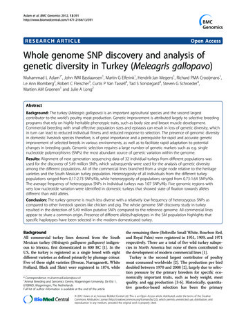
23were identified by hybridizing them to 32P-labeled02 DNA. Several of the resulting recombinantphage clones were cleaved with Bam HI that yieldedviral DNA inserts 15-16 kb in size which were subsequently recloned into the Bam HI site of the bacterial plasmid vectorpSP65 (Melton et al.; 1984). Oneof tbese recombinant plasmid clones tennedpSPHSRV-B-C11 (clone 11 in Fig. 3) was cbaracterized in detail by restriction enzyme analysis andSouthem blot. hybridizations (Flügel et al., 1987).In anotber set of experiments, a 2.2-kb BamHIDN A fragment tbat had been prepared from genomicDNA ofHSRV-infected HEL cells hybridized to theviral cDNA insert of clone 02. This BamHI fragment was directly cloned into the Bam Hl site of theplasmid vector pAT153, termed pHSRV-B-52(clone 52 in Fig. 3) and analyzed in detail.from mock-infected and HSRV-infected HEL cellsthat bad been digested with various restrictionenzymes. Fig. 2 sbows the result of these experiments using nick-translated [ 32P]DNA from clone02. Positive hybridization signals (Ianes 7 to 12)were clearly detectable in DNAs from HSRVinfected cells but not in genomes from uninfectedcells.(b) Ooning of HSRV DNAGenomic DNA from HSRV-infected HEL cellswas digested with BamHI and screened for HSRVspecific sequences by using 32P-labeled recombinantplasmid clones (02, C55) that bad been establishedfrom viral cDNA (Fig. 2). Hybridizing BamHIDNA fragments were isolated, purified by agarosegel electrophoresis, and Iigated to A.EMBL4 DNAthat bad been digested with BamHI tbereby removing a 19.3 kb fragment from the A.DNA. TheIigated DNA was packaged into phage particles andplated on the appropriate E. coli host. Two positiveplaques out of 30 000 recombinant phages screened{a)& s}'l( b)IJ,QU3., J,J.,Q. gJ,'AJ,QDNA was prepared from eacb ofthe recombinantplasmid clones as described in MATERIALS ANDMETHODS, section c. Purified DNAs were digested:zc:::::::::. *Q. Qo o 0lch'Q)( o11 -4, t 0':zs "'0I 0Q .yo .J,oe1:1. g*'x o oe J. K52gR USbSSI 552(c) Structural analysis of the recombinant cloneskb23456789101112Fig. 3. Restrietion maps oflinear HSRV DNA and ofviral DNA inserts ofrecombinant plasmid clones. {a) Diagram oflinear HSRVDNA. The boxes at the genome termini mark the LTR with the U3 region (hatched), the R region {blank) and the U5 region (blackened).{b) Restrietion maps oflinear HSRV DNA and ofviral DNA inserts ofrecombinant Clones 55, 2, 52, and 11. The symbols are: ,J., Hindlll;x , Eco RI; 0, Bam HI; e, Neo I; and D, Xba I. Open triangles below the lines mark deletions within viral nucleotide sequences. Thetwo thick upward arrows mark the 'integration site' of the 5' LTR into the pol gene of clone II. The map of clone 11 has been reversedto emphasize the colinearity with the other recombinant clones. Note that the scale of the restriction map of clone 11 is broken at the5' end. For further details f structure of clone II see RESULTS AND DISCUSSION, section d.
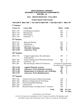
24with various restriction enzymes and analyzed byelectrophoresis on agarose gels. Subsequently, thegels were stained with EtdBr (Figs. 4A and 5A),transferred to nitrocellulose and hybridized with32P-labeled recombinant plasmids prepared fromcDNA (Fig. 4B,C and Fig. 5B,C). Hindilldigestionofclone C55 yields two fragments of5.4 and 3.6 kb;the 5.4-kb DNA fragment corresponds to theHSRV-specific DNA insert (Fig. 4B, lane 1). EcoRIcleaved the DNA of clone D2 into two large fragments (Fig. 4, lanes 2) of sizes of 6.4 (viral insert) and4.0 kb (vector) (Fig. 4). Further fme mappingrevealed that the overlapping regions of clones D2and C55 are located at the termini of the inserts ofboth recombinant plasmids as indicated schematically in Fig. 4C. The result was confrrmed by nucleotide sequence analysis which proved that the overlap regions encompassed 1225 and 651 nt (Flügelet al., 1987).12To determine the structure and the nucleotidesequence arrangements of those recombinant plasmid clones that contained even larger viral DNAinserts of 15.4 kb, further blot hybridization analyseswere performed using 32P-labeled DNA from clonesD2 and C55. The results shown in Fig. 5,A to C andFig. 6,A and B indicate that most of the recombinantclones harbor viral DNA inserts of 15 to 16 kb, sincethey gave positive hybridization signals with eitherD2 [ 32 P)DNA (Fig. SB) or with C55 [ 32P]DNA(Fig. 5C). Only one clone was negative (lane 8),whereas another recombinant clone bad a relativelysmall DNA insert of 2.8 kb that hybridized positively(lane 6). All other recombinant clones (lanes 1 to 5and 7) can be aligned with the HSRV restrictionmaps shown in Fig. 3. As a representative example,the restriction fra.gment cleavage pattem of theserecombinant clones obtained afterdouble digestionwith BamHI Hindill is shown in Fig. 6. The viralM2 M·-23.0Hind\1\--speiHindillHpallA- 2.32.0cBFig. 4. Blot hybridization of recombinant plasmid pHSRV-H-C55 to [ 32P]pyHSRV-E-D2 (panels A and B), both constructed fromcDNA. DNA ofC55 (lane 1) was cleaved with Hindill and DNA ofD2 (lane 2) was cleaved with Eco RI and separated electrophoreticallyon a 0.8% agarose gel. Phage Ä. DNA (0.25 1-'8 unlabeled and 0.021-'g labeled) cleaved with Hindill served as marker (M) and for controlof electrophoretic transfer to nitrocellulose paper. (A) EtdBr staining; (B) autoradiogram of the same gel after hybridization to32P-labeled pyHSRV-E-02 DNA. Sizes are given in kb. The EcoRI cleavage pattem of D2 DNA is the result of a partial digestion.(C) Schematic representation of the two recombinant · plasmids that harbor overlapping HSRV-specific sequences. Outer ring:pyHSRV-E-02, the thick line representing the 6.4-kb viral insert; inner ring: pHSRV -H-C55, the thick line represents a 5.4-kb viral insert.All restriction enzyroes shown produced DNA fragments ofthe same size (in bp) in the region overlapping both recombinant clones.The cross bars mark the viral DNA insert-vector boundaries.
25Fig. 5. Gel electrophoresis and Southem blot hybridization of spumaretroviral recombinant DNA clones. DNAs (5 pg) were digestedwith BamHl, separated electrophoretically on a 0.8% agarose gel, transferred to nitrocellulose and hybridized to 32P-labeledrecombinant clones from HSRV cDNA (D2 and C55). (A) EtdBr-stained gel ofclones C4, C5, C6, C7, C9, ClO, Cll, Cl2, and Cl3(lanes 1 to 9). (B) Autoradiograph ofthe same gel hybridized with 32P-labeled clone 02. (C) Autoradiograph ofthe same gel hybridizedwith 32P-labeled clone C55. The lanes Mare EcoRI fragments ofphage l DNA with the sizes given on the left-hand margin ofpanelA. The arrows indicate position of pSP65 vector DNA.DNA insert of clone 11 is 15.4 kb in size (Fig. 5, lane7) and a Hindiii-BamHl subfragment of this clonehybridized and co-migrated with the HindiiiBam HI DNA fragment of recombinant clone C55(Fig. 6A, lane 10 and Fig. 6B ). This result indicatesthat the Hindlll-BamHI DNA fragments of bothclones;Cll and C55, are colinear on the HSRV map(Fig. 3). This interpretation was verified by nucleotide sequence analysis (Flügel et al., 1987).Similar mapping experiments and nucleotide sequence analyses confrrmed the restriction maps of allrecombinant clones shown in Fig. 3. Clone 52 DNAis particularly interesting, since its viral DNA insertof 2.2 kb is located at the 3' end of the HSRV mapand, as revealed by nucleotide sequencing, containsthe complete 3' LTR. Fig. 1 shows that clone 52DNA hybridized to [ 32P]cDNA. Although recombinant clone 52 was isolated from the Bam HI digest ofgenomic D NA of virus-infected human cells andcloned into the BamHI site of pAT153, the viralD NA insert could not be cut out by digesting theclone B52 DNA with BamHI. Nucleotide sequenceanalysis ofthe recombinant clone demonstrated thatone ofthe BamHI sites (the 5' -BamHI site in Fig. 3)bad been retained but the second BamHI (at the 3'end) was not present (Flügel et al., 1987).(d) ConclusionsSpumaretroviral DNA has been molecularlycloned into various restriction enzyme sites of twodifferent .A. vectors and several bacterial plasmidvectors. Two completely independent sources ofHSRV DNA were used as starting material formolecular cloning cDNA and viral DNA from totalgenomic DNA of HSRV-infected cells. The viralDNA inserts of the resulting recombinant clone"hybridized to each other and displayed colinear restriction maps having LTRs at the ends of the genome. These results demonstrate that the completeHSRV genome is represented by the recombinantplasmid clones obtained.
26Fig. 6. Gel electrophoresis and Southem blot analysis ofspumaretroviral recombinant DNA clones. DNAs (5 J.g) were double-digestedwith BamHI Hindi li, separated electrophoretically on a 0.8% agarose gel, transferred to nitrocellulose and hybridized to 32P-labeledrecombinant clonesthat had been established from HSRV cDNA. (A) Etdßr-stained gel of clones C4, C5, C6, C7, C9, ClO, Cll, Cl2,and Cl3 (lanes 1 to 9). (8) Autoradiograph ofthe same gel hybridized to 32P-labeled clone C55. M marks the Mlul fragments from phageA. DNA. 32 P-labeled DNA from recombinant clone C55 was double-digested with BamHI Hindill (lane 10).However, some ofthe clonesbad sustained internal deletions. For example, clone 02 that was clonedfrom HSRV cDNA contains three deletions of different sizes as determined by nucleotide sequenceanalyses of different HSRV recombinant clones(Flügel et al., 1987). One deletion of 1230 bp wasfound to be located in the pol region (in the centralpart of the reverse transcriptase domain), the othertwo deletions were in the LTR. These studies cannotdetermine how these deletions were generated. Therehave been several reports on deletions that occurredduring molecular cloning of retroviral and speciallylentiviral DNA (Molineaux and Qements, 1983;Hahn et al., 1984). Since the deletions of clone 02in the pol gene and the LTR are flanked by a shortrepeat sequence, one can assume that they have oc-
27curred during molecular cloning. Altematively, theevent could have happened during in vitro cDNAsynthesis. This conclusion rests on the primarystructure of clone D2 DNA and in addition is basedon the assumption that during cDNA synthesis invitro, a full-length, double-stranded DNA was generated containing one LTR only which subsequentlyunderwent deletions. Proviral DNAs with one LTRhave been isolated and characterized previously (Juand Skalka, 1980).Recombinant clone 11 that was established fromviral DNA has an unusual and remarkable structure.Restrietion fine mapping of C 11 revealed that arecombination andjor integration event had occurredinto the pol regionoftbis clone. Nucleotide sequenceanalysis unequivocally showed that another HSRVDNA bad been inserted close to map position of 3.9kb (Fig. 3). Thus, the 5' LTR of the 'integrated'HSRV starts at this map position, the viral genomerunning backwards continues to the Bam HI site thatis located 5.6 kb upstream from this 'integration'point. The mechanism for this event is unknown.In conclusion, recombinant plasmid clones havebeen established for the complete HSRV genome.The recombinant cloneswill be useful for the analysisofthe structure of spumaretroviral genes and expression of their gene products. They will be invaluableas a new diagnostic sequence to probe patients withvarious diseases of suspected viral etiology.ACKNOWLEDGEMENTSWe thank Dr. Peter Gruss for critically reading themanuscript. This work was supported by a postdoctoral training stipend (627-1-1) from theDeutsche ForschungsgemeinschaftREFERENCESChang, A.C.Y. and Cohen, S.N.: Construction and characterization of amplifiable multicopy DNA cloning vehicles derivedfrom the PISA cryptic miniplasiJlid. J. 8acteriol. 134 (1978)1141-1156.Flügel, R.M., Rethwilm, A., Maurer, 8. and Darai, 0.: Nucleotide sequence analysis of the env gene and its flankingregions of the human spumaretrovirus reveals two novelgenes. EM80 J. 6 (1987) 2077-2084.Frischauf, A.-M., Lehrach, H., Poustka, A. and Murray, N.:Lambda replacement vectors carrying polylinker sequences.J. Mol. 8iol. 170 (1983) 827-842.Gajdusek, D.C.: Unconventional viruses and the origin anddisappearance of Kuru. Science 197 (1977) 943-960.Gubler, U. and Hoffman, 8J.: A simple and very efficientmethod for generating cDNA libraries. Gene 25 (1983)263-269.Hahn, 8.H., Shaw, G.M., Arya, S.K., Popovic, M., Gallo, R.C.and Wong-Staal, F.: Molecular cloning and characterizationof the HTLV-111 virus associated with AIDS. Nature 312(1984) 166-169.Hohn, 8.: In vitro packaging of Iambda and cosmid DNA.Methods Enzymol. 68 (1979) 299-310.Hooks, J.J. and Detrick-Hooks, B.: Spumavirinae: foamy virusgroup infections; comparative aspects and diagnosis. InKurstak, E. and Kurstak, C. ( s.) Comparative DiagnosisofViral Diseases, Vol. IV. Academic Press, New York, 1981,pp. 599-617.Ju, G., 8oone, L. and Skalka, A.-M.: Isolation and characterization ofrecombinant DNA clones of avian retroviruses:size heterogeneity and instability ofthe direct repeat. J. Virol.33 (1980) 1026-1033.Ju, G. and Skalka, A.-M.: Nucleotide sequence analysis of thelong terminal repeat (LTR) of avian retroviruses: structuralsimilarities with transposable elements. Cell 22 (1980)379-386.Loh, P.C. and Ang, K.S.: Replication of human syncytiumforming virus in human cells: effects of certain biological1factors and selective chemicals. J. Med. Virol. 7 (1981) 67-73.Maniatis, T., Fritsch, E.F. and Sambrook, J.: Molecular Ooning.A Laboratory Manual. Cold Spring Harbor Laboratory, ColdSpring Harbor, NY, 1982, pp. 194-195.Melton, D.A., Krieg, P.A., Rebagliati, M.R., Maniatis, T., Zinn,K. and Green, M.R.: Efficient in vitro synthesis ofbiologicallyactive RNA and RNA hybridization probes from plasmidscontaining a bacteriophage SP6 promoter. Nucl. Acids Res.12 ( 1984) 7035-7056.Molineaux, S. and Clements, J:E.: Molecular cloning of unintegrated visna viral DNA and characterization of frequentdeletions in the 3' terminus. Gene 23 (1983) 137-148.Muller, H.K., Ball, G., Epstein, M.A., Achong, B.O., Lenoir, G.and Levin, A.: The prevalence of naturally occurring anti·bodies to human syncytial virus in East African populations.J. Gen. Virol. 47 (1980)399-406.Murray, N.: Phage Iambda and molecular cloning. In Hendrix,R.W., Roberts, J.W., Stahl, F.W. and Weisberg, R.A. (Eds.)Lambda II. Cold Spring Harbor Laboratory, Cold SpringHarbor, NY, 1983, 395-432.Nara, P.L., Robey, W.G., Arthur, 0.0., Gonda, M.A., Asher,D.M., Yanagihara, R., Giss Jr., C.J., Gajdusek, D.C. andFischinger, P.J.: Simultaneous isolation ofsimian foamy virusaod HTLV-III/LAV from chimpanzee lymphocytes followingHTLV-III or LAV inoculation. Arch. Virol. 92 (1987)183-186.
28Neumann-Haefelin, 0., Rethwilm, A., Bauer, G., Gudat, F. andzur Hausen, H.: Characterization of a foamy virus isolatedfrom Cercopilhecw aethiop.r lymphoblastoid cells. Med.Microbiol. lmmunol. 172 (1983) 75-86.Obara, T., Bolognesi, D.P. and Bauer, H.: Ribosomal RNA inavian leukosis virus particles. lnt. J. Cancer 7 ( 1971)535-546.Rigby, P.W.J., Dieckmann, M., Rhodes, C. and Berg, P.: Labelingdeoxyribonucleic acid to high specific activity in vitro by nicktranslation with DNA polymerase I. J. Mol. Biol. 114 (1977)237-256.Rogers, N.G., Basnight, M., Gibbs Jr., CJ. and Gajdusek, D.C.:Latent viruses in chimpanzees with experimental Kuru.Nature 216 (1987) 446-449.Rothenberg, E. and Baltimore, D.: Synthesis oflong. representative DNA copies ofthe murine RNA tumor virus genome. J.Virol. 17 (1976) 168-174.Southern, E.M.: Detection of specific sequences among DNAfragments separated by gel electrophoresis. J. Mol. Biol. 98(1975) 503-517.Taylor, J.M., IUmensee, R. and Summers, J.: Efficient tran·scription of RNA into DNA by avian sarcoma virus polymerase. Biochim. Biophys. Acta 442 (1976) 324-330.Twigg, A. and Sheratt, 0.: Trans-complementable copy-numbermutants of plasmid ColEI. Nature 283 (1980) 216-218.Weiss, R., Teich, N., Varmus, H. and Coffin, J.: Taxonomy ofspumaviruses.ln Weiss, R., Teich, N., Varmus, H. and CoffinJ. (Eds.) RNA Tumor Viruses, 2nd ed. Cold Spring HarborLaboratory, Cold Spring Harbor, NY, 1982, pp. 157-207.Communicated by T. Hohn.
Cloning efficiency using Hindlll-digested cDNA was 10% {five out of 50 recombinant clones hybridized). DNAs from clones A.HSRV-EcoRI 1 to 30 thatgave strong hybridization signals, and DNA from clones A.HSRV-Hindlll 1 to 5 that hybridized positively were prepared according to standard protocols (Maniatis et al., 1982).