Transcription
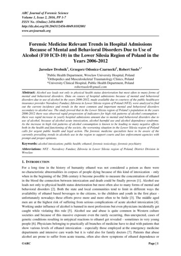
The First 21 DaysOanksiRd mystery su r ro u n dthe embr yo’s earliestd e ve l o p m e n t22Medicine at Michiganby sally pobojewskiYorgos Nikasne of life’s most profound mysteries— the creation of a new human being — starts with the union of egg and sperm and ends, ninemonths later, with a baby. It’s a process so intricate and inherently risky, with pitfalls at everystage, it’s amazing any of us survive long enough to be born at all. In fact, scientists estimate thatabout half of all human embryos die during the first two months of pregnancy when somethinggoes wrong in those critical early stages of development.It takes two months to form the human embryo, a process scientists call embryogenesis. Aftereight weeks, the embryo has all the organs and tissues found in a newborn baby — althoughmany exist in primitive form. During the last seven months of a human pregnancy, the fetus (nolonger called an embryo) continues to grow and develop, but the fundamental blueprint for thebaby-to-be is established during embryogenesis.
photo creditA human eggsurrounded by magazine
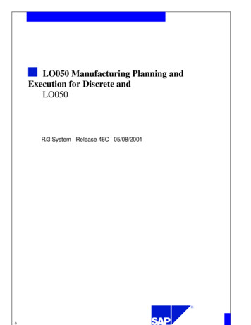
Many of the most important and least understood stagesof a human embryo’s development take place during the first21 days of pregnancy before the mother even knows she’spregnant and while the embryo is still incredibly small.At 21-days-old, a human embryo is less than twomillimeters long or roughly the size of the circle at the endof this sentence.But even at 21 days, the embryo is much more than justa simple ball of primitive cells. Its gender and basic bodyplan — top and bottom, front and back, left and right — arealready established and the developmental future of everycell in the embryo is already set.It’s an amazing journey. And it all starts with a single cell.Day 1 FertilizationIn the beginning is the eggInside the fallopian tube, a mature human egg waits in a stateof arrested development. Just released from the ovary, it isthe largest cell in the human body. The egg is packed withnutrients, growth factors, enzymes and proteins — nearlyeverything it needs to jump start the development of a humanembryo. Except for one little thing — it needs a sperm. If asperm doesn’t penetrate the egg’s tough outer membrane toactivate it within the next 24 hours, the egg will die.During fertilization and the first stage of embryonicdevelopment, the egg runs the show. All a mature humanegg really needs from a sperm is its DNA — the geneticcode stored in 23 chromosomes inherited from the father.When combined with 23 chromosomes in the egg fromthe mother, the new embryo has the full complement ofin-vitro fertilization (IVF) or other assisted reproductivetechnologies. In 2004, 49,458 babies were born in the U.S.from embryos created when sperm and egg got together ina Petri dish, rather than inside a woman’s body.Gary Smith, Ph.D., is an associate professor ofobstetrics/gynecology, urology and of molecular andintegrative physiology. He studies mouse, cow and humanembryos to learn what happens during the first few daysof development. Although everything happens faster inmice than in humans, the steps involved in the process areessentially the same in both species. Studying what happensto embryos outside the body is not ideal, however, becausethere are big differences between how embryos develop in aculture dish and how they develop inside a living animal.In the body, early embryos grow inside the moist,confined environment of the fallopian tube, where theyare in constant contact with proteins, nutrients and othercells. But in the laboratory, most embryos start out floatingin culture medium in a Petri dish — a situation that’s like “aperson floating out in Lake Michigan,” says Smith.To mimic conditions inside the fallopian tube wherefertilization and the initial stages of development take place,Smith worked with Shuichi Takayama, Ph.D., an associateprofessor in the College of Engineering, to develop a deviceabout half the size of a credit card that makes it possible toobserve an embryo and monitor its biological signals duringthe first days of development.Within the tight confines of narrow channels in themicrochip, scientists can keep an embryo moving, maintainthe flow of fresh nutrients and remove waste products.Smith calls it a dynamic culture, as opposed to the staticculture currently used to grow embryos in a Petri dish.Using a dynamic culture system that simulates naturalAt the eight-cell stage, the embryo somehow activatesits genes. From this point on, the embryo is on its ownto follow its unique genetic destiny.24genetic material required to make a human being.Much of what scientists know about fertilizationand what happens during the early days of an embryo’sdevelopment has come from research to help the 10percent to 15 percent of couples who need medicalassistance to have a baby. Many of these couples turn toMedicine at Michiganconditions, Smith and Takayama have found they gethealthier mouse and cow embryos that are more like thosegrown inside the animal’s body.Smith’s goal is to learn the body’s secrets for growinga healthy embryo and use that knowledge to help moreinfertile couples have a healthy baby.
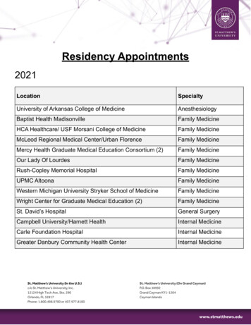
Yorgos NikasDay 2 to 3 Early Cleavagedevelopment past the eight-cell stage and some don’t,”Smith explains. “Most eggs will fertilize and the embryoswill start to divide normally, but when they reach theTurning on the embryo’s geneseight-cell stage, the system sometimes breaks down anddevelopment stops.”Inside the mother’s body, a fertilized egg moves through theExactly what an egg must have to make it possible forfallopian tube, pushed toward the uterus by filaments liningthe embryo to turn on its genes, and how to tell whichthe inside of the tube. Still dependent on nutrients andeggs have it, are two of the most important questions ingenetic instructions contributed by the egg, the embryodevelopmental biology today, according to Smith.divides to form two cells, then four cells, then eight. At“We know this developmental time point is verythe eight-cell stage, through a process scientists still don’timportant for the embryo’s survival and that gene activationunderstand, the embryo somehow activates its genes. Fromis regulated by maternal, or egg-derived, factors, but we don’tthis point on, the embryo is on its own to follow its uniqueunderstand how the transformation works,” Smith says.genetic destiny. If anything interferes with gene activation,Smith believes the factorsresponsible for activation of theembryo’s genome are programmedinto the egg while it develops insidecompartments in the ovary calledfollicles. It takes up to several hundreddays for a human follicle to producea mature egg. Before every menstrualcycle, several follicles begin to grow,but in the end, only one typicallyproduces an egg that is primed andready for fertilization.“Nature has developed this journeyto provide embryonic competence,”Smith says. “It’s naïve of us to thinkwe can just go in [to the ovary] andpluck out eggs. They may all lookthe same at a microscopic level, butdevelopmentally we know that not alleggs are created equal.”When embryos fertilized in a Petridish during in-vitro fertilization reachthe eight-cell stage, one or moreare chosen to be transferred to theThe embryo dividesto form more cells.mother’s uterus. Unfortunately, only(color-enhanced)about 20 percent of all transferredembryos will successfully implant inthe wall of the uterus and continue todevelop, according to Smith.the embryo will die.Biological tests or assays to determine in advance whichIn the language of reproductive biology, it’s calledeggs are capable of activating the embryo’s genome, andembryonic developmental competence, and it’s a criticalwhich embryos have the greatest potential to implant,point where development often stops for reasons scientistswould be a big step forward in improving the odds of adon’t understand.successful IVF pregnancy.“Some eggs have what it takes to support the embryo’s25www.medicineatmichigan.org/magazine
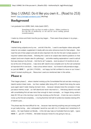
“Today all we can do is look at embryosunder a microscope and pick the prettiest oneswith the most time-appropriate cell number,”Smith says. “One of the goals of our research isdeveloping a culture and analysis system thatwill allow us to produce the healthiest embryosand select the embryo with the best chance ofimplantation.”Days 3 to 5 Blastocyst26By the time a three-day-old human embryoenters the uterus, it contains 16 identical cellspacked together to form a sphere called amorula. As it moves into the uterus, the embryoArtist’s depiction ofis preparing for a major transformation. Duringa human egg movingthrough the fallopian tube.the next 48 hours, the morula will become ablastocyst — a hollow oval with about 100 cellsdivided into two different types. Cells that willO’Shea is especially interested in teasing out the signalsform the placenta make up the outer layer of the blastocyst.required to transform human embryonic stem cells, andNestled inside is a small inner cell mass with about 50 cells.cells in the early embryo, into neurons — one of the firstThese 50 cells will give rise to the millions of specializedspecialized cells to develop in an embryo.cells, tissues and organs it takes to make a newborn baby.Scientists refer to cells in the inner cell mass as beingpluripotent, meaning they can become nearly any cell inthe human body. But this unlimited potential lasts forWeek 2 Implantationjust a few days. If pluripotent cells are removed from theblastocyst and cultured in the laboratory under the rightAt home in the uterine wallconditions, they form colonies of human embryonic stemcells. Otherwise, they are quick to get on with the businessWhen the human embryo is around six days old, it startsof becoming the specialized cells the embryo will need as itmaking a nest in the wall of the mother’s uterus. Cells ingrows and changes.the outer layer of the blastocyst stick to the uterine wall andSue O’Shea, Ph.D., a professor of cell and developmentalgrow long projections into it — the first step in developingbiology who directs the Michigan Center for Humana placenta with blood vessels linking mother and embryo.Embryonic Stem Cell Research, is trying to decipher theFrom now on, the embryo will depend on oxygen andflood of genetic signals involved in the transformationnutrients from the mother’s bloodstream to survive. As theof early embryonic cells into cells that are differentiated,placenta grows and embeds itself within the uterine wall,or specialized to perform specific tasks. Working withthe inner cell mass divides to form the amniotic cavity andmouse embryos, mouse embryonic stem cells and humana flat disc with two layers of cells that is the embryo.embryonic stem cell lines that have been approved by theOnce the embryo is embedded in the wall of the uterus, itfederal government for use in scientific research, O’Sheastarts preparing for a transformation called gastrulation thatand her graduate students Nicky Slawney and Lisa DeBoertakes place during the third week of development. Duringare identifying the genes and growth factors involved andthis process, every cell in the flat disc will migrate to a newlearning how they work.location and morph into one of three new cell types to formMedicine at MichiganSebastian Kaulitzki/Dreamstime.comCells make their first choice
the inner, middle and outer layers of the 21-day-old embryo.This intricate cellular choreography is regulated by growthfactors turned on and off at specific times and locations inresponse to signals from the embryo’s genes.“The embryo uses the same seven growth factor signalingpathways over and over,” O’Shea says. “But by varying signalstrength, turning cell receptors on or off, or turning growthfactors on or off at specific locations in the embryo, theprocess allows for a great deal of precision and complexity.”Directed by the node, cells move down and through thestreak to be reborn on the other side as either endoderm(the inner layer of the embryo) or mesoderm (the middlelayer). Once the two inner layers of the embryo are formed,the node sends a different signal to the remaining cells.Instead of going through the streak, these cells will fan outto form the outer layer of the embryo called the ectoderm.Once cells have passed through the primitive streakand gastrulation is complete, there is no turning back.To mimic conditions inside the fallopian tube,researchers in the Smith and Takayama laboratoriesdeveloped a device that makes it possible to observethe embryo and monitor its biological signalsduring the first days of development.Week 3 GastrulationThree cell layers and a body planDuring its third week, the human embryo goes througha developmental milestone. Gastrulation establishes theembryo’s basic body plan and seals the fate of its cells. Oncethe process is complete, the embryo will have three distinctlayers with a defined top and bottom, front and back, left andright. At no other time in its development will the embryoundergo such a radical transformation.Gastrulation begins with an indentation called theprimitive streak, which forms on top of the flat disc whenthe embryo is about 15 days old. At the top of the streak isa small structure called the node that churns out growthfactors signaling cells to break free from their neighbors,multiply and move toward the streak.Cells in the gastrulating embryo are exquisitely sensitiveto the strength of growth factors from the node. Embryoniccells closest to the node receive the strongest signal,inducing them to turn on genes that cause them to becomeone kind of cell. Cells on the edge of the embryo receivea weaker signal and this causes them to express differentgenes and become a different kind of cell.“Just being a couple cell diameters away can mean a bigdifference in gene expression,” O’Shea says.Each embryonic cell is now destined to follow a specificdevelopmental pathway. Endoderm cells will go on toform the liver, pancreas and gastrointestinal tract. Cellsin the mesoderm are fated to develop into the heart andblood vessels, bone, muscle and kidneys. The ectoderm willbecome the central and peripheral nervous systems, sensoryorgans, skin and hair.Because the blueprint for all the body’s organs isestablished during gastrulation, it’s the beginning of atime when exposure to alcohol, drugs, viruses and toxicchemicals can have a catastrophic effect on the embryo.From the third week to the eighth week while organsare still forming, the embryo will remain vulnerable todamage. Even if it’s not lethal, the result can be a lifetimeof disability for a child born with fetal alcohol syndrome orspina bifida.Knowing more about what happens to a human embryoduring its perilous journey from fertilization to gastrulationcould help researchers learn what causes birth defects andperhaps even find ways to prevent or correct them.Research to understand how embryos develop couldbenefit the health of adults, as well. Scientists are onlystarting to understand how mistakes during embryogenesiscan have life-long consequences in the form of diseases anddisorders like cancer, congenital heart defects, and Downsyndrome that begin when something goes wrong duringthe embryo’s first 21 days.More on the Web27www.medicineatmichigan.org/magazine
The First 21 Days ne of life's most profound mysteries — the creation of a new human being — starts with the union of egg and sperm and ends, nine . embryos to learn what happens during the first few days of development. Although everything happens faster in mice than in humans, the steps involved in the process are .