Transcription
Journal ofClinical MedicineArticleEfficacy of Removing Thermafil and GuttaCore from StraightRoot Canal Systems Using a Novel Non-Surgical Root CanalRe-Treatment System: A Micro-ComputedTomography AnalysisVicente Faus-Llácer 1 , Rubén Linero Pérez 1 , Ignacio Faus-Matoses 1 , Celia Ruiz-Sánchez 1 ,Álvaro Zubizarreta-Macho 2,3, * , Salvatore Sauro 4,5 and Vicente Faus-Matoses 112345 Citation: Faus-Llácer, V.; Pérez, R.L.;Faus-Matoses, I.; Ruiz-Sánchez, C.;Zubizarreta-Macho, Á.; Sauro, S.;Faus-Matoses, V. Efficacy ofRemoving Thermafil and GuttaCorefrom Straight Root Canal SystemsUsing a Novel Non-Surgical RootCanal Re-Treatment System:A Micro-Computed TomographyAnalysis. J. Clin. Med. 2021, 10, 1266.https://doi.org/10.3390/jcm10061266Academic Editor: Federico FoschiReceived: 31 January 2021Accepted: 15 March 2021Published: 18 March 2021Publisher’s Note: MDPI stays neutral*Department of Stomatology, Faculty of Medicine and Dentistry, University of Valencia, 46010 Valencia, Spain;fausvj@uv.es (V.F.-L.); rubenlinero@hotmail.com (R.L.P.); ignacio.faus@uv.es (I.F.-M.);ceruizsan@gmail.com (C.R.-S.); vicente.faus@uv.es (V.F.-M.)Department of Endodontics, Faculty of Health Sciences, Alfonso X El Sabio University, 28691 Madrid, SpainDepartment of Surgery, Faculty of Medicine and Dentistry, University of Salamanca, 37008 Salamanca, SpainDental Biomaterials and Minimally Invasive Dentistry, Department of Dentistry, Cardenal Herrera-CEUUniversity, 46115 Valencia, Spain; salvatore.sauro@uchceu.esDepartment of Therapeutic Dentistry, I.M. Sechenov First Moscow State Medical University,119146 Moscow, RussiaCorrespondence: amacho@uax.esAbstract: The present study aims to evaluate the effectiveness of an XP-endo non-surgical root canalre-treatment system in removing both GuttaCore and Thermafil gutta-percha carrier-based rootcanal filling materials from straight root canal systems using micro-computed tomography (microCT) analysis. The study was performed on 20 single-rooted upper teeth, which were randomlyallocated into the following study groups: Group A, Thermafil and AH Plus sealer (n 10); Group B,GuttaCore and AH Plus sealer (n 10). Before and after the non-surgical root canal re-treatmentprocedure, the samples were submitted for a micro-CT analysis. The volume of the root canal fillingmaterial (mm3 ), the volume of the remaining root canal filling material (mm3 ) and the time (minutes)needed to remove the root canal filling material were also recorded. Student’s t-test was used toanalyze the results. No statistically significant differences were found between the volume of theremaining root canal filling material in the GuttaCore and Thermafil root canal filling systems atthe coronal third (p 0.782), middle third (p 0.838) or apical third (p 0.882) of the straight rootcanal systems; however, the GuttaCore required a statistically significant (p 0.037) shorter amountof time (4.72 0.76 min) to be removed than the Thermafil carrier-based root canal filling material(5.92 1.42 min). The XP-endo Finisher non-surgical endodontic re-treatment system removes bothGuttaCore and Thermafil gutta-percha carrier-based root canal filling materials from straight rootcanal systems, although removal of the GuttaCore gutta-percha carrier-based root canal fillingmaterial required less time.with regard to jurisdictional claims iniations.Keywords: endodontics; GuttaCore; micro-computed tomography; non-surgical root canalre-treatment; root canal filling material; ThermafilCopyright: 2021 by the authors.1. IntroductionLicensee MDPI, Basel, Switzerland.At present, root canal treatment has a reported survival rate of up to 84.1% [1] to97.3% [2]; however, intraoperative complications during the endodontic therapy can potentially lead to treatment failure [3], continued primary endodontic infection [4,5] andthe possible need for subsequent treatment options including non-surgical root canalre-treatment, endodontic surgery, autotransplantation, intentional replantation or toothextraction [6–9]. Root canal treatment failure is frequently linked to insufficient disinfectionpublished maps and institutional affil-This article is an open access articledistributed under the terms andconditions of the Creative CommonsAttribution (CC BY) license (https://creativecommons.org/licenses/by/4.0/).J. Clin. Med. 2021, 10, 1266. i.com/journal/jcm
J. Clin. Med. 2021, 10, 12662 of 14of the root canal system, the root canal filling material overextending through the apical foramen, inadequate obturation, unsuitable coronal restoration, untreated canals andiatrogenic procedural errors due to natural cavity design with limited access or instrumentation complications (ledges, perforations or fractured instruments) [3,10–13]. Therefore,non-surgical root canal re-treatment is recommended as a first treatment approach forthe removal of root canal filling material, enabling thorough cleaning, disinfection andre-instrumentation of the root canal system [14–16]. Numerous non-surgical root canalre-treatment techniques and materials have been proposed, including stainless steel handfiles, nickel titanium (NiTi) endodontic rotary and reciprocating instruments, specializedNiTi endodontic re-treatment files [17], ultrasonic appliances, Gates Glidden burs or acombination thereof. In addition, the adjuvant use of gutta-percha solvent agents or heatapplication has also been proposed for facilitating access to the root canal system forremoval of the root canal filling material [18] in order to enable proper cleaning, shaping and disinfection of the root canal system. Although many non-surgical root canalre-treatment techniques can be used to remove root canal filling material within the rootcanal system, none of them are capable of completely removing all the aforementionedfilling material [17]; therefore, novel non-surgical root canal re-treatment systems have beenproposed, such as the XP-Endo Retreatment System (FKG Dentaire, La-Chaux-de-Fonds,Switzerland), which is composed of three endodontic rotary files: the DR1 endodonticrotary file has been designed with alternating cutting edges and a triangular cross-sectionand the XP-endo Shaper and XP-endo Finisher R endodontic rotary files are made of NiTiMaxWire alloy (Martensite-Austenite Electropolish Flex), which increases the efficacy ofroot canal filling removal due to its shape memory and austenite phase conversion [19].Many root canal filling materials and techniques for sealing the root canal system havebeen described; however, lateral condensation remains the most commonly used techniquefor root canal filling [20,21]. The lack of homogeneity in the root canal system has led to thedevelopment of new root canal filling techniques, including warm gutta-percha techniquessuch as gutta-percha carrier-based root canal filling techniques and vertical condensationroot canal filling techniques [22,23]. Gutta-percha carrier-based root canal filling materialshave become widely used [24,25] due to easy handling [26] and high marginal sealingcapacity [27]; however, the rigid carrier of these root canal filling techniques’ results isdifficult to completely remove, especially the Thermafil carrier-based root canal fillingsystem. Therefore, a novel carrier-based root canal filling system has been developed,with a special carrier based on a cross-linked gutta-percha core; this should be easier toremove compared to the Thermafil carrier-based root canal filling system [28].The present study aims to analyze the efficacy of the XP-endo non-surgical rootcanal re-treatment system in removing two types of gutta-percha carrier-based root canalfilling materials, Thermafil and GuttaCore, from straight root canal systems using a microcomputed tomography (micro-CT) analysis. The null hypothesis (H0 ) holds that there isno difference between the gutta-percha carrier-based root canal filling materials duringremoval from straight root canal systems.2. Materials and Methods2.1. Study DesignTwenty (20) single-rooted anterior teeth (upper canine) were studied. The teeth wereextracted due to periodontal concerns. The studied teeth had curvatures of 10 , measuredusing Schneider’s method [29], mature roots and no prior root canal filling materials,root resorptions or calcium metamorphosis. The sample teeth were chosen from the Department of Stomatology at the University of Valencia (Valencia, Spain) during the period fromSeptember to October 2020. An ANOVA one-way test was used to calculate the samples.A randomized controlled experimental trial was carried out following the principles outlined in the statement by the German Ethics Committee (Zentrale Ethikkommission, 2003)on using organic tissues for the purposes of medical research. The Ethics Committee of the
J. Clin. Med. 2021, 10, 12663 of 14University of Valencia approved the study (Process no. 19/2020). All patients providedinformed consent for their teeth to be used in the study.2.2. Experimental ProcedureDigital radiographs were taken of the teeth in the buccolingual and mesiodistaldirections so as to ensure that the root axes were straight and the root canal sections wereslightly oval. The teeth were all decoronated using a diamond disc (Brasseler, GA, USA,Savannah, GA, USA) to obtain a standardized root length of 16 mm. To ascertain the apicalpatency, a 10-K file (Dentsply Maillefer, Baillagues, Switzerland) was used until the filecould be seen at the apical foramen when magnified (OPMI pico, Zeiss Dental Microscopes,Oberkochen, Germany). A 15-K file was used to confirm the glide path (Dentsply Maillefer,Baillagues, Switzerland). Afterwards, root canal procedures were carried out using theProTaper Gold endodontic rotary system (Dentsply Maillefer, Baillagues, Switzerland)set to a 25.08 F2 file with a 6:1 reduction handpiece (X-Smart plus, Dentsply Maillefer,Baillagues, Switzerland). The torque-controlled motor was set to continuously rotate at300 rpm and 2 N/cm torque, following the recommendations provided by the manufacturer.The root canal systems were then irrigated using 5 mL of a solution with 5.25% sodiumhypochlorite (NaOCl; Clorox, Oakland, CA, USA). The final irrigation consisted in 5 mL of5.25% NaOCL (Clorox, Oakland, CA, USA), 5 mL of 17% ethylenediaminetetraacetic acid(EDTA) (SmearClear, SybronEndo, CA, USA), 5 mL of 5.25% NaOCl (Clorox, Oakland, CA,USA) and 5 mL of sterile saline solution (Braun , Melsungen, Germany). A specializedendodontic needle (Miraject Endo Luer, Hager and Werken, Duisburg, Germany) wasinserted up to 1 mm of the working length. The root canal systems were then driedusing sterile paper points (Dentsply Maillefer, Ballaigues, Switzerland). Next, the rootcanal systems were randomly sealed (Epidat 4.1, Galicia, Spain) into one of the followingwarm gutta-percha carrier-based root canal filling systems: Group A, Thermafil (DentsplyMaillefer, Ballaigues, Switzerland) along with an epoxy-amine resin-based sealer (AHPlus, Dentsply DeTrey, Konstanz, Germany) (n 10); or Group B, GuttaCore (DentsplyMaillefer, Ballaigues, Switzerland) along with an epoxy-amine resin-based sealer (AHPlus, Dentsply DeTrey, Konstanz, Germany) (n 10). This was achieved by heating aGuttaCore obturator (size 25) in a ThermaPrep heater obturator oven (Dentsply Maillefer,Ballaigues, Switzerland) before inserting it in the root canal system up to the workinglength. A periapical radiograph of the buccolingual projection was taken to assess thequality of the root canal filling; if any voids were detected, these specimens were discardedand replaced. Lastly, CavitTM (3M ESPE, Saint Paul, MN, USA) was used to temporarilyseal the access cavity, and the specimens were stored in a 37 C atmosphere with 100%humidity for 14 days.The gutta-percha carrier-based root canal filling materials were removed, while thetime needed was registered with a chronometer, under magnification (OPMI Pico, Zeiss Dental Microscope, Oberkochen, Germany) using the XP-Endo Retreatment System (FKGDentaire, La-Chaux-de-Fonds, Switzerland), starting with continuous rotation with theDR1 endodontic rotary file (FKG Dentaire, La-Chaux-de-Fonds, Switzerland) at 800 rpmand 1.5 N/cm torque inserted in the gutta-percha carrier-based root canal filling materialsso as to create a 3–4-mm space from the cementoenamel junction. Afterwards, an XP-endoShaper endodontic rotary file with 300 µm apical diameter and 4% taper (FKG Dentaire,La-Chaux-de-Fonds, Switzerland) was used at 1000 rpm and 1.0 N/cm torque until theroot canal filling material was fully removed. Finally, the XP-endo Finisher R endodonticrotary file with 300 µm apical diameter and 0% taper (FKG Dentaire, La-Chaux-de-Fonds,Switzerland) was used at 1000 rpm and 1.0 N/cm torque until the working length as asupplementary cleaning approach to the root canal system, as detailed in the manufacturer’s recommendations. All procedures were performed inside a cabinet with a heatermaintained at 37 C (800-Heater; PlasLabs, Lansing, MI, USA).Finally, the root canal system was irrigated using a solution consisting of 5 mL of5.25% NaOCl (Clorox, Oakland, CA, USA), 5 mL of 17% EDTA (SmearClear, SybronEndo,
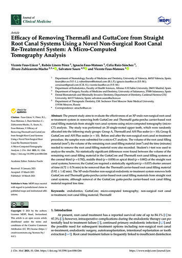
J. Clin. Med. 2021, 10, 12664 of 14CA, USA), 5 mL of 5.25% NaOCl (Clorox, Oakland, CA, USA) and 5 mL of sterile salinesolution (Braun , Melsungen, Germany), with a 0.3-mm endodontic needle inserted 1 mminto the working length. A sonic handpiece driver (EndoActivator , Dentsply Maillefer, Ballaigues, Switzerland) was used to help increase contact between the irrigationsolution and the surface of the root canal walls. The non-surgical root canal retreatment was considered finished when the gutta-percha was not visible inside the rootcanal system under the microscope (OPMI Pico, Zeiss Dental Microscope, Oberkochen,Germany); thereby, the chronometer stopped and the time required to remove the guttapercha carrier-based root canal filling materials was recorded. Each endodontic rotary filewas used in two root canal systems. One clinician performed all of the aforementionedendodontic procedures.2.3. Micro-CT ScanningAll samples underwent a micro-CT scan (Micro-CAT II, Siemens Preclinical Solutions,Knoxville, TN, USA) both prior to and following the non-surgical root canal re-treatment.The following exposure parameters were used: 80 kV, 500 mA, 21 µm isotropic resolution and 360 rotation. An initial micro-CT scan (Micro-CAT II, Siemens PreclinicalSolutions, Knoxville, TN, USA) was carried out after root canal treatment with one of therandomly assigned gutta-percha carrier-based root canal filling materials, either GuttaCore(Dentsply Maillefer, Ballaigues, Switzerland) (Figure 1A) or Thermafil (Dentsply Maillefer, Ballaigues, Switzerland) (Figure 1B) (study groups A and B, respectively). A secondmicro-CT scan (Micro-CAT II, Siemens Preclinical Solutions, Knoxville, TN, USA) wascarried out after non-surgical root canal re-treatment with GuttaCore (Dentsply Maillefer, Ballaigues, Switzerland) (Figure 1C) and Thermafil (Dentsply Maillefer, Ballaigues,Switzerland) (Figure 1D). Finally, the remaining root canal filling material was measuredand expressed in thirds (Figure 1E,F).2.4. Measurement ProcedureCobra v.7 was used to automatically reconstruct the micro-CT images (Exxim Computing Corporation, Livermore, CA, USA), and the root canal system’s volume was renderedand the remaining root canal filling material analyzed using Amira 3D software v.6.0(Thermo Scientific, Agawam, MA, USA). This micro-CT created digital files written instandard tessellation language (STL); this was accomplished using a scattering of pointsthat combined to a tessella network, creating 3D polygonal objects made up of tessellasin the shape of equilateral triangles. These STL digital files were then imported to theFIJI program (National Institutes of Health, Bethesda, MD, USA). The teeth were thenaligned by superimposing the STL digital files of each tooth from both before and after thenon-surgical root canal re-treatment onto each other. The external surface of the root wasused as a reference, and the images were superimposed using the best fit algorithm. Next,the same individual analyzed the volume of the carrier-based root canal filling materialwithin the root canal systems (mm3 ), the volume of any remaining carrier-based root canalfilling material (mm3 ), the proportion (%) between the volume of the root canal system andthe remaining carrier-based root canal filling material from the coronal, middle and apicalthirds of the straight root canal systems and the time taken to perform the non-surgicalroot canal re-treatment (min).
J. Clin. Med. 2021, 10, x FOR PEER REVIEWJ. Clin. Med. 2021, 10, 12665 of 155 of 14Figure 1. Reconstructed three-dimensional micro-computed tomography (CT) images of the GuttaCore (A) and Thermafil root canal filling materials (B), after non-surgical root canal re-treatment ofFigure 1. Reconstructed three-dimensional micro-computed tomography (CT) images of the GutGuttaCore (C) and Thermafil root canal filling materials (D). The results were expressed in thirdstaCore (A) and Thermafil root canal filling materials (B), after non-surgical root canal re-treatmentboth in GuttaCore(E) and Thermafilrootcanalfilling materialsof GuttaCore(C) and Thermafilroot canalfillingmaterials(D). The(F).results were expressed inthirdsin GuttaCore2.5.bothStatisticalTests (E) and Thermafil root canal filling materials (F).SAS 9.4 was used for the statistical analysis (SAS Institute Inc., Cary, NC, USA).2.4. Measurement ProcedureThe descriptive analysis included the mean and standard deviation (SD) of quantitativeCobraused ofto comparativeautomaticallyreconstructthe micro-CTimages (Exximdata.Forv.7thewaspurposesstatisticsthe volumeof the remainingroot rootcanalsystem’svolume3filling material (mm ) and the proportion (%) between the volume of the rootcanal wassystemrenderedandthe remainingroot canalmaterialanalyzedAmira middle3D soft-andand theremainingcarrier-basedroot fillingcanal fillingmaterialfromusingthe coronal,apical thirds of the straight root canal systems were compared with the time (minutes)taken to remove the carrier-based root canal filling material from the coronal, apical and
J. Clin. Med. 2021, 10, 12666 of 14middle thirds of the straight root canal systems. The comparative statistics were calculatedusing Student’s t-test, given that the variables were normally distributed. The cut-off forstatistical significance was determined to be p 0.05.3. ResultsTable 1 and Figure 2 show the mean and SD values of the pre-operative root canal filling material volume (mm3 ) and remaining post-operative root canal filling material volume(mm3 ) at the coronal third of the straight root canal systems. One NiTi endodontic rotaryfile (XP-endo Finisher R) was fractured during the non-surgical root canal re-treatmentof a tooth in the GuttaCore root canal filling material study group, and this sample was,therefore, withdrawn from the study.Table 1. Descriptive analysis of the pre-operative root canal filling material volume (mm3 ) and theremaining post-operative root canal filling material volume (mm3 ) at the coronal third of the rootcanal systems.Study GroupGuttaCoreJ. Clin. Med. 2021, 10, x FOR PEER 0.202.550.215.530.005.160.009.630.12 a8.98 a0.11 aMaximum14.870.6413.097 of 1500.69Statistically significant differences between groups (p terialmaterialvolumevolume(mm(mm33)) andand thethe remainingremainingFigure3) at the coronal third of the straight root ost-operative root canal filling material volume (mm3 ) at the coronal third of the straight root canalnal systems, regarding the root canal filling material systems.systems, regarding the root canal filling material systems.Table2 andFigure3 showthe mean andSD valuesfor the pre-operativeroot canalThepairedt-testfoundno statisticallysignificantdifferences(p 0.782) betweenthe3filling materialvolumeand remainingpost-operativerootcanalcanalfilling inmaterialvolumeof remainingroot(mmcanal) fillingmaterial inthe straight rootsystemsteeth3 at the middle third of the straight root canal systems.volume(mmsealedwiththe) GuttaCoreroot canal filling system (0.12 0.20 mm3 ) and the teeth sealedwith the Thermafil root canal filling system (0.11 0.21 mm3 ), at the coronal third of theTable 2. Descriptiveof (Figurethe pre-operativeroot canal filling material volume (mm3) and therespectiveroot canalanalysissystems2).remaining post-operative root canal filling material volume (mm3) at the middle third of the rootcanal systems.Study ean3.56 a0.02 aSD0.940.03Minimum Maximum2.365.140.000.09
J. Clin. Med. 2021, 10, 12667 of 14Table 2 and Figure 3 show the mean and SD values for the pre-operative root canalfilling material volume (mm3 ) and remaining post-operative root canal filling materialvolume (mm3 ) at the middle third of the straight root canal systems.Table 2. Descriptive analysis of the pre-operative root canal filling material volume (mm3 ) and theremaining post-operative root canal filling material volume (mm3 ) at the middle third of the rootcanal systems.Study GroupGuttaCoreThermafilJ. Clin. Med. 2021, 10, x FOR PEER .032.360.001.920.003.560.02 a3.67 a0.01 aMaximum5.140.095.690.09 8 of 15Statistically significant differences between groups (p terialmaterialvolumevolume(mm(mm33)) andand thethe htrootca3post-operative root canal filling material volume (mm ) at the middle third of the straight rootcanalnal systems, categorized by root canal filling material system.systems, categorized by root canal filling material system.Table3 andFigure4 showthe mean significantand SD valuesof the pre-operativeroot canalThepairedt-testfoundno statisticallydifferences(p 0.838) betweenthe3) and the remaining post-operative root canal filling matefillingmaterialvolume(mmremaining root canal filling material volume of the straight root canal systems in the teeth3) at the apical third of the straight root canal systems.rial volume(mmsealedwith theGuttaCoreroot canal filling system (0.02 0.03 mm3 ) and the teeth sealedwith the Thermafil root canal filling system (0.01 0.03 mm3 ), at the middle third of theTable 3. Descriptiveof (Figurethe pre-operativeroot canal filling material volume (mm3) and therespectiveroot canalanalysissystems3).remaining post-operative root canal filling material volume (mm3) at the apical third of the rootTable 3 and Figure 4 show the mean and SD values of the pre-operative root canalcanal systems.filling material volume (mm3 ) and the remaining post-operative root canal filling material3 ) at the apical third of the straight root canal systems.volumeStudy (mmGroupTimeMeanSDMinimum MaximumnPre-operative101.38 0.17Pre-operative101.34 00.11aStatistically significant differences between groups (p 0.05).The paired t-test revealed no statistically significant differences (p 0.882) betweenthe remaining root canal filling material volume of the straight root canal systems in theteeth sealed with the GuttaCore root canal filling system (0.06 0.06 mm3) and the teethsealed with the Thermafil root canal filling system (0.05 0.04 mm3), at the apical third
J. Clin. Med. 2021, 10, 12668 of 14Table 3. Descriptive analysis of the pre-operative root canal filling material volume (mm3 ) and theremaining post-operative root canal filling material volume (mm3 ) at the apical third of the rootcanal systems.Study GroupGuttaCoreJ. Clin. Med. 2021, 10, x FOR PEER 10a0.520.060.720.040.690.000.010.002.250.172.869 of 150.111.380.06 a1.34 a0.05 aStatistically significant differences between groups (p 0.05).3) and the remainingFigure 4.4. BoxBox plotfillingmaterialvolume(mm3 ) and the remainingFigureplot ofof fillingmaterialvolume(mm3post-operative root canal filling material volume (mm ) 3at the apical third of the straight root canalpost-operative root canal filling material volume (mm ) at the apical third of the straight root canalsystems, categorized by root canal filling material system.systems, categorized by root canal filling material system.Table 4 and Figure 5 show the proportion (%) between the volume of the root canalThe paired t-test revealed no statistically significant differences (p 0.882) betweensystem and the remaining carrier-based root canal filling material from the coronal,the remaining root canal filling material volume of the straight root canal systems in themiddle and apical thirds of the straight root canal systems.teeth sealed with the GuttaCore root canal filling system (0.06 0.06 mm3 ) and the teeth3 at the apical third ofsealedthe Thermafilrootfilling(%)system(0.050.04 mmTable 4.withDescriptiveanalysis ofthecanalproportionbetweenthe volumeof the),rootcanal systemthecanal systems(Figure4).canal filling material volume from the coronal, middle andandrootthe remainingcarrier-basedrootapicalTablethirds.4 and Figure 5 show the proportion (%) between the volume of the root canalsystem and the remaining carrier-based root canal filling material from the coronal, middleStudy GroupRoot ThirdEstimateSDand apical thirds of the straight root canal systems.Coronal1.114 a1.491GuttaCoreMiddle0.473 a1.491Table 4. Descriptive analysis of the proportion (%) between the volume of the root canal system and theApical4.084 a1.491remaining carrier-based root canal filling material volume from the coronal, middle and apical thirds.Coronal1.070 a1.415Thermafill0.184 .0111.415Coronal1.1141.491Statisticallysignificant differencesMiddlebetween groups (p 0.05).0.473 a1.491GuttaCoreApical4.084 a1.491The paired t-test did notCoronalshow statistically significant1.070 a differences related1.415to theaMiddle0.184proportion(%) of the volume ofroot canal system andremainingGuttaCore 1.415root canalThermafillaApical7.0111.415filling system and Thermafil root canal filling system at the coronal (p 0.983), middle (paasignificantdifferences(p 0.05). Statistically0.888) andapical thirds(p between0.163) ofgroupsthe straightroot canal systems (Figure 5).
J. Clin. Med. 2021, 10, 1266J. Clin. Med. 2021, 10, x FOR PEER REVIEW9 of 1410 of 15Area ApicalArea CoronalArea gure5.5. Boxbetweenthethevolumeof therootrootcanalsystemand theFigureBox nvolumeof dsoftheremaining carrier-based root canal filling material from the coronal, middle and apical thirds of thestraight root canal systems.straight root canal systems.Table5 andFigureshowthestatisticallymean andsignificantSD valuesdifferencesfor the time(min)toneededtoThepairedt-testdid 6notshowrelatedthe htrootcanalsystems.portion (%) of the volume of root canal system and remaining GuttaCore root canal fillingsystem and Thermafil root canal filling system at the coronal (p 0.983), middle (p 0.888)Table5. Descriptive(min) rootused canalto removethe rootcanal5).filling materials inandapicalthirds (p analysis 0.163)ofofthethetimestraightsystems(Figureside the root canal systems.Table 5 and Figure 6 show the mean and SD values for the time (min) needed toremovetheGrouproot canal fillingfrom theSDstraight rootcanal systems. MaximumStudyMeanMinimumn materialsGuttaCore94.72 a0.763.775.90used to remove the root canal filling materials insideTable 5. Descriptive analysis of the time (min)bThermafill105.921.424.409.02theroot canal systems.a,b Statisticallysignificant differences between groups (p 0.05).Study GroupnMeanSDMinimumMaximumThe paired t-test found statisticallysignificant differences (p 0.037) between theGuttaCore94.72 a0.763.775.90time needed to remove the root canal bfilling material volume from the straight root canalThermafill101.424.409.025.92systems of teeth sealed with the GuttaCore root canal filling system (4.72 0.76 min) anda,b Statistically significant differences between groups (p 0.05).the teeth sealed with the Thermafil root canal filling system (5.92 1.42 min) (Figure 6).The paired t-test found statistically significant differences (p 0.037) between thetime needed to remove the root canal filling material volume from the straight root canalsystems of teeth sealed with the GuttaCore root canal filling system (4.72 0.76 min) andthe teeth sealed with the Thermafil root canal filling system (5.92 1.42 min) (Figure 6).
J. Clin. Med. 2021, 10, 1266J. Clin. Med. 2021, 10, x FOR PEER REVIEW10 of 1411 of thethestraightrootroot )) thatthat therethere isis basedrootcanalfillingmaterials
canal systems; however, the GuttaCore required a statistically significant (p 0.037) shorter amount of time (4.72 0.76 min) to be removed than the Thermafil carrier-based root canal filling material (5.92 1.42 min). The XP-endo Finisher non-surgical endodontic re-treatment system removes both