Transcription
Hindawi Publishing Corporation e Scientific World JournalVolume 2014, Article ID 423259, 10 pageshttp://dx.doi.org/10.1155/2014/423259Research ArticleSanguis draconis, a Dragon’s Blood Resin, Attenuates HighGlucose-Induced Oxidative Stress and Endothelial Dysfunctionin Human Umbilical Vein Endothelial CellsYi Chang,1,2,3 Ting-Chen Chang,4 Jie-Jen Lee,5,6,7 Nen-Chung Chang,8 Yung-Kai Huang,9Cheuk-Sing Choy,10,11 and Thanasekaran Jayakumar3,51Department of Anesthesiology, Shin Kong Wu Ho-Su Memorial Hospital, 95 Wen-Chang Road, Taipei 101, TaiwanSchool of Medicine, Fu-Jen Catholic University, 510 Zhong-Zheng Road, Taipei 205, Taiwan3Graduate Institute of Medical Sciences, College of Medicine, Taipei Medical University, Taipei 110, Taiwan4Department of Lymphatic Vascular Surgery, Wan Fang Hospital, Taipei Medical University, Taipei 110, Taiwan5Department of Pharmacology, Taipei Medical University, Taipei 110, Taiwan6Department of Surgery, Mackay Memorial Hospital, Taipei 104, Taiwan7Mackay Junior College of Medicine, Nursing, and Management, Taipei 112, Taiwan8Department of Cardiology, School of Medicine, Taipei Medical University, Taipei 110, Taiwan9School of Oral Hygiene, College of Oral Medicine, Taipei Medical University, Taipei 110, Taiwan10Emergency and Intensive Care Department, Min-Sheng Hospital 168, Taoyuan 330, Taiwan11Department of Medicine, Taipei Medical University, Taipei 110, Taiwan2Correspondence should be addressed to Cheuk-Sing Choy; prof.choy@gmail.comand Thanasekaran Jayakumar; tjaya 2002@yahoo.co.inReceived 11 April 2014; Accepted 10 May 2014; Published 2 June 2014Academic Editor: Duen-Suey ChouCopyright 2014 Yi Chang et al. This is an open access article distributed under the Creative Commons Attribution License, whichpermits unrestricted use, distribution, and reproduction in any medium, provided the original work is properly cited.Hyperglycaemia, a characteristic feature of diabetes mellitus, induces endothelial dysfunction and vascular complications bylimiting the proliferative potential of these cells. Here we aimed to investigate the effect of an ethanolic extract of Sanguis draconis(SD), a kind of dragon’s blood resin that is obtained from Daemonorops draco (Palmae), on human umbilical vein endothelialcells (HUVEC) under high-glucose (HG) stimulation and its underlying mechanism. Concentration-dependent (0–50 𝜇g/mL)assessment of cell viability showed that SD does not affect cell viability with a similar trend up to 48 h. Remarkably, SD (10–50 𝜇g/mL) significantly attenuated the high-glucose (25 and 50 mM) induced cell toxicity in a concentration-dependent manner.SD inhibited high glucose-induced nitrite (NO) and lipid peroxidation (MDA) production and reactive oxygen species (ROS)formation in HUVEC. Western blot analysis revealed that SD treatments abolished HG-induced phosphorylation of extracellularsignal-regulated kinase 1/2 (ERK 1/2), nuclear transcription factor, 𝜅B (NF-𝜅B), VCAM-1, and E-selectin, and it also blocked thebreakdown of PARP-116 kDa protein in a dose-dependent manner. Furthermore, we found that SD increased the expression ofBcl-2 and decreased Bax protein expression in HG-stimulated HUVEC. Thus, these results of this study demonstrate for the firsttime that SD inhibits glucose induced oxidative stress and vascular inflammation in HUVEC by inhibiting the ERK/NF-𝜅B/PARP1/Bax signaling cascade followed by suppressing the activation of VCAM-1 and E-selectin. These data suggest that SD may have atherapeutic potential in vascular inflammation due to the decreased levels of oxidative stress, apoptosis, and PARP-1 activation.1. IntroductionVascular disorders through overexpression of adhesionmolecules are thought to play in the pathogenesis of atherosclerosis. Adhesion molecules are proteins which regulatethe interaction between endothelium and leukocytes. Anincrease in their expression on the endothelial surface causesincreased adhesion of leukocytes. Endothelial cells in humanatherosclerotic lesions have been shown to overexpressintercellular adhesion molecule-1 (ICAM-1), vascular cell
2adhesion molecule-1 (VCAM-1), and E-selectin [1], and adhesion molecules are reported to activate by nuclear transcription factor-𝜅B (NF-𝜅B). Previous studies have shown thathigh glucose activates NF-𝜅B, one of the transcription factorsfor proinflammatory genes. NF-𝜅B is present in the cytoplasm as an inactive form bound to its inhibitor molecule,inhibitory factor of NF-𝜅B-𝛼 (I𝜅B-𝛼). Translocation of NF-𝜅Bfrom the cytoplasm to the nucleus is preceded by the phosphorylation, ubiquitination, and proteolytic degradation ofI𝜅B-𝛼 [2]. Moreover, during the oxidative stress, endothelialcells generate ROS, such as superoxides and peroxynitrite,leading to low-density lipo protein (LDL) oxidation, andthe formation of ROS together with inflammatory factorsincluding chemokines, cytokines, and adhesion moleculeshas been shown to be increased in atherosclerotic lesions[3]. Hyperglycemia induces mitochondrial superoxide production and prevents activity and expression of endothelialnitric oxide synthase (eNOS) in endothelial cells [4]. ROS canmodify endothelial function by a variety of mechanisms, suchas peroxidation of membrane lipids, activation of NF-𝜅B, anddecreasing the availability of nitric oxide (NO) [5].Poly (ADP-ribose) polymerase (PARP), an abundantnuclear enzyme, initiates an energy consuming cellularmetabolic cycle by transferring ADP-ribose units fromNAD and ATP to nuclear proteins that leads to cellularmetabolic disturbances and culminates in endothelial dysfunction [6]. It has been demonstrated that hyperglycemiainduces PARP activation in endothelial cells in culture aswell as in the vasculature of diabetic animals [6]. Recentstudies have also established that activation of PARP playsa pivotal role in the overexpression of adhesion moleculesand cytokines [7] and also on peroxynitrite formation [8]. Inaddition, a study has suggested that activation of PARP-1 isreported to be associated with hyperglycemia-induced ROSformation, as it is evidenced that PARP inhibitors blockedROS production [9]. These observations lead to the suggestion that PARP-1, CAMs, ROS, and NF-𝜅B inhibitors couldbe used as a therapeutic strategy in diabetic complications.Sanguis draconis (SD) is a kind of red resin that is obtainedfrom several botanical origins, and most SD that is tradedinternationally is from Daemonorops [10]. SD has long beenused as a traditional Chinese medicine for improving bloodcirculation, stopping hemorrhages, and healing wounds andcuts and is also used as an antiseptic [11]. Hou et al. havereported that SD can ameliorate the progress of insulinresistance and enhance insulin sensitivity [12], and it hasbeen elucidated that SD could efficiently reduce diabeticsby inhibiting high plasma lipid level [13] and intestinalcarbohydrate absorption [14]. On the other hand, SD has alsofound to inhibit platelet aggregation, thrombus formation,and myocardial ischemia [15]. Our previous studies havedescribed that SD inhibits streptozotocin-induced iNOSprotein expression, pancreatic injury, and lipid peroxidationvia the inhibition of NF-𝜅B activation [16], and it also inhibitsthe production of NO and prostaglandin E2 (PGE2) bydownregulating iNOS and COX-2 gene expression via thesuppression of NF-𝜅B (p65) activation [17]. We postulatedthat SD can retreat the effects induced by high glucoseconcentration in endothelial cells; therefore, we evaluatedThe Scientific World Journalthe effect of SD on ROS, NO, and MDA production andexpression of adhesion molecules NF-𝜅B, PARP-1, ERK, andon Bax-Bcl2 in HUVEC treated with high concentrations ofglucose.2. Materials and Methods2.1. Preparation of the Ethanolic Extract of S. draconis.Commercially available plant material (Sanguis draconis) waspurchased from a traditional Chinese medicine drug storeand the authenticity of SD was confirmed by Professor ChingChiung Wang of the School of Pharmacy, Taipei MedicalUniversity. A certificate of source and specimen is kept at ourlab. Other details of preparation of SD are described in ourprevious paper [17].2.2. Chemicals and Reagents. Cell culture reagents includingM-199 medium, L-glutamine, penicillin, streptomycin, andfetal bovine serum were obtained from Gibco BRL (GrandIsland, NY, USA). Anti-mouse and anti-rabbit immunoglobulin G-conjugated horseradish peroxidase (HRP) was purchased from Amersham Biosciences (Sunnyvale, CA, USA)and/or Jackson-ImmunoResearch (West Grove, PA, USA).Anti-eNOS, anti-p-NF-𝜅B, anti-cleaved PARP, anti-VCAM1, anti-Bax, and Bcl2 were all purchased from Santa CruzBiotechnology (Santa Cruz, CA, USA). The anti-p42/p44ERK (Thr202/Tyr204) was from Cell Signaling (Beverly,MA, USA). The Hybond-P polyvinylidene difluoride (PVDF)membrane, enhanced chemiluminescence (ECL) Westernblotting detection reagent, and analysis system were obtainedfrom Amersham (Buckinghamshire, UK). All other chemicals used in this study were of reagent grade.2.3. Isolation and Culture of HUVEC. Human umbilical cordswere obtained from the Hospital of National Taiwan University, Taipei, Taiwan, and human umbilical vein endothelialcells were isolated by enzymatic digestion as described previously [18]. After 15-min incubation with 0.1% collagenaseat 37 0.5 C, umbilical cord vein segments were perfusedwith 30 mL of medium 199 containing 10 U/mL penicillinand 100 𝜇g/mL streptomycin for the collection of cells.After centrifugation for 8 min at 900 g, the cell pellet wasresuspended in previous medium supplemented with 20%heat-inactivated fetal bovine serum, 30 𝜇g/mL endothelialcell growth supplement, and 90 𝜇g/mL heparin. Confluentprimary cells were detached by trypsin, EDTA (0.05% : 0.02%,v/v), and passages between three and five were used in theexperiments. Cultures had typical cobblestone morphologyand stained uniformly for human von Willebrand factor(vWF) [19] as assessed by indirect immunofluorescence.2.4. Cell Viability Assay. The viability of HUVECs upontreatment of glucose, SD alone, and combined together wasmeasured by a colorimetric MTT assay. Briefly, HUVECs(2 105 cells/well) were seeded on 24-well plates and cultured in DMEM containing 10% FBS for 24 h. HUVECswere treated with glucose at concentrations of (5.5–150 𝜇M)
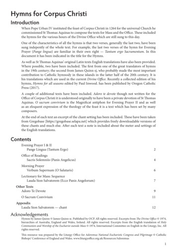
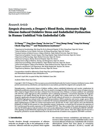
The Scientific World Journaland SD (10–50 𝜇g/mL) alone and pretreated with SD (10–50 𝜇M) in glucose (25 and 50 mM) induced cells or anisovolumetric solvent control (0.1% DMSO) for 24 or 48 h.The cell number was measured based on the ability ofmitochondria in viable cells to reduce MTT as previously described [17]. The cell number index was calculated as the absorbance of treated cells/control cells 100%.2.5. Measurement of Intracellular ROS. Starved HUVECs (2 105 cells/well) were loaded with DCF-DA (20 𝜇M) for 20 min.After treatment with SD (50 𝜇g/mL) for 2 hr, cells werestimulated with glucose (25–75 mM) for 24 hr, washed withPBS, and then detached using trypsin. Levels of intracellularROS were detected by flow cytometry (Beckman Coulter).All experiments were repeated at least four times to ensurereproducibility.2.6. Determination of Nitrite Production. HUVECs culturedin 12-well plates were washed twice with Hanks balanced saltsolution (HBSS) and then incubated at 37 0.5 C in thesame buffer for 30 min with various concentrations of SD.Acetylcholine (30 𝜇M) was used as a positive control. Supernatants were collected and then injected into a nitrogen purgechamber containing vanadium(III) chloride in hydrochloricacid at 91 0.5 C. All NO metabolites can be liberatedas gaseous NO and reacted with ozone to form activatednitrogen dioxide that is luminescent in red and infraredspectra. The chemiluminescence was detected using a nitricoxide analyzer (NOA280, Sievers Instruments, Boulder, CO,USA) [20]. For calibration, the area under the curve wasconverted to nanomolar NO using a NaNO3 standard curve,and the final data were expressed as pmol/mg protein.2.7. Lipid Peroxidation Assay. Lipid peroxidation was assayedby the thiobarbituric acid (TBA) reaction method. The cellswere homogenized in ice-cold 1.15% KCl. The samples wereused to measure the malondialdehyde (MDA) formed in aperoxidizing lipid system. The amount of thiobarbituric acidreactive substance (TBARS) was determined using a standardcurve of 1,1,3,3-tetramethoxypropane.2.8. Western Blot Analysis. Western blot analysis was performed as previously described [21]. Lysates from eachsample were mixed with 6 sample buffer (0.35 M Tris,10% w/v SDS, 30% v/v glycerol, 0.6 M DTT, and 0.012% w/vbromophenol blue, pH 6.8) and heated to 95 C for 5 min. Proteins were separated by electrophoresis and transferred ontopolyvinylidene difluoride (PVDF) membranes for pERK1/2,p-eNOS, pNF-𝜅B, VCAM-1, PARP, Bax, and Bcl2. Themembranes were blocked with 5% nonfat milk in TBS-0.1%Tween 20 and sequentially incubated with primary antibodies and HRP-conjugated secondary antibodies followed byenhanced chemiluminescence (ECL) detection (AmershamBiosciences). BIO-PROFIL Bio-1D light analytical software(Vilber Lourmat, Marne La Vallee, France) was used for thequantitative densitometric analysis. Data of specific protein3levels are presented as relative multiples in relation to thecontrol.2.9. Statistical Analyses. The experimental results areexpressed as the mean SEM and are accompanied by thenumber of observations. For analysis of the results, a one-wayanalysis of variance (ANOVA) test was performed usingSigma Stat v3.5 software. When group comparisons showeda significant difference, the Student-Newman-Keuls test wasused. A 𝑃 value of 0.05 was considered to be statisticallysignificant.3. Results3.1. Effects of SD on HUVEC Cell Viability. The cytotoxiceffect of various concentrations of SD and glucose onHUVECs cells was measured by MTT assay. The resultsindicated that SD did not affect the viability of HUVECat the used concentrations of 0–50 𝜇g/mL for 24 and 48 h(Figures 1(a) and 2(b)). However, treatment of cells withglucose (5.5–150 𝜇M) decreased the cell viability of HUVECin a concentration dependent manner. Interestingly, cellssimultaneously incubated with glucose (25 and 50 mM) andSD (0–50 𝜇g/mL) increased cell viability in a concentrationdependent manner (Figures 1(c), 1(d), 1(e), and 1(f)).3.2. SD Inhibited HG-Induced ROS and NO Production. Toinvestigate the effectiveness of SD in inhibiting HG-inducedROS formation in HUVECs, a cell-permeative ROS-sensitivedye, DCFDA (nonfluorescent in a reduced state but fluorescent upon oxidation by ROS), was used. The intracellular levelof ROS in HUVECs increased concentration dependentlyfollowing incubation with high glucose (25–75 mM) compared with 5.5 mM glucose. Incubation of HUVECs with SDresulted in a marked reduction of HG-induced intracellularROS generation (Figure 2(a)). Contrary to our expectations,glucose increased NO production instead of reducing it;however, when HUVECs were treated with SD plus glucose,SD completely abrogated the production of NO induced byglucose (Figure 2(b)).3.3. SD Inhibited HG-Induced MDA Production. Studieshave indicated that damaging cell membranes may cause adecrease of cell viability through peroxidation of membranelipids. Therefore, we projected to estimate the levels of MDAin the present study. As shown in Figure 2(c), MDA, a markerof lipid peroxidation, was markedly elevated in HG-inducedHUVEC. However, treatment of SD (50 𝜇g/mL) significantlyattenuated the elevation of MDA concentration in HGstimulated HUVEC.3.4. SD Enhanced the Phosphorylation of eNOS. The resultshowed that SD (30 and 50 𝜇g/mL) alone had no influence onthe protein expressions of phosphorylated eNOS at Ser1177,whereas high glucose leads to a significant decrease in theexpression of eNOS (Figure 3(a)). Treatment with SD significantly attenuated the decreased level of eNOS expression.
4The Scientific World Journal120HUVEC, cell viability (% of control)HUVEC, cell viability (% of control)12010080604020001020305010080604020001024 h48 h 80 60402005.5255075100150120100 302010Glu 50 mM24 hHUVEC, cell viability (% of control)HUVEC, cell viability (% of control) 8040Glu 25 mM24 h402005.52550751001201101009080706040 150 3020100Glu 25 mM48 hGlu 50 mM48 hGlu 5.5 mMSD 30 𝜇g/mLGlu 5.5 mMSD 30 𝜇g/mLSD 0 𝜇g/mLSD 50 𝜇g/mLSD 0 𝜇g/mLSD 50 𝜇g/mLSD 10 𝜇g/mL (d)900 Glucose (mM)48 h120 60(c)100 80Glucose (mM)24 h11050(b)HUVEC, cell viability (% of control)HUVEC, cell viability (% of control)(a) 30SD (𝜇g/mL)12010020SD (𝜇g/mL)SD 10 𝜇g/mL(e)(f)Figure 1: Effects of SD on the cell viability of human umbilical vein endothelial cells (HUVEC): ((a), (b)) the viability of HUVECs duringtreatment with various concentrations (10–50 𝜇g/mL) of SD for 24 and 48 h; ((c), (d)) the viability of HUVECs during treatment with variousconcentrations (25–150 mM) of glucose for 24 and 48 h; ((e), (f)) the viability of HUVECs upon treatment with various concentrations (0–50 𝜇g/mL) of SD in glucose-induced (25 and 50 mM) HUVECs for 24 and 48 h. Data are shown as the mean SEM of three independentexperiments. 𝑃 0.05; 𝑃 0.01, compared with the glucose treated group.
The Scientific World Journal5160160140 140120######1008060 120Nitrite (ng/mL)Intercellular ROS (% of control)180100#####8060404020200Glucose (mM) 5.52550752.52550750Glucose (mM) 5.52550752.5255075 SD (50 𝜇g/mL) SD (50 𝜇g/mL)(a)(b)10 MDA (nmol/mg)8 64######20Glucose (mM) 5.52550752.5255075 SD (50 𝜇g/mL)(c)Figure 2: Effects of SD on HG-induced formation of ROS, NO, and LPO in HUVECs: (a) ROS production was determined as described inMaterials and Methods; (b) the nitrite concentration in the culture medium was determined by Griess reagent; and (c) lipid peroxidation wasassayed by measuring the amount of TBARS formation (malondialdehyde, MDA); Data are shown as the mean SEM of three independentexperiments. 𝑃 0.05; 𝑃 0.01, compared with the normal group; # 𝑃 0.05; ## 𝑃 0.01, compared with the HG-treated group.3.5. SD Inhibited the Phosphorylation of ERK and NF-𝜅B. Todetermine whether SD affects the activation of the MAPKpathway, we analyzed the phosphorylation levels of pERKMAPK. First, HUVECs were pretreated with SD (30 and50 𝜇g/mL) for 30 min and then stimulated with 50 mMglucose for 1 h. The HG-induced increased phosphorylationof pERK was inhibited by SD in a concentration-dependentmanner (Figure 3(b)) and restored it to the level in cellsexposed to 5.5 mM glucose. Moreover, we measured NF𝜅B activation in HG-induced HUVECs, as it is suggestedthat increased ROS production in HG-induced HUVEC maypartially cause the activation of NF-𝜅B. As we expected, theexpression of NF-𝜅B was increased markedly in HUVEC cellstreated with high glucose (50 mM). In addition, pretreatmentwith SD (30 and 50 𝜇g/mL) markedly inhibited the HGinduced expression of NF-𝜅B concentration dependently(Figure 3(c)).3.6. Effects of SD on HG-Induced Endothelial Cell AdhesionMolecules. To examine whether glucose induces expression of VCAM-1 and E-selectin in HUVECs, we cultured HUVECs at normal glucose (5.5 mM) and high glucose (50 mM) concentrations for 24 h. Immunoblot analysisshowed that stimulation of HUVECs with high concentrations of glucose increased the production of VCAM-1 andE-selectin (Figure 4(a)). To further determine whether SDcan inhibit the expression of endothelial adhesion molecules,HUVECs were pretreated with SD at concentrations of 30and 50 𝜇g/mL and stimulated with 50 mM glucose for 24 h.As shown in Figure 4(a), pretreatment of SD in HUVECsignificantly inhibited the HG-induced expression of ICAM-1and E-selectin in a concentration manner.3.7. Effects of SD on HG-Induced PARP-1 Overactivation. Aspreviously described, the full PARP-1 protein is 116 kDa, and
6The Scientific World JournalVCAM-1p-eNOSE-selectin𝛽-ActinGlucose (mM)5.55.55.5505050SD (𝜇g/mL) 3050 3050(a)𝛽-ActinGlucose (mM)5.55.5SD (𝜇g/mL) 305.550505050 e (mM)5.55.55.5505050SD (𝜇g/mL) 3050 3050(b)𝛽-ActinGlucose (mM)5.5505.55050SD (𝜇g/mL) 503050(b)p-NF𝜅B𝛽-ActinBcl2Glucose (mM)5.5505.55050SD (𝜇g/mL) 503050(c)Figure 3: Effects of SD on HG-induced phosphorylation of eNOS,ERK, and NF-𝜅B in HUVECs: HUVECs (2 105 cells/well) werepretreated with SD (30 and 50 𝜇g/mL) for 2 h and then treated withglucose (50 mM) for 30 min to detect the phosphorylation of (a)eNOS, (b) ERK1/2, and (c) NF-𝜅B. The 𝛽-actin was used as aninternal control.the 85 kDa cleavage product is an early marker of apoptosis.In this experiment, we detected both the 116 kDa full PARPprotein and the 85 kDa breakdown product. The resultsrevealed that exposure of HUVEC to high glucose concentrations (50 mM) for 24 h resulted in a significant decrease in theamount of PARP-116 kDa protein and increased their breakdown product of 85 kDa protein. Interestingly, treatment ofSD alone significantly enhanced the amount of PARP-116full protein in HUVEC. Moreover, the amount of 85 kDaPARP breakdown product induced by high glucose (50 mM)was markedly reduced in SD treated cells in a concentrationdependent manner (30 and 50 𝜇g/mL) (Figure 4(b)).3.8. Effects of SD on HG-Induced Bcl-2/Bax Expression. Asshown in Figure 2(a), the inhibition of HG-induced ROS bySD led us to evaluate any possible effects of SD on othermediators of oxidative stress-induced apoptosis. The resultsrevealed that the ratio between the anti- and proapoptoticmediators Bcl2 and Bax was not much affected by SDtreatment alone, especially that Bcl-2 did not alter betweenBox𝛽-ActinGlucose (mM)5.55.55.5505050SD (𝜇g/mL) 3050 3050(c)Figure 4: Effects of SD on HG-induced expression of VCAM1, E-selectin, PARP-1, and Bax/Bcl2 in HUVEC: HUVECs (2 105 cells/well) were pretreated with SD (30 and 50 𝜇g/mL) for 2 hand then treated with glucose (50 mM) for 30 min to detect theexpression of (a) VCAM-1 and E-selectin, (b) PARP-1, and (c)Bax/Bcl2. The 𝛽-actin was used as an internal control.normal and SD treated cells (Figure 4(c)). Moreover, HGinduced Bax was moderately reversed by SD, which indicatedthat Bax might play a role in this effect.4. DiscussionIn diabetes, hyperglycemia causes vascular complicationsthat are produced mainly by the overproduction of ROS [22].Dysfunction and activation of the endothelium are regardedas important factors in the pathogenesis of vascular diseasein diabetes mellitus [23]. In this work, we evaluated theprotective effect of Sanguis draconis (SD), a kind of red resin,against the dysfunction and activation of endothelial cellsinduced by high concentrations of glucose. The major findingof this study is that SD was capable of attenuating the increaseof ROS, NO, and MDA and inhibits overactivation of PARPs,VCAM-1, NF-𝜅B, and Bcl2 in cells exposed to high glucoseconcentration, and our data contribute in part to elucidate themolecular mechanisms involved in this effect.
The Scientific World JournalOxidative stress, a hallmark of high glucose-inducedendothelial dysfunction, was significantly attenuated by SDtreatment in the current study. There is growing evidence thatoxidative stress is involved in the pathogenesis of diabeticcomplications [24] and an acute increase of glycemia isreported to be accompanied by oxidative stress generation[25]. Lipid peroxidation (LPO), a process induced by freeradicals, leads to oxidative deterioration of polyunsaturatedlipids. Malondialdehyde (MDA), a secondary product oflipid peroxidation, is used as an indicator of tissue or celldamage. In previous studies, it was found that MDA levelwas increased in HUVECs when cells were incubated withglycated protein and iron [26]. An increased production ofROS has been reported in inflammatory responses, and itacts as a potential mediator of diabetes mellitus-associatedvascular diseases. ROS is also considered to be of the important mediators of several biologic responses, including cellproliferation and extracellular matrix deposition. A recentstudy showed that hyperglycemia stimulates the generationof free radicals and oxidative stress in various cell types [27].In this study, an elevation in HG-mediated production ofcellular MDA and ROS indicate that oxidative stress inducedby high glucose in HUVEC is important in determiningthe character of diabetic complication as well as vascularinflammation. Interestingly, we found that SD treatmentpotentially inhibited the high glucose-induced MDA andROS formation, suggesting a role of protecting oxidativeinsults in HG-induced HUVEC.Bioavailability of endothelial NO observed in individuals with hyperglycemia is considered to be a critical andinitiating factor in the pathogenesis of diabetic vascularcomplications [28]. NO is synthesized in endothelial cellsfrom the substrate L-arginine via endothelial NO synthase(eNOS) [26], and this enzyme plays an important role forthe maintenance of cardiovascular function by producingNO synthesis. Nevertheless, this enzyme can be detached,leading to the generation of superoxide instead of NOunder certain pathological circumstances and oxidative stressconditions [29]. Our previous study showed that SD inhibitsNO production induced by IL-1𝛽/IFN-𝛾 in endothelial cells[17]. The results of this study revealed that high glucosecauses a significant degree of oxidative stress, which leads toincreases in the formation of NO; convincingly pretreatmentof SD could reverse the effect of high glucose by suppressingNO production. These results indicate that SD may have aprotective effect on HUVEC by preventing or decreasing theinjury of endothelial cells by interfering with NO synthaseand NO.In HUVEC, high glucose-induced cellular damage couldlead to cell apoptosis probably viaERK activation. To makethis hypothesis solid, we next aimed to gain insight intothe cell signaling pathways mediating the action of SDon HG-induced human endothelial cells. We found thatHG potentiated the activation of both ERK1/2 and NF𝜅B, indicating that overactivation of endothelial signalingmolecules can be triggered by HG stimulations. ERK isthe signal cascade involved in the protection of oxidativedamage and its activation is generally thought to mediatecell survival [30]. In agreement with our study, a previous7study has shown that high D-glucose activates the ERK1/2,NF-𝜅B, and iNOS when this signaling pathway is formerlytriggered by an exogenous inflammatory stimulus in cultured HUVEC [31]. Chronic activation of NF-𝜅B is associated with various pathological conditions, including insulinresistance. Human subjects with type 2 diabetes exhibitincreased activity of NF-𝜅B in muscle that directly correlateswith impaired insulin mediated glucose disposal. Enduringhyperglycemia and/or extreme perturbations in glycemiaare common generators of oxidative stress, which has beenshown to induce insulin resistance through activity of NF𝜅B. Interestingly, administration of the common salicylateaspirin (7.0 g/day) in patients with type 2 diabetes enhancesglucose homeostasis and peripheral insulin sensitivity, at leastin part through the inhibition of NF-𝜅B nuclear activity[32]. Targeted manipulation or ablation of NF-𝜅B, therefore,remains at the forefront of innovative treatments for diabetesand inflammatory pathologies. Our study also shows thathigh glucose elevated the expression of NF-𝜅B in HUVECand these effects were blocked by SD treatment. Changes inexpression of activated ERK and NF-𝜅B closely reflect thecell damage; such results suggest that high glucose-inducedHUVEC cell damage is protected, at least partly, by inhibitingERK and NF-𝜅B activation.In agreement with others [33], we have further found thatERK1/2 and NF-𝜅B activation was responsible for endothelialVCAM-1 and E-selectin induction. Other signaling molecule,like PARP-1, has been reported as promoters of ICAM-1expression in different vascular cell types [34] and mighttherefore be involved in the induction of ICAM-1 in HGinduced HUVEC. Adhesion induced by glucose was almostcompletely abrogated by SD when added before the addition of glucose, indicating that SD can have a preventive and protective effect against the endothelial activationinduced by hyperglycemia. SD treatment was also capableof decreasing the glucose-induced expression of VCAM1 and E-selectin. Similar results have been described in aprevious study in endothelial cells, where they have reportedthat dehydroepiandrosterone (DHEA), an adrenal steroidabrogated expression of adhesion molecules in HUVEC,activated with either TNF-𝛼 or oxidized low density proteins[35]. Recently, various phytochemicals have been shown toinhibit the expression of adhesion molecules in endothelial cells. For instance, epigallocatechin-3-O-gallate (EGCG)inhibits angiotensin II-induced adhesion molecule expression by inhibiting p38 MAPK and ERK1/2 phosphorylation[36]. Anthocyanins inhibited TNF-𝛼-induced ICAM-1 andVCAM-1 expressions via the NF-𝜅B-dependent pathway[37]. Phloretin inhibited the TNF-𝛼-stimulated expressionof adhesion molecules without activating NF-𝜅B [38]. Grapeseed proanthocyanidin extract inhibited VCAM-1 expression in HUVECs via the NF-𝜅B-independent pathway [39].In this study, the observed effect of SD on suppressingHG-induced expression of VCAM-1 and E-selectin mayperhaps via inhibition of the ERK1/2 phosphorylation andNF-𝜅B-dependent pathway. Thus, development of therapeutic drugs for diabetic complications targeting CAMsexpression may prove useful in the prevention of vascularinflammation.
8The Scientific World JournalMoreover, recent studies have demonstrated that activation of PARP is associated with the pathogenesis of diabetesand diabetic complications, including cardiovascular dysfunction. PARP has been shown to act as a coactivator in NF𝜅B-mediated transcription [40]. Some studies have indicatedthat PARP inhibitor prevents the diabetes-induced elevation in circulating nitrite levels in streptozotocin-induceddiabetes. A study has reported that PARP deficiency suppresses
Department of Surgery, Mackay Memorial Hospital, Taipei , Taiwan Mackay Junior College of Medicine, Nursing, and Management, Taipei , Taiwan Department of Cardiology, School of Medicine, Taipei Medical University, Taipei , Taiwan . and/or Jackson-ImmunoResearch (West Grove, PA, USA).