Transcription
Molecular Vision 2011; 17:1231-1238 http://www.molvis.org/molvis/v17/a138 Received 5 July 2010 Accepted 1 May 2011 Published 6 May 2011 2011 Molecular VisionAsymmetric phenotype of Axenfeld-Rieger anomaly and aniridiaassociated with a novel PITX2 mutationSimon K. Law, Maha Sami, Natik Piri, Anne L. Coleman, Joseph CaprioliJules Stein Eye Institute, David Geffen School of Medicine, University of California, Los Angeles, CAPurpose: To evaluate the asymmetry of the anterior segment phenotype between the two eyes of a patient with AxenfeldRieger syndrome (ARS).Methods: The entire database of a tertiary glaucoma practice was screened for patients with ARS. The medical recordsof patients with ARS were reviewed. The clinical characteristics of ocular examination of the two eyes of each patientwere recorded and compared. Dental and medical information were also reviewed where available. The anterior segmentphenotype was tabulated to assess asymmetry. Asymmetric anterior segment characteristics of patients with ARS werecompared with reported cases in the literature.Results: Eight patients with ARS were identified from screening of more than 5,000 patients of a tertiary glaucomapractice. All patients had Axenfeld-Rieger anomaly in both eyes except one patient presented with an asymmetricphenotype of the anterior segment with features of Axenfeld-Rieger anomaly in one eye, but aniridia in the other eye. Thispatient had non-ocular findings including flat midface, hypodontia with lack of an upper incisor, and redundantperiumbilical skin, typical for ARS. A heterozygous C T nucleotide substitution was identified in exon 4 of the pituitaryhomeobox 2 (PITX2) gene, resulting in the replacement of a glutamine codon (CAG) with a stop codon (TAG) at aminoacid position 67. This mutation is denoted c.199C T at the cDNA level or p.Gln67Stop (or Q67X) at the protein level.Only three cases with asymmetric anterior segment phenotype between the two eyes of a patient with AGS have beenreported in the literature.Conclusions: Variability in phenotype may occur between the two eyes of an individual affected by ARS. The currentcase undermines the advantage of genetic testing to correctly diagnose a rare disease.Axenfeld-Rieger syndrome (ARS) is characterized by avariable combination of anterior segment dysgenesis, dentalanomalies, and umbilical hernia. Reported anterior segmentfeatures in ARS include iris stromal hypoplasia (poordevelopment of the anterior stroma of the iris),iridogoniodysgenesis (hypoplastic iris with abnormal“wooly” iridocorneal angle tissue), corectopia (eccentricpupil), polycoria (iris tears), ectropion uveae (eversion of thepupillary margin), posterior embryotoxon (prominent andcentrally displaced Schwalbe’s line), and iris strands bridgingthe iridocorneal angle to the trabecular meshwork [1]. Abouthalf of the patients develop secondary angle closure glaucoma[2]. Although there are a few cases of an asymmetricphenotype between fellow eyes of a patient with ARS, similaranterior segment characteristics of fellow eyes are the usualpresentation [3,4]. The purpose of this study was to evaluatethe asymmetry of phenotype of the anterior segment betweenthe two eyes of a patient with Axenfeld-Rieger syndrome(ARS), and present a case of ARS with asymmetric anteriorsegment phenotype and the associated genotype.Correspondence to: Simon K. Law, M.D., 100 Stein Plaza #2-235,Jules Stein Eye Institute, Los Angeles, CA, 90095; Phone: (310)794-1477; FAX: (310) 794-6616; email: law@jsei.ucla.eduMETHODSThe Institutional Review Board at University of CaliforniaLos Angeles, Los Angeles, CA approved the current study andit was performed in accordance with the Declaration ofHelsinki. The entire database of a tertiary glaucoma practicewas screened to identify patients with ARS. The medicalrecords of patients with ARS were retrospective reviewed.The medical data collected included the eye examination, anddental and medical records. The clinical characteristics of thetwo eyes of each patient with ARS were recorded andcompared. The anterior segment phenotype was tabulated toassess asymmetry. A literature review of asymmetricphenotypes of the anterior segment of patients with ARS wasconducted.In review of the preliminary data collected, one patienthad markedly asymmetric anterior segment phenotypebetween the two eyes. Genetic testing was ordered for thispatient. PCR and DNA sequencing was performed by GeneDx(Gaithersburg, MD). Primer sequences and PCR conditionscannot be disclosed here since GeneDx has proprietary rightson this information.RESULTSEight patients with ARS were identified from screening themedical records of more than five thousand patients of atertiary glaucoma practice. All patients had symmetric1231
1232MaleFemale2003M.A.TABLE 1. CLINICAL PRESENTATIONS OF THE TWO EYES OF AN INDIVIDUAL WITH AXENFELD-RIEGER SYNDROME.AnteriorAnteriorAdditionalEye withCentral cornealDentalsegmentsegmentocular findingsGlaucomathicknessAbnormalitiesphenotype ofphenotype of(Right/Left eye)first eyesecond eyein micronAxenfeld &Axenfeld &Both eyes560 / erOptic nerveBoth eyes616 / 616Unknownanomalyanomalycoloboma (righteye)AxenfeldAxenfeldMicrocornea andNone576 / 606Presenceanomalyanomalyscleral thinning(both eyes)Axenfeld &Axenfeld &Corneal scar andBoth eyes571 / left cornealPresenceRiegerRiegerscleral thinningscaranomaliesanomalies(left eye)Axenfeld &Axenfeld &Both gerCataract andBoth eyesUnknownanomalyanomalycorneal scar (botheyes)Axenfeld &Axenfeld &Both eyes626 / sAxenfeld &AniridiaBoth eyes664 / alitiesSigns of Axenfeld anomaly include: prominent schwalbe line, iris adhesion to cornea and trabecular meshwork. Signs of Rieger anomaly include: iris hypoplasia,correctopia or polycoria, ectropion uveae. *J.B. and M.B. had a mother-and-son .C.Year ofBirthPatientMolecular Vision 2011; 17:1231-1238 http://www.molvis.org/molvis/v17/a138 2011 Molecular Vision
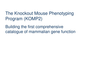
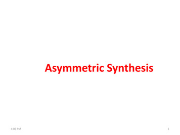
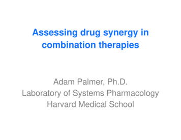
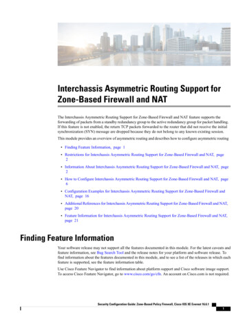
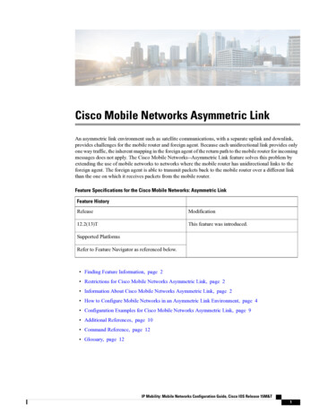
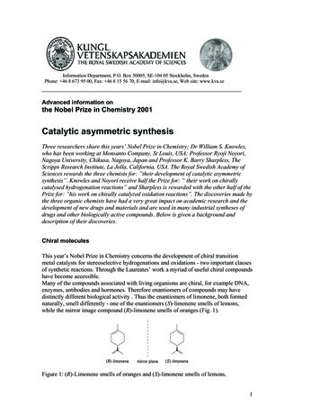
Molecular Vision 2011; 17:1231-1238 http://www.molvis.org/molvis/v17/a138 Axenfeld anomaly and/or Rieger anomaly in both eyes, exceptfor one patient who presented with an asymmetric phenotypeof the anterior segment with Axenfeld-Rieger anomaly in oneeye, but aniridia in the other eye. Table 1 summarizes theclinical characteristics of the ocular examination of the eightpatients. Except for the case (Patient MA) with a markedlyasymmetric phenotype between the two eyes, the rest of thepatients had iris stromal hypoplasia, corectopia, polycoria,ectropion uveae, and posterior embryotoxon in both eyes.Seven of the eight patients had glaucoma in both eyes. Onepatient who had mild Axenfeld anomaly had no glaucoma ineither eye. Six patients had central corneal thickness (CCT)measured for at least one eye. The average CCT (standarddeviation) of the right eye was 602 (40) microns and left eyewas 613 (57) microns.In this retrospective review, the seven patients that hadbilateral ocular findings typical for ARS received no geneticwork up. However, the patient with the unusual asymmetricphenotype with features of Axenfeld-Rieger anomaly in oneeye and aniridia in the other eye posed a diagnostic uncertaintyand systemic and genetic evaluation were performed. Detailsof the clinical presentation and evaluation of this patient arereported as follows. 2011 Molecular Vision20/40 in the left, with moderate hyperopia in the right and mildmyopia in the left. The right eye was amblyopic, and wasundergoing occlusion therapy. She has no strabismus ornystagmus. Slit lamp examination of the right eye revealedpolycoria, adhesion of iris strands to the peripheral cornea,and posterior embryotoxon. (Figure 1) Examination of the lefteye revealed a small iris stub resembling aniridia, and a similarappearance was noted on an old photograph taken when shewas 3 months old (Figure 2 and Figure 3).Corneal pachymetrywas 664 microns in the right and 683 microns in the left. Thehorizontal corneal diameter measured 11 mm in both eyes.Axial length by ultrasonic measurement was 21.78 mm in theright eye and 24.46 mm in the left eye. Gonioscopy revealedextensive iris strands bridging the iridocorneal angle to thetrabecular meshwork in the right and complete angle closureby the stub of the iris in the left. Optic disc examinationrevealed a cup/disc ratio of 0.30 in the right eye and 0.95 inCase (Patient M.A.): The patient was referred to ourinstitute when she was four years old with an uncertaindiagnosis of ARS versus aniridia. Shortly before the referral,she was found to have an increased intraocular pressure (IOP)in both eyes: 27 mmHg in the right and 46 mmHg in the left,and was started on glaucoma medications. When she wasusing timolol and dorzolamide as fixed combination andbimatoprost eye drops in both eyes, the IOP measured byGoldmann tonometry was 14 mmHg in the right and 32 in theleft. Her spectacle-corrected vision was 20/80 in the right andFigure 2. Anterior segment photograph of the left eye shows aniridia.Figure 1. Anterior segment photograph of the right eye showspolycoria, adhesion of iris strands with peripheral cornea, andposterior emblyotoxon.Figure 3. Old photograph of patient when she was 3 months oldshows similar anterior segment appearance.1233
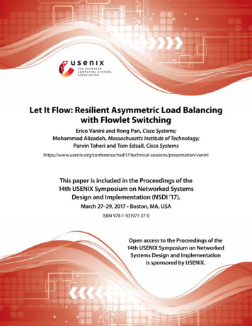
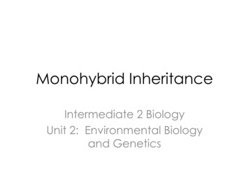
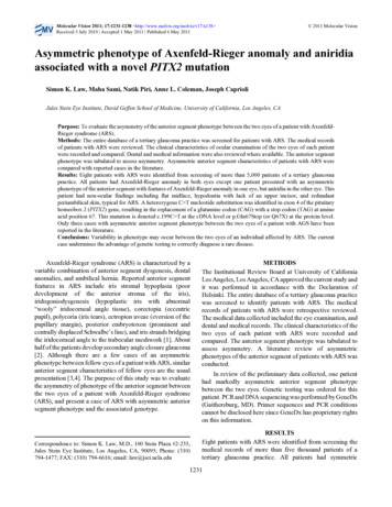
Molecular Vision 2011; 17:1231-1238 http://www.molvis.org/molvis/v17/a138 the left, and there was no retinal pathology (Figure 4 andFigure 5). Further physical examination revealed a flatmidface, hypodontia with lack of an upper incisor, andredundant periumbilical skin (Figure 6 and Figure 7).Ultrasonic examination of kidneys was normal and there wasno cardiac abnormality. She has normal intelligence. Otherfamily members including both parents and a younger brotherwere examined and no similar anomaly was noted. However,the maternal uncle has Russell-Silver syndrome and was notavailable to be examined.Parents of the patient underwent genetic counseling andconsented to the genetic testing of the patient. Genomic DNAFigure 4. Optic disc photograph of the right eye shows normal neuralrim.Figure 5. Optic disc photograph of the left eye shows severe loss ofneural rim. 2011 Molecular Visionfrom the patient’s blood sample was PCR-amplified andscreened for mutations in pituitary homeobox 2 (PITX2),forkhead box C1 (FOXC1), and paired box 6 (PAX6) genesthat are known to be associated with ARS. Coding exons ofPITX2b (NM 153426) were analyzed in this study. Aheterozygous C T nucleotide substitution (a nonsensemutation) was identified in exon 4 of PITX2, resulting in thereplacement of a glutamine codon (CAG) with a stop codon(TAG) at amino acid position 67. This mutation is denoted c.199C T at the cDNA level or p.Gln67Stop (or Q67X) at theprotein level (Figure 8). No sequence alterations weredetected in FOXC1 and PAX6.She subsequently received a tube shunt procedure in theleft eye and an additional tube shunt procedure 2 years laterFigure 6. Facial photograph of patient shows flat midface andhypodontia lacking of an upper incisor.Figure 7. Abdominal photograph of patient shows redundantperiumbilical skin.1234
Molecular Vision 2011; 17:1231-1238 http://www.molvis.org/molvis/v17/a138 2011 Molecular Visionwith pressure of the left eye controlled at 20–22 mmHg onminimum topical medication.in exon 4 of PITX2 identified has not previously been reported,to our knowledge [7].Literature review: A literature review identifiedasymmetric phenotypes of the anterior segment in threepatients with ARS (Table 2). In 1883, Vossius [5] describeda nine-year-old girl with bilateral iris defects associated withdental anomalies. One eye had an ectopic pupil (corectopia)with full-thickness iris stromal defects (polycoria) while thefellow eye had a near-total absence of iris tissue (aniridia).The other two patients were reported by Perveen et al. [4] in2000. These two patients had unilateral Peters anomaly in oneeye but the other eye had Rieger anomaly or iris hypoplasia[4].PITX2 mutations are primarily responsible for ARS buthave also been associated with Peters anomaly, irishypoplasia/iridogoniodysgenesis syndrome, aniridia, and ringdermoid of the cornea [4,7,12]. Intragenic mutations ofPITX2 have been described in more than 40 ARS patients todate and include missense, nonsense, splice-site mutations,and deletions/insertions/duplications. There is no apparentcorrelation between the mutation location in PITX2 and theseverity of the clinical phenotype. However, thecharacterization of several missense mutations indicates thatthe severity of the phenotype can be correlated to theremaining function of the mutant PITX2. In heterozygotes,the function of one wild-type PITX2 allele alone results in thesevere ARS form. Contribution of the mutant allele wouldresult in the milder iris hypolasia or iridogoniodysgenesissyndrome forms, depending on the degree of residual activity[13]. A nonsense mutation in PITX2 found in patient M.A. ispredicted to result in degradation of the mutant mRNAthrough the nonsense-mediated decay pathway that preventthe synthesis of the truncated protein [14]. Even if thetruncated 66 amino acid (aa) long protein is synthesized andstable, it would be shorter than the native protein by 251 aaand would lack helix 2 and helix 3/4 of the DNA bindinghomeodomain involved in transcriptional regulation and theOAR domain in the COOH-terminal end that has beenproposed to increase the DNA binding and transcriptionalactivity of PITX2 during development [15]. The absence ofclinical and genetic abnormalities in other family memberssuggests that this may be a de novo genomic alteration [3].Finally, since PITX2 is known to inhibit FOXC1 activity[16], the mechanism for PITX2-related severity of the ocularphenotype in this patient may be the simultaneousconsequence of PITX2 haploinsufficiency and a gain offunction of FOXC1.DISCUSSIONAlthough tremendous variability in phenotype within a singlefamily with ARS has been observed, phenotypes between thetwo eyes of an affected individual are usually similar [1].Seven of the eight patients with ARS in our tertiary glaucomapractice presented with a similar anterior segment phenotypebetween the two eyes. However, one patient presented withanterior segment features of Axenfeld-Rieger anomaly in oneeye, and aniridia in the other eye. In 1883, Vossius [5]described a nine-year-old girl with Rieger anomaly in one eyeincluding an ectopic pupil with full-thickness iris stromaldefects, while the fellow eye had a near-total absence of iristissue (aniridia). To our knowledge, description of theasymmetric phenotype of Axenfeld-Rieger anomaly in oneeye and severe iris hypoplasia resembling aniridia in the othereye has not been previously reported.It is known that mutations in the transcription factorPITX2 and FOXC1 genes lead to ARS, while mutations of thePAX6 gene underlies many cases of aniridia [6,7]. However,Henkind and associates [8] described a family with ARS thatincluded two members who appeared to have aniridia. Inaddition, cases of ARS have been described associated with adeletion of PAX6 [6,9,10], and cases of bilateral aniridia withproven PITX2 and FOXC1 mutations [4,11]. Because of themixed anterior segment anomalies between the two eyes ofthis patient and the association of Wilm’s tumor in sporadiccases of aniridia, genetic testing was conducted for PITX2,FOXC1, and PAX6 [6]. The nonsense mutation, Q67X,Figure 8. Sequencing analysis of the Axenfeld-Rieger syndromepatient’s PITX2 gene with the C T mutation.Mutations of PITX2 are usually associated with fullspectrum ARS phenotypes that include both ocular andnonocular features, as in patient M.A [1]. Intriguingly, PITX2is also involved in left-right polarity determination, yetasymmetry defects are not a usual feature of ARS [3,4,17].Iris abnormalities are usually stationary, but rarely have beendemonstrated to change over time [18]. In our patient, theaniridia in the left eye appears to remain unchanged sinceinfancy by comparison with old photographs. Aniridia is apanocular disease with associated ocular and visual defectssuch as corneal opacification, cataract, foveal dysplasia, opticnerve hypoplasia, and nystagmus, but none of which is foundin this patient [6]. It is possible that the different phenotypesof iris abnormality in the two eyes of this patient fall within aspectrum of iris defect in ARS.The major clinical concern in ARS is the risk ofdeveloping sight-threatening glaucoma, which is estimated to1235
1236Signs of Axenfeld anomaly include: prominent schwalbe line, iris adhesion to cornea and trabecular meshwork. Signs of Rieger anomaly include: iris hypoplasia,correctopia or polycoria, ectropion uveae. Signs of Peters anomaly were not explained in the report by Perveen et al. [4].TABLE 2. CLINICAL PRESENTATIONS AND MUTATIONS OF CASES WITH ASYMMETRIC ANTERIOR SEGMENT PHENOTYPE BETWEEN THE TWO EYES OF AN INDIVIDUAL WITH AXENFELD-RIEGER SYNDROME alitiesabnormalitiesphenotype ofphenotype offindingsfirst eyesecond eye[5]Near totalfull thickness irisUnknownUnknownUnknownUnknownUnknownabsence of irisstromal defects(aniridia)[4]Peters anomaly Rieger anomaly,Mild unilateralAbsencePresencePresencePITX2 Ivs 3 ( 2); A Tanterior polarfovealcataracthypoplasia[4]AxenfeldPeters anomalyNoneAbsencePresenceUnknownPITX2 C ins 1083anomaly, irishypoplasiaCurrent ITX2 c. 199 C T (Q67X)anomaly,AxenfeldanomalyMolecular Vision 2011; 17:1231-1238 http://www.molvis.org/molvis/v17/a138 2011 Molecular Vision
Molecular Vision 2011; 17:1231-1238 http://www.molvis.org/molvis/v17/a138 occur in 50% of the patients [1,2]. Although physicalocclusion of the angle structure is not prerequisite for elevatedIOP and glaucoma in ARS, the severity of glaucoma doescorrelate with the level of iris insertion into the angle [2]. Inthe seven patients of this series who had glaucoma in botheyes, the eye with more extensive areas of angle closurepresented with higher IOP and was more resistant to medicaland surgical management.Patients in this series had a thicker central corneacompared to normal adults, and a thick central cornea isassociated with artificially higher IOP estimation byapplanation. Pachymetry of patients with ARS is rarelyreported in the literature. In mouse models withoverexpressing PITX2A isoform in the cornea, cornealhypertrophy was noted together with iridocorneal adhesion,gray and tearing eyes, and severe apoptosis-associated retinaldegeneration [19]. In human with ARS-causing PITX2mutation, V45L, elevated transactivating properties in cellculture were found [20]. It is suggestive that increased as wellas reduced PITX2 activities are deleterious duringdevelopment [17]. Further functional studies may clarify if thenovel mutation of PITX2 found in our patient is associatedwith a gain-of-function leading to an increase of thickness ofthe cornea.In summary, although phenotypic presentation of ARS ishighly variable within a single family, only a few cases in theliterature had different phenotypes between the two eyes ofan individual. We presented a case of an asymmetricphenotype of Axenfeld-Rieger anomaly in one eye andaniridia in another of a patient with a novel nonsense pointmutation of PITX2. The current case undermines thevariability in phenotype even between the two eyes of anindividual affected by ARS and the advantage of genetictesting in correctly diagnosing a rare disease.ACKNOWLEDGMENTSThis study is supported in part by an unrestricted grant fromResearch to Prevent Blindness. No author has a proprietaryinterest in the development or marketing of any of the productsor devices mentioned in the study. 2011 Molecular CES1.2.3.4.Alward WL. Axenfeld-Rieger syndrome in the age of moleculargenetics. Am J Ophthalmol 2000; 130:107-15. [PMID:11004268]Shields MB. Axenfeld-Rieger syndrome: a theory ofmechanism and distinctions from the iridocorneal endothelialsyndrome. Trans Am Ophthalmol Soc 1983; 81:736-84.[PMID: 6676983]Hjalt TA, Semina EV. Current molecular understanding ofAxenfeld-Rieger syndrome. Expert Rev Mol Med 2005;7:1-17. [PMID: 16274491]Perveen R, Lloyd IC, Clayton-Smith J, Churchill A, vanHeyningen V, Hanson I, Taylor D, McKeown C, Super M,19.20.1237Kerr B, Winter R, Black GC. Phenotypic variability andasymmetry of Rieger syndrome associated with PITX2mutations. Invest Ophthalmol Vis Sci 2000; 41:2456-60.[PMID: 10937553]Vossius A. Congenitale abnormalien der iris. Klin MonatsblAugenheilkd 1883; 21:233-7.Prosser J, van Heyningen V. PAX6 mutations reviewed. HumMutat 1998; 11:93-108. [PMID: 9482572]Tümer Z, Bach-Holm D. Axenfeld-Rieger syndrome andspectrum of PITX2 and FOXC1 mutations. Eur J Hum Genet2009; 17:1527-39. [PMID: 19513095]Henkind P, Siegel IM, Carr RE. Mesodermal dysgenesis of theanterior segment: Rieger’s anomaly. Arch Ophthalmol 1965;73:810-7. [PMID: 14302514]Riise R, Storhaug K, Brondum-Nielsen K. Rieger syndrome isassociated with PAX6. Acta Ophthalmol Scand 2001;79:201-3. [PMID: 11284764]Hanson IM. PAX6 and congenital eye malformation. PediatrRes 2003; 54:791-6. [PMID: 14561779]Ito YA, Footz TK, Berry FB, Mirzayans F, Yu M, Khan AO,Walter MA. Severe molecular defects of a novel FOXC1W152G mutation result in aniridia. Invest Ophthalmol Vis Sci2009; 50:3573-9. [PMID: 19279310]Alward WL, Semina EV, Kalenak JW, Héon E, Sheth BP, StoneEM, Murray JC. Autosomal dominant iris hypoplasia iscaused by a mutation in the Rieger syndrome (RIEG/PITX2)gene. Am J Ophthalmol 1998; 125:98-100. [PMID: 9437321]Kozlowski K, Walter MA. Variation in residual PITX2 activityunderlies the phenotypic spectrum of anterior segmentdevelopmental disorders. Hum Mol Genet 2000; 9:2131-9.[PMID: 10958652]Culbertson MR. RNA surveillance. Unforeseen consequencesfor gene expression, inherited genetic disorders and cancer.Trends Genet 1999; 15:74-80. [PMID: 10098411]Amendt BA, Semina EV, Alward WL. Rieger syndrome: aclinical, molecular, and biochemical analysis. Cell Mol LifeSci 2000; 57:1652-66. [PMID: 11092457]Berry FB, Lines MA, Oas JM, Footz T, Underhill DA, Gage PJ,Walter MA. Functional interactions between FOXC1 andPITX2 underlie the sensitivity to FOXC1 gene dose inAxenfeld-Rieger syndrome and anterior segment dysgenesis.Hum Mol Genet 2006; 15:905-19. [PMID: 16449236]Lines MA, Kozlowski K, Walter MA. Molecular genetics ofAxenfeld-Rieger malformations. Hum Mol Genet 2002;11:1177-84. [PMID: 12015277]Judisch GF, Phelps CD, Hanson L. Rieger’s syndrome: a casereport with a 15-year follow-up. Arch Ophthalmol 1979;97:2120-2. [PMID: 508179]Holmberg J, Liu CY, Hjalt TA. PITX2 gain-of-function inRieger syndrome eye model. Am J Pathol 2004;165:1633-41. [PMID: 15509533]Priston M, Kozlowski K, Gill D, Letwin K, Buys Y, Levin AV,Walter MA, Héon E. Functional analyses of two newlyidentified PITX2 mutants reveal a novel molecularmechanism for Axenfeld-Rieger syndrome. Hum Mol Genet2001; 10:1631-8. [PMID: 11487566]
Molecular Vision 2011; 17:1231-1238 http://www.molvis.org/molvis/v17/a138 2011 Molecular VisionArticles are provided courtesy of Emory University and the Zhongshan Ophthalmic Center, Sun Yat-sen University, P.R. China.The print version of this article was created on 3 May 2011. This reflects all typographical corrections and errata to the articlethrough that date. Details of any changes may be found in the online version of the article.1238
Jules Stein Eye Institute, David Geffen School of Medicine, University of California, Los Angeles, CA Purpose: To evaluate the asymmetry of the anterior segment phenotype between the two eyes of a patient with Axenfeld-Rieger syndrome (ARS). Methods: The entire database of a tertiary glaucoma practice was screened for patients with ARS.