Transcription
ANRV255-CB21-25ARI8 September 200517:11Annu. Rev. Cell Dev. Biol. 2005.21:605-631. Downloaded from arjournals.annualreviews.orgby Stanford University Robert Crown Law Lib. on 03/01/09. For personal use only.Stem Cell Niche:Structure and FunctionLinheng Li and Ting XieStowers Institute for Medical Research, Kansas City, Missouri 64110;email: lil@stowers-institute.org, tgx@stowers-institute.orgAnnu. Rev. Cell Dev. Biol.2005. 21:605–31First published online as aReview in Advance onJuly 1, 2005The Annual Review ofCell and DevelopmentalBiology is online athttp://cellbio.annualreviews.orgdoi: 10.1146/annurev.cellbio.21.012704.131525c 2005 byCopyright Annual Reviews. All rightsreserved1081-0706/05/11100605 20.00Key Wordsadult stem cell, self-renewal, differentiation, multipotentiality,signalingAbstractAdult tissue-specific stem cells have the capacity to self-renewand generate functional differentiated cells that replenish lost cellsthroughout an organism’s lifetime. Studies on stem cells from diverse systems have shown that stem cell function is controlled byextracellular cues from the niche and by intrinsic genetic programswithin the stem cell. Here, we review the remarkable progress recently made in research regarding the stem cell niche. We comparethe differences and commonalities of different stem cell niches inDrosophila ovary/testis and Caenorhabditis elegans distal tip, as well asin mammalian bone marrow, skin/hair follicle, intestine, brain, andtestis. On the basis of this comparison, we summarize the commonfeatures, structure, and functions of the stem cell niche and highlightimportant niche signals that are conserved from Drosophila to mammals. We hope this comparative summary defines the basic elementsof the stem cell niche, providing guiding principles for identificationof the niche in other systems and pointing to areas for future studies.605
ANRV255-CB21-25ARI8 September 200517:11ContentsAnnu. Rev. Cell Dev. Biol. 2005.21:605-631. Downloaded from arjournals.annualreviews.orgby Stanford University Robert Crown Law Lib. on 03/01/09. For personal use only.INTRODUCTION . . . . . . . . . . . . . . . . .Stem Cell Behavior isRegulated by Both ExtrinsicSignals and Intrinsic Programs .The Hypothesis of and Evidencefor the Stem Cell Niche . . . . . . .STEM CELL NICHES INDROSOPHILA OVARY ANDTESTIS . . . . . . . . . . . . . . . . . . . . . . . . .Germ Line Stem Cell andSomatic Stem Cell Niches in theDrosophila Ovary . . . . . . . . . . . . . .The Germ Line Stem Cell Nichein the Drosophila Testis . . . . . . . . .THE GERM LINE STEM CELLNICHE IN C. ELEGANS . . . . . . . .KNOWN STEM CELL NICHESIN MAMMALIAN SYSTEMS . . .The Hematopoietic Stem CellNiche . . . . . . . . . . . . . . . . . . . . . . . . .The Epithelial Stem Cell Nichein Skin . . . . . . . . . . . . . . . . . . . . . . . .606606607608608610610613613615INTRODUCTIONStem Cell Behavior isRegulated by Both ExtrinsicSignals and Intrinsic ProgramsStem cells are a subset of cells that havethe unique ability to replenish themselvesthrough self-renewal and the potential to differentiate into different types of mature cells.These characteristics therefore play essentialroles in organogenesis during embryonic development and tissue regeneration. There aretwo main types of stem cells: embryonic andadult. The pluripotent embryonic stem cellis derived from the inner cell mass of blastocysts and has the ability to give rise to allthree embryonic germ layers—ectoderm, endoderm, and mesoderm (Chambers & Smith2004, Thomson et al. 1998). As developmentproceeds, the need for organogenesis arises,606Li·XieThe Intestinal Stem Cell Niche . . .The Neural Stem Cell Niche . . . . .The Germ Line Stem Cell Nichein Mice . . . . . . . . . . . . . . . . . . . . . . .CONCLUSION ANDPROSPECTIVE . . . . . . . . . . . . . . . . .Common Features, Structures, andFunctions of the Stem CellNiche . . . . . . . . . . . . . . . . . . . . . . . . .FUTURE DIRECTIONS . . . . . . . . . . .Cellular and MolecularComponents of the Stem CellNiche . . . . . . . . . . . . . . . . . . . . . . . . .Asymmetric Versus SymmetricStem Cell Division . . . . . . . . . . . .Stem Cell Maintenance andReversion from CommittedDaughter Cells . . . . . . . . . . . . . . . .Normal Stem Cells and CancerStem Cells: Niche-Dependentor Niche-Independent . . . . . . . . .CLOSING REMARKS . . . . . . . . . . . . .617618619622622622623623623623624and the embryo proper forms germ line stemcells (GSCs) for reproduction and somaticstem cells (SSCs) for organogenesis. Althoughdiversified, GSCs and SSCs retain the feature of self-renewal. They either are progressively restricted in development, giving riseto multiple lineages (including tissue-specificcells), or are unipotent, giving rise to singlelineage cells destined for certain tissues (Fuchset al. 2004, Rossant 2004, Weissman 2000).After birth, adult stem cells, including bothGSCs and SSCs, reside in a special microenvironment termed the “niche,” which varies innature and location depending on the tissuetype. These adult stem cells are an essentialcomponent of tissue homeostasis; they support ongoing tissue regeneration, replacingcells lost due to natural cell death (apoptosis)or injury. To sustain this function throughout the organism’s life span, a delicate balance
Annu. Rev. Cell Dev. Biol. 2005.21:605-631. Downloaded from arjournals.annualreviews.orgby Stanford University Robert Crown Law Lib. on 03/01/09. For personal use only.ANRV255-CB21-25ARI8 September 200517:11between self-renewal and differentiation mustbe maintained. The underlying mechanismsthat control this delicate balance are fundamental to understanding stem cell regulation,the nature of cancer/tumor formation, andthe therapeutic use of stem cells in humandisease.There are various intrinsic programs thatcontrol stem cell self-renewal and potency(Morrison et al. 1997). For example, HoxB4is sufficient to induce and expand hematopoietic stem cells (HSCs) when introduced intoembryonic stem cells (Kyba et al. 2002,Sauvageau et al. 1995). Bmi, a member ofthe polycomb family, is required for selfrenewal of stem cells in the hematopoietic andneural systems (Lessard & Sauvageau 2003,Molofsky et al. 2003, Park et al. 2003). Toensure appropriate control of stem cell behavior, these intrinsic genetic programs mustbe subject to environmental regulation. Thisis supported by many studies, some of whichare discussed later. Therefore, both environmental regulatory signals and intrinsic programs are required to maintain stem cell properties and to direct stem cell proliferation anddifferentiation.The Hypothesis of and Evidencefor the Stem Cell NicheIn 1978, Schofield proposed the “niche”hypothesis to describe the physiologicallylimited microenvironment that supports stemcells (Schofield 1978). The niche hypothesishas been supported by a variety of cocultureexperiments in vitro and by bone marrowtransplantation, in which the niche is first“emptied” through irradiation or drug treatments (Brinster & Zimmermann 1994, Dexteret al. 1977, Moore et al. 1997, Rios & Williams1990, Roecklein & Torok-Storb 1995,Sitnicka et al. 1996). However, these studiesdid not resolve the issue of the exact stem celllocation and niche structure in vivo (Simmonset al. 2001, Verfaillie et al. 1999).Although locating and further identifyingstem cell niches in mammals has been dif-ficult owing to their extremely complicatedanatomic structures, studies regarding stemcells and their location/niche in other geneticmodel systems, including those of Drosophilaand Caenorhabditis elegans, have been fruitful. In Drosophila, GSCs were located in theanterior region of ovary germarium on thebasis of lineage tracing and laser ablation(Lin & Spradling 1993, Wieschaus & Szabad1979). In 2000, the germarial tip adjacent toGSCs was defined as the niche supportingGSCs in the Drosophila ovary (Xie & Spradling2000), whereas the hub, located at the tip ofDrosophila testis, served this function in testis(Kiger et al. 2001, Tulina & Matunis 2001). InC. elegans, a distal tip cell (DTC) located at thetip of the germ line organization region wasfound to function as the niche in supportingGSCs (Crittenden et al. 2002).In mammals, the epithelial stem cell location was successfully identified in the bulgearea of hair follicles, and the intestinal stemcell location was identified near the crypt base.These were based on the adult stem cell’s ability to retain the BrdU or 3 H-thymidine labels(Cotsarelis et al. 1990, Potten et al. 2002). Recently, there has been significant progress regarding stem cells and their surrounding microenvironments in a variety of mammalianmodels. In 2003, two independent, simultaneous studies using genetic mutant mousemodels led to the identification of osteoblastic cells, primarily those lining the trabecular bone surface, as the key component of theHSC niche (Calvi et al. 2003, Zhang et al.2003). In the neural system, the stem cell nichewas found in endothelial cells located at thebase of the subventricular zone (SVZ) andsubgranular zone (SGZ) (Doetsch et al. 1999,Palmer et al. 1997, Shen et al. 2004).Historically, “niche” is generally used todescribe the stem cell location. In our view,however, “niche” is composed of the cellular components of the microenvironment surrounding stem cells as well as the signals emanating from the support cells. In this review,we summarize the research defining thestem cell niche in Drosophila and mammals;www.annualreviews.org Stem Cell Niche607
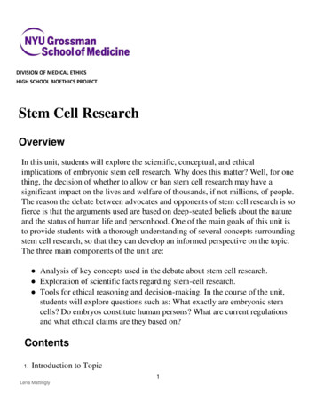
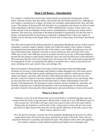
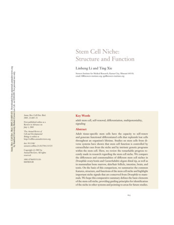
ANRV255-CB21-25ARI8 September 200517:11compare the differences and commonalitiesof stem cell niches in these different systems;and use this information to define the basicfeatures, structures, and functions of the stemcell niche.STEM CELL NICHES INDROSOPHILA OVARY ANDTESTISAnnu. Rev. Cell Dev. Biol. 2005.21:605-631. Downloaded from arjournals.annualreviews.orgby Stanford University Robert Crown Law Lib. on 03/01/09. For personal use only.Germ Line Stem Cell andSomatic Stem Cell Niches in theDrosophila OvaryTwo or three GSCs are located at the tip ofthe ovariole in the structure referred to asthe germarium. These GSCs are surroundedby three types of somatic cells: terminal filament, cap cells, and inner germarial sheath(IGS) cells (Figure 1). The stem cells areeasily identified by their direct contact withcap cells and the presence of a spectrosome(Lin 2002, Xie & Spradling 2001). Normally,a GSC divides to generate two daughter cells:one daughter that stays in association with capcells and another daughter that moves awayfrom the cap cells to form a cystoblast, whicheventually becomes, through incomplete cytokinesis, an interconnected 16-cell cyst. Genetic and cell biological studies demonstratethat cap cells are the niche for GSCs (Xie &Spradling 2000). The anchorage of GSCs tocap cells through E-cadherin-mediated celladhesion is essential for maintaining GSCs(Song & Xie 2002). Also, the number of GSCscorrelates with the number of cap cells (Xie& Spradling 2000). Finally, cap cells expressgenes, such as dpp, gbb, hh, piwi, and Yb, thatare known to be important for maintainingGSCs (Cox et al. 2000, King et al. 2001,Song et al. 2004, Xie & Spradling 1998, 2000)(Figure 1).BMP-, Hh-, and Piwi-mediated signalingpathways play an important role in the control of ovarian GSC self-renewal (Figure 1).Two BMP-like genes, dpp and gbb, are expressed in niche cells, and GSCs mutant fordpp, gbb, and their downstream components608Li·Xieare lost prematurely (Song et al. 2004, Xie& Spradling 1998, 2000). Dpp overexpression completely prevents GSC differentiationand thereby causes GSC-like tumor formation (Song et al. 2004, Xie & Spradling 1998).BMP signaling was recently shown to exertcontrol of GSC self-renewal by repressing expression of bam (Chen & McKearin 2003,Song et al. 2004), which is necessary and sufficient for cystoblast differentiation (Ohlstein& McKearin 1997).Piwi- and Yb-mediated signaling is alsorequired for controlling ovarian GSC selfrenewal (Cox et al. 2000, King et al. 2001,Lin & Spradling 1997). Interestingly, Yb regulates expression of piwi and hh in TF/capcells; these genes in turn control GSC selfrenewal (King et al. 2001). Yb-mediated signaling is also involved in repressing bam expression in GSCs (Chen & McKearin 2005,Szakmary et al. 2005). It would be interestingto know the relationship between BMP signaling and Piwi-mediated signaling in controlling GSC self-renewal. Zero populationgrowth (a Drosophila homolog of mammalianinnexin-4) is expressed in GSCs and is alsorequired for GSC maintenance, although theunderlying molecular mechanism for suchmaintenance is largely unknown (Gilboa et al.2003, Tazuke et al. 2002).Two or three SSCs located in the middleof the germarium are responsible for generating somatic follicle and stalk cells (Figure 1).The follicle cells encapsulate 16-cell cysts,whereas the stalk cells connect adjacent eggchambers. Although the ovarian SSCs lack aunique marker, they can be identified usinglineage tracing (Margolis & Spradling 1995,Song & Xie 2002, 2003, Zhang & Kalderon2001). SSCs have low levels of Fasciclin III(Fas 3) expression, whereas differentiated follicle cells have high levels of Fas 3 expression.Loss of adhesion between SSCs and IGS jeopardizes SSC self-renewal, suggesting that theproximal IGS cells are at least a part of theSSC niche, anchoring the SSCs (Song et al.2002). Although cap cells are not physicallyassociated with SSCs, they produce two
ARI8 September 200517:11Annu. Rev. Cell Dev. Biol. 2005.21:605-631. Downloaded from arjournals.annualreviews.orgby Stanford University Robert Crown Law Lib. on 03/01/09. For personal use only.ANRV255-CB21-25Figure 1Drosophila germarium cross section showing the locations of germ line stem cells (GSCs), somatic stemcells (SSCs), and their niches. Two or three GSCs (red cells, left) are situated in their niche, composed ofcap cells (green cells, left) and terminal filament cells (light blue cells, left tip), whereas their differentiatedprogeny, including cystoblasts and differentiated cysts (yellow cells, middle), are surrounded by innersheath cells (purple cells and green cells, bottom and top). Two or three SSCs (red cells, bottom and top)directly contact the posterior group of inner sheath cells (green cells, bottom and top) forming their niche,whereas their differentiated progeny, also known as follicle progenitor cells (orange cells on right), furtherproliferate and generate differentiated follicle cells. Two inserts depict major signaling pathwayscontrolling GSC (top and left) and SSC (top and right) self-renewal and proliferation; these inserts alsodepict niche cells (green) and stem cells (pink).www.annualreviews.org Stem Cell Niche609
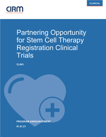
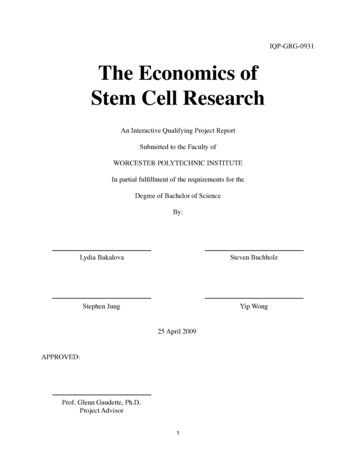
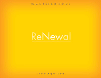
ANRV255-CB21-25ARI8 September 200517:11diffusible growth factors, Hh and Wg, thatare required for controlling SSC maintenanceand proliferation (Forbes et al. 1996, Kinget al. 2001, Song & Xie 2003). This supportsthe hypothesis that these cap cells are alsoa part of the SSC niche (Forbes et al. 1996;King et al. 2001, Song & Xie 2003, Zhang &Kalderon 2001).Annu. Rev. Cell Dev. Biol. 2005.21:605-631. Downloaded from arjournals.annualreviews.orgby Stanford University Robert Crown Law Lib. on 03/01/09. For personal use only.The Germ Line Stem Cell Niche inthe Drosophila TestisIn the apical tip of the Drosophila testis, twotypes of stem cells, GSCs and SSCs (the latter are also known as cyst progenitor cells),are responsible for producing differentiatedgerm cells and somatic cyst cells, respectively(Fuller 1993, Kiger et al. 2001) (Figure 2).Seven to nine GSCs, each containing a spectrosome, are attached to the hub (Hardyet al. 1979, Lindsley & Tokuyasu 1980, Yamashita et al. 2003). A male GSC dividesasymmetrically, giving rise to one stem cellthat remains in contact with the hub andone gonialblast that moves away from thehub and differentiates (Hardy et al. 1979,Lindsley & Tokuyasu 1980, Yamashita et al.2003). As a GSC divides to produce a gonialblast, the neighboring SSCs also divideto generate two cyst cells, which envelopthe gonialblast. This process leads to production of 64 sperm (Gonczy & DiNardo1996, Hardy et al. 1979). The hub generates signals, including Unpaired (Upd) andBMP, to control GSC self-renewal (Kawaseet al. 2004, Kiger et al. 2001, Shivdasani& Ingham 2003, Tulina & Matunis 2001)(Figure 2).Upd from the hub activates the JAK-STATpathway in GSCs and promotes their selfrenewal (Kiger et al. 2001, Tulina & Matsunis2001). Additionally, the activation of JAKSTAT signaling can reprogram mitotic germcysts into GSCs (Brawley & Matunis 2004).As in the ovary, BMP signaling is requiredfor controlling GSC self-renewal in the testis(Kawase et al. 2004, Schulz et al. 2004,610Li·XieShivdasani & Ingham 2003). Hub cells andsomatic cyst cells express gbb at high levelsand dpp at much lower levels; consequently,BMP downstream components are essentialfor controlling testicular GSC self-renewal(Kawase et al. 2004). Because dpp overexpression fails to suppress completely spermatogonial cell differentiation, BMP signaling likelyplays a permissive role in controlling maleGSC self-renewal. BMP and JAK-STAT signaling pathways are required for controllingmale GSC self-renewal; thus, they must somehow interact with each other. The integration between these two pathways in maleGSCs is an important area in need of futureexploration.Gonialblast differentiation is tightly controlled by unknown signals from SSCs and somatic cyst cells (Kiger et al. 2001, Tran et al.2000). In somatic cells mutant for Egfr and raf,GSC- and gonialblast-like single germ cellsare greatly increased in number and remainactive longer than do wild type cells.One mechanism ensuring that only oneof the two stem cell daughters self-renews iscontrol of the spindle orientation of the stemcell so as to place one self-renewing daughterin the niche and the other daughter destinedto differentiate outside the niche (Figure 2).Cnn and APC1, centrosomal components inGSCs, control orientation of the spindle perpendicular to the hub. Mutation in these components leads to an increase in GSC numberand subsequent crowding in the niche. APC2,which is concentrated at the junction between GSCs and hub cells, also controls correct GSC spindle orientation (Yamashita et al.2003).THE GERM LINE STEM CELLNICHE IN C. ELEGANSIn the C. elegans hermaphrodite gonad, onlythe 225 germ cells closest to the distal tipcell (DTC) are mitotic; those further proximalare arrested in meiotic pachytene (Crittendenet al. 1994) (Figure 3). Specific stem cellswithin the mitotic region have not been
ARI8 September 200517:11Annu. Rev. Cell Dev. Biol. 2005.21:605-631. Downloaded from arjournals.annualreviews.orgby Stanford University Robert Crown Law Lib. on 03/01/09. For personal use only.ANRV255-CB21-25Figure 2Cross section of the apical tip of the Drosophila testis, showing the locations of germ line stem cells(GSCs), somatic stem cells (SSCs), and their niches. Hub cells (green) at the apical tip of the testis formniches for both GSCs (red) and SSCs (gray, left), which generate, respectively, spermatogonial cells(yellow) and somatic cyst cells (light gray) encapsulating differentiated spermatogonial cells. The insert ontop describes major signaling pathways involved in communication between GSCs and the niche cells forcontrolling self-renewal and proliferation.www.annualreviews.org Stem Cell Niche611
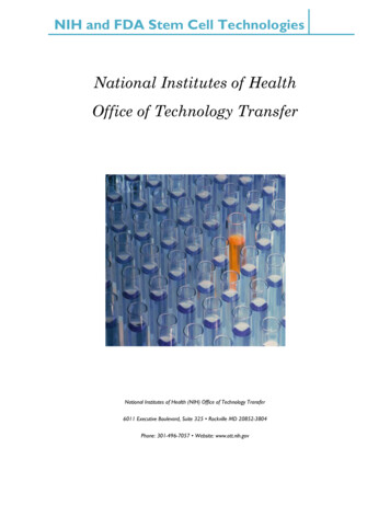
ARI8 September 2005Annu. Rev. Cell Dev. Biol. 2005.21:605-631. Downloaded from arjournals.annualreviews.orgby Stanford University Robert Crown Law Lib. on 03/01/09. For personal use only.ANRV255-CB21-2517:11Figure 3Cross section of the C. elegans hermaphrodite gonad. The putative germ line stem cells (GSCs) (red ) aredirectly associated with their distal tip cell (DTC) niche cell (green), whereas their differentiated progeny(light yellow) move away from the DTC, progressing from the mitotic phase to the meiotic phase. TheGLP-1 (Notch-like) signaling pathway is involved in communication between the DTC and GSCs andrepresses functions of differentiation-promoting gene products, such as Gld-1, Gld-2, and Nos-3, whichregulate entry into meiosis (insert).identified. The somatic DTC is required formaintaining these cells in mitosis (Kimble &White 1981). Although mitotic and meioticgerm cells in the tube share a central core ofcytoplasm, only those mitotic germ cells located at the most distal tip (i.e., GSCs) adjacent to the DTC behave like stem cells, capa612Li·Xieble of self-renewing and generating differentiated gametes. The proximal mitotic neighbors behave more like transient amplifyingcell populations, described in other systems.As germ cells move further away from theDTC, they terminate their mitotic activitiesand commit meiosis. Only those germ cells
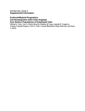
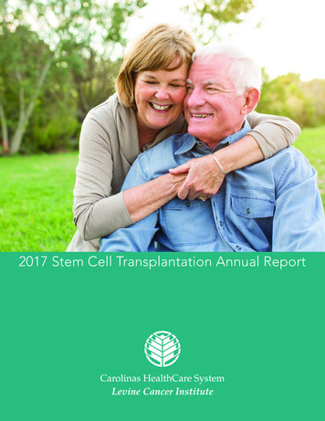
Annu. Rev. Cell Dev. Biol. 2005.21:605-631. Downloaded from arjournals.annualreviews.orgby Stanford University Robert Crown Law Lib. on 03/01/09. For personal use only.ANRV255-CB21-25ARI8 September 200517:11that physically interact with the DTC maintain their GSC identity; thus, the signal fromthe DTC either must be short ranging or mediated by a direct cell-cell interaction.Signaling from the DTC to control GSCself-renewal is through a Notch-like cascade. The mitotic germ cells express theNotch-type receptor, GLP-1, which is activated by the Delta-like signal from DTC,LAG-2 (Crittenden et al. 1994, Hendersonet al. 1994). Constitutive GLP-1 activitydownregulates the meiosis-promoting genesgld-1, gld-2, and nos-3 and thereby causes expansion of germ cell numbers (Berry et al.1997). Because individual stem cells have notbeen identified in the mitotic region, it remains unclear whether they are maintainedthrough a population mechanism or an asymmetric division mechanism.KNOWN STEM CELL NICHESIN MAMMALIAN SYSTEMSThe stem cell and the niche hypothesis, firstdeveloped in the hematopoietic system inmammals, has provided the conceptual background for stem cell studies in Drosophilaand C. elegans (Schofield 1978, Weissman1994). Conversely, studies in Drosophila on themolecular pathways controlling the stem cellniche have provided important insight intoidentification of the stem cell niche in mammalian systems (Lin 2002, Spradling et al.2001). In this section, we describe and compare the location and physical organization(if known) of adult stem cells in bone marrow, skin/hair follicle, intestine, neuron, andtestis.The Hematopoietic Stem Cell NicheBone marrow serves as the pioneer system forstudying stem cells; the concept and basic features of stem cells were defined from studying hematopoietic stem cells (HSCs) (Orkin2000, Till & McCulloch 1961, Weissmanet al. 2001). However, the way in which HSCsinteract with their local environment to pro-mote stem cell maintenance has not beenclear. Most studies of HSCs have examinedtheir behavior in cell populations obtainedfrom their natural niche in the bone marrow.Thus far, however, only limited culture systems exist that allow sustained maintenanceand expansion of HSCs in vitro, attesting tothe importance of as-yet poorly defined interactions in the bone marrow niche. Two independent studies recently 1) identified a subset of osteoblastic cells (N-cadherin CD45 )to which HSCs physically attach in thebone marrow, 2) identified an N-cadherin/βcatenin adherens complex between HSCs andosteoblastic cells, 3) showed that Jagged1,generated from osteoblasts, influences HSCsby signaling through the Notch receptor,and 4) demonstrated that the number of Ncadherin osteoblastic lining cells controlsthe number of HSCs (Calvi et al. 2003, Zhanget al. 2003). Homing studies to trace the location of GFP-labeled HSCs after transplantation also pointed to the endosteal surface as apossible stem cell niche (Nilsson et al. 2001).In vitro coculture of HSCs with osteoblastscan expand the HSC population (Taichman &Emerson 1998), and depletion of osteoblastsleads to loss of hematopoietic tissue (Visnjicet al. 2004). In addition, N-cadherin is a keytarget of Angiopoietin-1 (Ang-1)/Tie-2 signaling that maintains HSC quiescence (Araiet al. 2004) (Figure 4).A primary function of the niche is toanchor stem cells. In addition to N-cadherin,other types of adhesion molecules, includingintegrin, play an important role in themicroenvironment/stem cell interaction(Simmons et al. 1997). Stromal cell-derivedfactor-1 (SDF-1) and its receptor CXCR4are involved in homing of HSCs (Lapidot &Kollet 2002) (Figure 4).Although the analysis of the signals generated by the niche has just begun, gene expression profiling studies of HSCs have revealed which signals HSCs potentially receivefrom the niche. The components of evolutionally conserved and developmentally regulated pathways are prominent in stem cellswww.annualreviews.org Stem Cell Niche613
ANRV255-CB21-25ARI8 September 200517:11Annu. Rev. Cell Dev. Biol. 2005.21:605-631. Downloaded from arjournals.annualreviews.orgby Stanford University Robert Crown Law Lib. on 03/01/09. For personal use only.and are indeed involved in the regulation ofstem cell self-renewal or maintenance. Thesecomponents include the Shh, Wnt, Notch,and TGF-β/BMP pathways (Akashi et al.2003, Gomes et al. 2002, Ivanova et al. 2002,Park et al. 2002, Ramalho-Santos et al. 2002).614Li·XieFor example, the Wnt/β-catenin pathway isimportant for self-renewal of HSCs (Reyaet al. 2003). The Notch pathway is requiredfor maintaining HSCs in the undifferentiatedstate (Calvi et al. 2003, Duncan et al. 2005,Li et al. 1998, Varnum-Finney et al. 2000).
ANRV255-CB21-25ARI8 September 200517:11The BMP signal plays a role in control ofHSC number (Zhang et al. 2003). The Shhsignal mediated by the BMP pathway is ableto maintain stem cells in vitro (Bhatia et al.1999). (Figure 4).Annu. Rev. Cell Dev. Biol. 2005.21:605-631. Downloaded from arjournals.annualreviews.orgby Stanford University Robert Crown Law Lib. on 03/01/09. For personal use only.The Epithelial Stem Cell Nichein SkinSkin, with its appendix hair follicle structure,has well-organized architecture (Figure 5)and provides an excellent system for studyingthe molecular mechanisms that regulate stemcell self-renewal, proliferation, migration, andlineage commitment (Fuchs & Segre 2000).Each hair follicle is composed of a permanent portion, which includes sebaceous glandsand the underlying bulge area, and a dynamicrenewing portion, which undergoes cycles ofanagen phase (a period of active growth), catagen phase (apoptosis-driven retraction), andtelogen phase (a short period of rest) (Hardy1992). The bulge area functions as a niche,where epithelial stem cells (Niemann & Watt2002) are located and maintained (Cotsareliset al. 1990, Sun et al. 1991). Epithelial stemcells are multipotent, giving rise to daughter cells that either migrate upward to serveas epidermal progenitors for generating epidermal cells during wound repair or migratedownward to convert to hair-matrix progenitors, which further give rise to the hair shaft(Niemann & Watt 2002, Oshima et al. 2001,Taylor et al. 2000).During the early anagen phase, the dermal papilla region may provide the dynamicsignals that activate stem cells; however, thecellular components of the niche in the bulgeare yet to be defined other than as stem cellsper se. The dermal sheath derived from mesenchymal cells adjacent to the epithelial stemcells in the bulge area most likely providesthe niche function. The recent identificationof markers for epithelial stem cells, such asCD34, will be helpful in further identifyingthe adjacent niche cells and the related nichestructures, including adhesion molecules (i.e.,α6 integrin) (Blanpain et al. 2004).Recent studies showed that label-retainingcells can regenerate the entire HF structurein transplantation experiments, thus demonstrating that these cells are bona fide epidermal stem cells (Blanpain et al. 2004, Braunet al. 2003). Molecular analysis of epithelial stem cells has revealed the following features: 1) the expression of adhesion moleculesknown to be involved in stem cell-niche interaction, 2) the presence of growth inhibitionfactors such as TGFβ/BMP molecules andcell cycle inhibitors, and 3) the components ofWnt signaling pathways, including receptorsand inhibitors such as Dkk, sFRP, and WIF.Taken together, these molecular features indicate that the epithelial stem cell niche isa growth- and differentiation-restricted environment (Tumbar et al. 2004). This conclusion is, in general, consistent with themany previous studies that have used genetic Figure 4Illustration of the hematopoietic stem cell (HSC) niche. The HSC niche is located primarily on thesurface of trabecular bone, where a small subset of spindle-shaped N-cadherin-positive osteoblastic cells(indicated as SNO cells) are the key component of the HSC niche. N-cadherin and β-catenin form anadherens complex at the interface between stem cells and niche cells, assisting stem cells in attaching tothe niche. Multiple growth factors and cytokines are involved in stem-niche interaction. These includeSCF/Kit, Jagged/Notch, SDF-1/CXCR4, and Ang1/Tie2. BMP4 is expressed in osteoblastic cells, butthe type of receptor expressed in HSCs is unknown. The Wnt signal is important for stem cellself-renewal, but the Wnts present in the niche are unknown. The same is true for FGF and hedgehog.In vitro data suggest they affect HSC behavior; however, whether they are present as niche signals isunknown. Different types of stromal cells (illustrated as different colors and shapes) may regulate stemcell activation, proliferation, and differentiation by secreting different microenvironmental signals.Finally, maturated blood cells migrate and infiltrate into blood vessel.www.annualreviews.org Stem Cell Niche615
ARI8 September 2005Annu. Rev. Cell Dev. Biol. 2005.21:605-631. Downloaded from arjournals.annualreviews.orgby Stanford University Robert Crown Law Lib. on
Stem Cell Niche: Structure and Function Linheng Li and Ting Xie Stowers Institute for Medical Research, Kansas City, Missouri 64110; email: lil@stowers-institute.org, tgx@stowers-institute.org Annu. Rev. Cell Dev. Biol. 2005. 21:605-31 First published online as a Review in Advance on July 1, 2005 The Annual Review of Cell and Developmental .