Transcription
J Orthop Traumatol (2017) 18:343–348DOI 10.1007/s10195-017-0460-0ORIGINAL ARTICLESegond’s fracture: a biomechanical cadaveric study usingnavigationE. Monaco1 Daniele Mazza1 A. Redler1 D. Lupariello1 R. Lanzetti1M. Guzzini1 A. Ferretti1 Received: 2 May 2017 / Accepted: 8 May 2017 / Published online: 13 July 2017Ó The Author(s) 2017. This article is an open access publicationAbstractBackground Segond’s fracture is a well-recognised radiological sign of an anterior cruciate ligament (ACL) tear.While previous studies evaluated the role of the anterolateral ligament (ALL) and complex injuries on rotationalstability of the knee, there are no studies on the biomechanical effect of Segond’s fracture in an ACL deficientknee. The aim of this study was to evaluate the effect of aSegond’s fracture on knee rotation stability as evaluated bya navigation system in an ACL deficient knee.Materials and methods Three different conditions weretested on seven knee specimens: intact knee, ACL deficientknee and ACL deficient knee with Segond’s fracture. Staticand dynamic measurements of anterior tibial translation(ATT) and axial tibial rotation (ATR) were recorded by thenavigation system (2.2 OrthoPilot ACL navigation systemB. Braun Aesculap, Tuttlingen, Germany).Results Static measurements at 30 showed that the meanATT at 30 of knee flexion was 5.1 2.7 mm in the ACLintact condition, 14.3 3.1 mm after ACL cut(P 0.005), and 15.2 3.6 mm after Segond’s fracture(P 0.08). The mean ATR at 30 of knee flexion was20.7 4.8 in the ACL intact condition, 26.9 4.1 inthe ACL deficient knee (P [ 0.05) and 30.9 3.8 afterSegond’s fracture (P 0.005). Dynamic measurementsduring the pivot-shift showed that the mean ATT was7.2 2.7 mm in the intact knee, 9.1 3.3 mm in theACL deficient knee(P 0.04) and 9.7 4.3 mm in theACL deficient knee with Segond’s fracture (P 0.07).& Daniele Mazzadaniele.mazza@hotmail.it1Sant’Andrea Hospital, University of Rome La Sapienza, Viadi Grottarossa 1035, Rome, ItalyThe mean ATR was 9.6 1.8 in the intact knee,12.3 2.3 in the ACL deficient knee (P [ 0.05) and19.1 3.1 in the ACL deficient knee with Segond’slesion (P 0.016).Conclusion An isolated lesion of the ACL only affectsATT during static and dynamic measurements, while theaddition of Segond’s fracture has a significant effect onATR in both static and dynamic execution of the pivot-shifttest, as evaluated with the aid of navigation.Keywords Anterior cruciate ligament Segond’s fracture Navigation Pivot-shiftIntroductionIn 1879 Paul Segond described a resistant fibrous band inthe lateral compartment of the knee, whose traction injuryresulted in a cortical avulsion of the lateral proximal tibialplateau [1].The precise pathogenesis of Segond’s fracture hasbeen the subject of debate, partially due to the complexity of the anterolateral ligamentous anatomy.Segond demonstrated that an internal rotation and varusstress applied to the knee causes tension on the lateraljoint capsule at its midpoint; he believed that a resistantband of tissue produces an avulsion fracture of the lateraltibial plateau, posterior to the insertion of the ileo-tibialtract (ITT).This injury was named Segond’s fracture and numerousstudies [2–4] have demonstrated an association of Segond’sfracture with tears of the anterior cruciate ligament (ACL)(75–100% of patients), meniscal tears (66–75% ofpatients), damage to the structures of the posterolateralcorner of the knee, and other avulsion injuries.123
344Today, the presence of a Segond’s fracture is consideredas an indirect radiological sign of an ACL tear. In a recentdescriptive study on pattern and prevalence of injuries ofthe lateral capsule in acute ACL surgery, Ferretti et al. [5]found that a lesion was found in about 90% of cases, whileSegond’s fracture was found in only 10% of cases. Theyconcluded that this avulsion fracture represents the tip ofthe iceberg of lesions of the anterolateral capsule andligament, possibly affecting the rotational stability of theknee.Moreover, recently Claes et al. [6] described thepresence of a constant thickening of the anterolateralcapsule named the anterolateral ligament (ALL) in theanterolateral compartment of the knee inserting in theregion on the proximal tibia from where Segond’s fractures consistently avulse, thus suggesting that Segond’sfracture is actually a bony avulsion of the ALL [7].However, even if many anatomical and biomechanicalstudies, dating back to the early 1960s, have focused onanatomy and biomechanics of the anterolateral capsuleand ALL [8–10], their role as secondary restraints of theACL in controlling internal tibial rotation and the pivotshift phenomenon is still debated. Surprisingly, all previous studies focused on anatomy and function of softtissues, including ligaments and capsule, whose dissection was performed in different ways by various authors,leading to conflicting results [11–13]. Until now, nostudy has actually been made to evaluate the biomechanical role of a true Segond’s fracture on an ACLdeficient knee.The purpose of this study was to evaluate the effect ofprogressive lesions of the ACL and Segond’s fracture onknee rotation stability as evaluated by a navigation systemwith software that enables the anterior tibial translation(ATT) and axial tibial rotation (ATR) to be precisely calculated statically and dynamically during a pivot-shift testin a cadaveric model.J Orthop Traumatol (2017) 18:343–348Computer navigation systemThe navigation system (2.2 OrthoPilot ACL navigationsystem B. Braun Aesculap, Tuttlingen, Germany) used wasa wireless system which does not require computedtomography. Two 2.5-mm K-wires were placed in thedistal portion of the femur and in the proximal portion ofthe tibia, respectively, and rigid bodies with reflectivemarkers were attached. Extra-articular landmarks wereregistered by the system using an instrumented pointer. Theextra-articular landmarks included the tibial tuberosity, theanterior edge of the tibia, and medial and lateral points ofthe tibial plateau. Kinematics of the knee joint weredynamically registered by the navigator during a range ofmotion from full extension to 90 of knee flexion.This system provided a real-time calculation of ATT andATR at all degrees of knee flexion, while the surgeonmanipulated the knee joint. According to the manufacturer,the error rate of the navigation system is estimated to beless than 1 mm or less than 1 . All navigation processeswere performed by the same single surgeon (AF). Threedifferent conditions were tested: intact knee, ACL deficientknee and ACL injury with Segond’s fracture. Static measurements of the ATT and ATR at 30 of knee flexion wereperformed in the three conditions. All measurements wererecorded under a manual maximum force applied by thesame surgeon, who made every effort to apply a similarload to the knee, to minimize intra-observer variability.Dynamic measurements of ATT and ATR during thepivot-shift test were always performed in the same threedifferent conditions. The pivot-shift test was performed bythe same surgeon three times at each step, and the datawere averaged by the navigation system. The pivot-shifttest was performed using manual loads applied by the samesurgeon to minimize intra-observer variability. ATT wasexpressed in millimetres and ATR in degrees during thepivot shift, and a diagram showing the curve of ATT andATR during the test was visualized and saved with ascreen-shot at the end of each step of the procedure.Materials and methodsSpecimen sectioning and procedureFor this study four entire whole fresh-frozen cadavers wereused. Cadavers included 3 men and 1woman with anaverage age at death of 65 years (range 48–72). Thecadavers were stored at -20 C and thawed at room temperature for 24 h before testing. Samples were excludedfrom the study if they showed signs of ligamentous injuries, severe osteoarthritis, bony abnormalities, or previoussurgical intervention. The cadavers remained fully intactand no soft tissue was cut or removed from around the kneeor adjacent joints to most closely match a normal humanknee. Saline solution was used to keep the specimens moistthroughout testing.123Firstly the knee was tested intact, and static and dynamicmeasurements recorded. After the intact knee was tested,the ACL was entirely cut, using an open technique througha minimally invasive medial parapatellar arthrotomy. Thearthrotomy was then closed and both static and dynamicmeasurements recorded.Afterwards, with the knee at 90 of flexion, the lateralcompartment was approached with a hockey stick incision.After the dissection of subcutaneous tissue, an exposure ofthe lateral compartment was performed by splitting the ileotibial tract along its fibers. A fracture of the lateral tibial
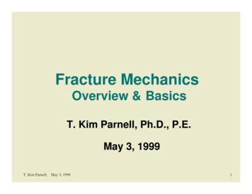
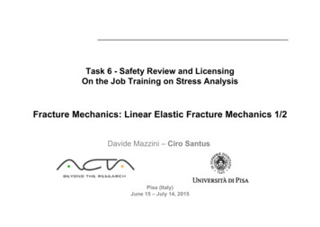
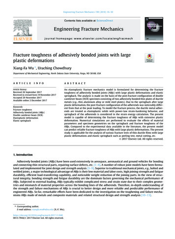
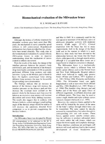
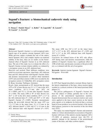
J Orthop Traumatol (2017) 18:343–348plateau at a site of the distal insertion of the anterolateralligament (reproducing Segond’s lesion) was produced usinga 1.5-cm-width standard scalpel (Fig. 1). The site selectedwas midway between the fibular head and Gerdy’s tubercle.The lateral approach was then closed and all static anddynamic measurements were recorded in the ACL deficientknee condition with Segond’s fracture.All the biomechanical tests were performed at theLaboratoire de Anatomie, Universitè of Nice. IRB approvalwas not required.Statistical analysisAll the data were analysed by a single researcher. To assesswhether data were normally distributed and homogeneouswe used Kolmogorov–Smirnov’s test and Levene’s test,respectively.A Kruskal–Wallis H test was conducted to determinewhether there were differences in antero-posterior (AP) androtational (ROT) scores between groups. Subsequently, pairwise comparisons were performed using Dunn’s procedure.Statistical Package for Social Sciences (SPSS) version22 was used for calculations. Statistical significance wasset at P \ 0.05.ResultsOne knee was excluded because an ACL tear was found atthe initial inspection, leaving a total of seven lowerextremities eligible for the study.Static measurements at 30 The mean ATT at 30 of knee flexion was 5.1 2.7 mm(ACL intact condition), 14.3 3.1 mm after ACL cutFig. 1 Segond’s fracture on cadaver model345(P 0.005), and 15.2 3.6 mm after Segond’s fracture(P 0.08). The mean ATR at 30 of knee flexion was20.7 4.8 (ACL intact), 26.9 4.1 in the ACL deficient knee (P 0.06), and 30.9 3.8 after Segond’sfracture (P 0.005) (Fig. 2, 3).Dynamic pivot-shiftThe mean ATT was 7.2 2.7 mm in the intact knee,9.1 3.3 mm in the ACL deficient knee (P 0.04) and9.7 4.3 mm in the ACL deficient knee with Segond’sfracture (P 0.07). The mean ATR was 9.6 1.8 in theintact knee, 12.3 2.3 in the ACL deficient knee(P 0.06) and 19.1 3.1 in the ACL deficient knee withSegond’s lesion (P 0.016) (Tables 4, 5).DiscussionTo the authors’ knowledge, this is the first study evaluatingthe biomechanical effect of Segond’s fracture.The most important finding of this study is that a combined lesion of ACL and Segond’s fracture resulted in asignificant increase in ATR when compared both to theintact knee and ACL deficient knee in static and dynamicmeasurements.It is well known that the pivot-shift test is the mostreliable test to evaluate rotational stability in the ACLdeficient knee, and its disappearance should be the goal ofmodern ACL surgery [14]. As a matter of fact, the persistence of rotational instability is related to poor functionaloutcomes, with a higher risk of revision surgery [15]. Thepivot-shift test is a complex multi-planar phenomenon thatinvolves translation and rotation of the tibia on the femurwith the knee subjected to valgus stress and internal rotation ranging from full extension to 30 –40 of flexion.However, its major limitation is that, as it is manuallyperformed by the clinician, it is strongly correlated to theexperience of the examiner, with an inter-observer variability. Several instruments, such as electromagnetic sensors, accelerometers and image analysis have beendeveloped to evaluate and quantify the pivot-shift test, butsurgical navigation is one of the best tools to objectivelymeasure the pivot-shift phenomenon. The software used inthe present study, which had been already used and validated in previous studies, allowed us to simultaneously anddynamically assess ATT in millimetres and ATR indegrees during the execution of the pivot-shift test.Pearle et al. [16] evaluated the reliability of navigatedknee stability in a cadaveric study comparing this systemwith the robotic/UFS testing system, showing that thesurgical navigation system has an overall accuracy ofapproximately 1 mm or 1 . For these reasons, we could123
346Fig. 2 Anterior tibialtranslation during pivot-shift(static measurements)J Orthop Traumatol (2017) 18:343–348ATT during static measurementat30 knee flexion2018P 0.0051614.315.2141210865.1420ACL intactFig. 3 Intra- and extra-tibialrotation (static measurements)40ACL deficientSegondATR during static measurement at 30 knee flexionP 0.0053530.93026.92520.720151050ACL intactFig. 4 Anterior tibialtranslation during pivot-shift(dynamic measurements)ACL deficientSegondATT during dynamic pivot shift14P 0.04121089.19.77.26420Intact Kneespeculate that surgical navigation should be considered thebest way to study the pivot-shift phenomenon.It has been well known since the 1970s that anterolateralstructures are an important restraint to internal rotation ofthe knee and they have been described in differentanatomical studies and named in different ways (middleone third of lateral capsule; anterolateral femurotibial123ACL deficientSegondligament, ALFTL; anterolateral ligament, ALL; anterolateral complex, ALC) [6, 17–19]. Lesions of these structureshave been recently found to be strongly correlated to thepivot-shift phenomenon in ACL tears [20].Monaco et al. [20] evaluated tibial rotation, after cuttingthe ACL and lateral capsule, using an older software version of the same navigation system used in the present
J Orthop Traumatol (2017) 18:343–348Fig. 5 Intra- and extra-tibialrotation (dynamicmeasurements)347ATR during dynamic pivot shift25P 0.01619.1201512.3109.650Intact Kneestudy. No significant rotational instability was seen untilafter the lesion to the lateral capsule, suggesting thatrotational instability of the knee may be due to secondaryinjuries in conjunction with injuries to the ACL, rather thanto injury of only the ACL. A lesion to the lateral capsularligament in the previously sectioned ACL resulted in asignificant increase in combined rotation at 30 , 45 , and60 of knee flexion (P 0.03). Moreover, a high correlation was shown between these lesions of the anterolateralstructures and the pivot-shift phenomenon.Sonnery-Cottet et al. [21], in a recent navigated study onknee specimens, evaluated the effect of progressive lesionsof ACL, ALL and ileo-tibial tract (ITT) on 12 fresh frozencadaveric knees, using a navigation system at 20 and 90 of flexion under a simulated pivot-shift test. They showedthat cutting the ALL after sectioning the ACL produced anincrease in internal rotation as compared with the ACLdeficient knee, concluding that ALL is involved in therotational control of the knee during a simulated pivot shift.These finding are in agreement with the results of Parsonset al. [22], reporting that ALL had a greater contribution intibial internal rotation at higher degrees of flexion. However, this assessment was based on the evaluation with arobotic testing system of the in situ forces acting on ACLand ALL during anterior drawer and internal tibial rotation.In a recent paper, Bonazinga et al. [8] used a differentnavigation system (Polaris, NDI, Waterloo, Ontario,Canada) to evaluate the effect of progressive lesions ofACL and ALL during a pivot-shift test. They found that,while an isolated lesion of the ACL did not significantlyaffect acceleration, ALL plays a significant role in controlling acceleration during a pivot-shift test, its resectionhaving no effect on anterior displacement.The results of this navigated study seem to confirm thesefindings. In fact an isolated lesion of the ACL resulted in anincreased ATT in static at 30 of flexion and dynamicACL deficientSegondconditions during the pivot-shift in comparison to the intactknee. We could speculate that an isolated lesion of theACL is not sufficient to elicit a clinically detectable pivotshift, while the additional Segond’s fracture has a significant effect on tibial rotation and is responsible for theappearance of a high grade of pivot-shift phenomenon. Inactual fact the biomechanical effect of a Segond’s fractureclosely resembles that of a complete tear of the lateralcapsule, as previously reported. Therefore, the definition ofSegond’s fracture as a bony injury of the ALL [6] seems tobe reliable, and its repair could be justified to help ACLreconstruction in controlling the pivot-shift phenomenon.This study has several limitations. First, it is a cadavericstudy with a limited number of samples tested. Second, theage of the donor specimens may not well represent thepopulation typically undergoing ACL reconstruction. Furthermore, despite the fact that the pivot-shift test wasalways performed by the same examiner to avoid interobserver variability, the results may be dependent on theload applied by the surgeon during its execution, even ifnavigation was used for measurements. Another limitationof this study is that we were unable to reproduce the possible stretching of the whole lateral capsule on the cadaveroccurring in vivo, along with the bony avulsion, as reportedto occur in the majority of cases of Segond’s fracture [5].This possible plastic deformity of the anterolateral capsuleoccurring in vivo would lead to an underestimate of theactual role of Segond’s fracture as evaluated in the presentstudy.However, this study also presents some strengths. In theliterature no studies have evaluated the biomechanicaleffect of Segond’s fracture during the pivot-shift phenomenon. The study was performed using a real-timecomputer navigation system with dedicated software andwith wires strongly fixed in the bone to precisely assess theeffect of the lesion. Moreover, an entire cadaveric model123
348was used and all soft tissues were left intact to betterreproduce the clinical scenario of the pivot-shift test.In conclusion, while an isolated complete tear of ACLhas a mild, if any, effect on rotational stability of the knee,the addition of Segond’s fracture has a significant effect onATR in both static and dynamic conditions during theexecution of the pivot-shift test, as evaluated with the aidof navigation. Therefore, repair of Segond’s fracture couldbe justified to help ACL reconstruction in controlling thepivot-shift phenomenon.Compliance with ethical standardsConflict of interest Prof. Andrea Ferretti and Edoardo Monaco areconsultant for Arthrex.Patient consent Not applicable.Ethical approval Ethics approval is not required for this study in ourUniversity IRB (University La Sapienza).Funding For this study, there was no source of funding.Open Access This article is distributed under the terms of the CreativeCommons Attribution 4.0 International License (http://creativecommons.org/licenses/by/4.0/), which permits unrestricted use, distribution, and reproduction in any medium, provided you give appropriatecredit to the original author(s) and the source, provide a link to theCreative Commons license, and indicate if changes were made.References1. Segond P. Recherches cliniques et experimentales sur lesepanchements sanguins du genou par entorse. Progres Medical.Paris, 1879 (accessible from http://www.patrimoine.edilivre.com)2. Woods GW, Stanley RF, Tullos HS (1979) Lateral capsular sign:X-ray clue to a significant knee instability. Am J Sports Med7:27–333. Goldman AB, Pavlov H, Rubenstein D (1988) The Segondfracture of the lateral tibia: a small avulsion that reflects majorligamentous damage. AJR Am J Roentgenol 151:1163–11674. El-Khoury GY, Daniel WW, Kathol MH (1997) Acute andchronic avulsive injuries. Ra- diol Clin North Am 35:747–7665. Ferretti A, Monaco E, Fabbri M, Maestri B, De Carli A (2016)Prevalence and classification of injuries of anterolateral complexin acute anterior cruciate ligament tears. Arthroscopy33(1):147–1546. Claes S, Vereecke E, Maes M, Victor J, Verdonk P, Bellemans J(2013) Anatomy of the anterolateral ligament of the knee. J Anat223(4):321–328123J Orthop Traumatol (2017) 18:343–3487. Claes S, Luyckx T, Vereecke E, Bellemans J (2014) The Segondfracture: a bony injury of the anterolateral ligament of the knee.Arthroscopy. 30(11):1475–14828. Amis AA, Scammell BE (1993) Biomechanics of intra-articularand extra-articular reconstruction of the anterior cruciate ligament. J Bone Joint Surg Br 75(5):812–8179. Bonanzinga T, Signorelli C, Grassi A, Lopomo N, Bragonzoni L,Zaffagnini S, Marcacci M (2016) Kinematics of ACL andanterolateral ligament. Part I: combined lesion. Knee Surg SportsTraumatol Arthrosc 25(4):1055–106110. Caterine S, Litchfield R, Johnson M, Chronik B, Getgood A(2014) A cadaveric study of the anterolateral ligament: re-introducing the lateral capsular ligament. Knee Surg Sports TraumatolArthrosc 23(11):3186–319511. Daggett M, Ockuly AC, Cullen M et al (2016) Femoral origin of theanterolateral ligament: an anatomic analysis. Arthroscopy32(5):835–84112. Dodds AL, Halewood C, Gupte CM, Williams A, Amis AA (2014)The anterolateral ligament: anatomy, length changes and association with the Segond fracture. Bone Joint J. 96-B(3):325–33113. Helito CP, Demange MK, Bonadio MB et al (2014) Radiographiclandmarks for locating the femoral origin and tibial insertion ofthe knee anterolateral ligament. Am J Sports Med 42:2356–236214. Kennedy MI, Claes S, Fuso FA, Williams BT, Goldsmith MT,Turnbull TL, Wijdicks CA, LaPrade RF (2015) The anterolateralligament: an anatomic, radiographic, and biomechanical analysis.Am J Sports Med 43(7):1606–161515. Ferretti A, Monaco E, Vadalà A (2014) Rotatory instability of theknee after ACL tear and reconstruction. J Orthop Traumatol.15(2):75–7916. Pearle AD, Solomon DJ, Wanich T et al (2007) Reliability ofnavigated knee stability examination: a cadaveric evaluation.AmJ Sports Med 35:1315–132017. Hughston JC, Andrews JR, Cross MJ, Moschi A (1976) Classification of knee ligament instabilities. Part II. The lateral compartment. J Bone Joint Surg Am 58(2):173–17918. Muller W (1983) The knee. Springer Verlag, New York19. Vincent JP, Magnussen RA, Gezmez F et al (2012) The anterolateral ligament of the human knee: an anatomic and histologicstudy. Knee Surg Sports Traumatol Arthrosc 20(1):147–15220. Monaco E, Ferretti A, Labianca L, Maestri B, Speranza A, KellyMJ, D’Arrigo C (2012) Navigated knee kinematics after cuttingof the ACL and its secondary restraint. Knee Surg Sports Traumatol Arthrosc 20(5):870–87721. Sonnery-Cottet B, Lutz C, Daggett M, Dalmay F, Freychet B,Niglis L, Imbert P (2016) The involvement of the anterolateralligament in rotational control of the knee. Am J Sports Med44(5):1209–121422. Parsons EM, Gee AO, Spiekerman C, Cavanagh PR (2015) Thebiomechanical function of the anterolateral ligament of the knee.Am J Sports Med 43(3):669–674
knee and ACL deficient knee with Segond's fracture. Static and dynamic measurements of anterior tibial translation (ATT) and axial tibial rotation (ATR) were recorded by the navigation system (2.2 OrthoPilot ACL navigation system B. Braun Aesculap, Tuttlingen, Germany). Results Static measurements at 30 showed that the mean ATT at 30 of knee flexion was 5.1 2.7 mm in the ACL intact .