Transcription
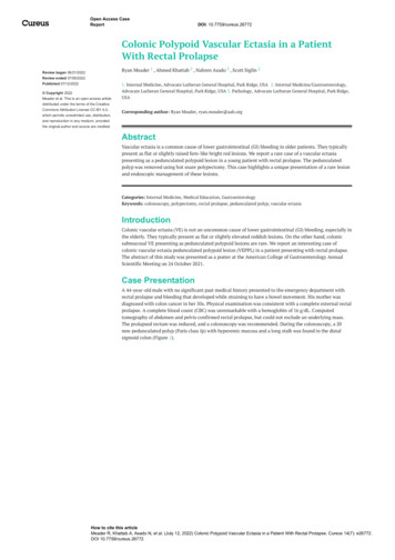
Open Access CaseReportDOI: 10.7759/cureus.26772Colonic Polypoid Vascular Ectasia in a PatientWith Rectal ProlapseRyan Meader 1 , Ahmed Khattab 2 , Nahren Asado 3 , Scott Siglin 2Review began 06/21/2022Review ended 07/05/2022Published 07/12/2022 Copyright 2022Meader et al. This is an open access article1. Internal Medicine, Advocate Lutheran General Hospital, Park Ridge, USA 2. Internal Medicine/Gastroenterology,Advocate Lutheran General Hospital, Park Ridge, USA 3. Pathology, Advocate Lutheran General Hospital, Park Ridge,USAdistributed under the terms of the CreativeCommons Attribution License CC-BY 4.0.,which permits unrestricted use, distribution,Corresponding author: Ryan Meader, ryan.meader@aah.organd reproduction in any medium, providedthe original author and source are credited.AbstractVascular ectasia is a common cause of lower gastrointestinal (GI) bleeding in older patients. They typicallypresent as flat or slightly raised fern-like bright red lesions. We report a rare case of a vascular ectasiapresenting as a pedunculated polypoid lesion in a young patient with rectal prolapse. The pedunculatedpolyp was removed using hot snare polypectomy. This case highlights a unique presentation of a rare lesionand endoscopic management of these lesions.Categories: Internal Medicine, Medical Education, GastroenterologyKeywords: colonoscopy, polypectomy, rectal prolapse, pedunculated polyp, vascular ectasiaIntroductionColonic vascular ectasia (VE) is not an uncommon cause of lower gastrointestinal (GI) bleeding, especially inthe elderly. They typically present as flat or slightly elevated reddish lesions. On the other hand, colonicsubmucosal VE presenting as pedunculated polypoid lesions are rare. We report an interesting case ofcolonic vascular ectasia pedunculated polypoid lesion (VEPPL) in a patient presenting with rectal prolapse.The abstract of this study was presented as a poster at the American College of Gastroenterology AnnualScientific Meeting on 24 October 2021.Case PresentationA 44-year-old male with no significant past medical history presented to the emergency department withrectal prolapse and bleeding that developed while straining to have a bowel movement. His mother wasdiagnosed with colon cancer in her 50s. Physical examination was consistent with a complete external rectalprolapse. A complete blood count (CBC) was unremarkable with a hemoglobin of 16 g/dL. Computedtomography of abdomen and pelvis confirmed rectal prolapse, but could not exclude an underlying mass.The prolapsed rectum was reduced, and a colonoscopy was recommended. During the colonoscopy, a 20mm-pedunculated polyp (Paris class Ip) with hyperemic mucosa and a long stalk was found in the distalsigmoid colon (Figure 1).How to cite this articleMeader R, Khattab A, Asado N, et al. (July 12, 2022) Colonic Polypoid Vascular Ectasia in a Patient With Rectal Prolapse. Cureus 14(7): e26772.DOI 10.7759/cureus.26772
FIGURE 1: Endoscopic image in the distal sigmoid colonThis image demonstrates a 20 mm-pedunculated polyp with hyperemic mucosa and a long stalk.The polyp was removed with hot-snare polypectomy without immediate or delayed complications.Histopathologic examination showed colonic mucosa with erosion, reactive epithelial change, andunderlying submucosal dilated vessels with extensive thrombosis (Figures 2, 3). The diagnosis of colonicvascular ectasia presenting as a pedunculated polyp was made.2022 Meader et al. Cureus 14(7): e26772. DOI 10.7759/cureus.267722 of 5
FIGURE 2: Histopathologic examination imageLow-power photograph showing colonic type mucosa with surface erosion, reactive changes, and underlyingsubmucosal dilated vascular spaces (vascular ectasia) in addition to a large thrombosed vascular space. 1Xmagnification.FIGURE 3: Histopathologic examination imageLow-power photograph showing colonic type mucosa with underlying submucosal abnormally dilated congestedvascular spaces consistent with vascular ectasia. 1X magnification.DiscussionThe incidence of VE discovered on colonoscopy was reported as 1-6% [1]. Morphologically, VE are composed2022 Meader et al. Cureus 14(7): e26772. DOI 10.7759/cureus.267723 of 5
of dilated thin-walled vessels which are lined by endothelium only or endothelium along with small amountsof smooth muscle [2]. Histologically, VE represent dilated vessels covered by surface epithelium [2]. Thesedilated vessels can occur in the mucosa or submucosa which leads to the creation of a mucosal orsubmucosal vascular ectasia. The pathogenesis of these lesions are still not well understood. The leadingproposed mechanism is that these lesions develop with aging due to intermittent and recurrent obstructionof mucosal and submucosal veins as a result of increased contractility at the level of the muscularis propria[3-4]. The recurrent obstruction results in increased pressure and congestion in venules and capillaries thuscausing vascular dilatation and arteriovenous shunts [3-4]. Endoscopically typical mucosal and submucosalvascular ectasia present as a flat or slightly raised fern like lesion. Vascular ectasia have also been found tooccur in association with a variety of systemic diseases such as renal failure, cirrhosis and aortic stenosis [5].Endoscopically a submucosal vascular ectasia presenting as a pedunculated polypoid lesion is rare.Of the 16 previously reported VEPPL cases, these lesions were typically removed endoscopically via snarepolypectomy or mucosal resection. During one reported case, the polyp was removed with snarepolypectomy that resulted in excessive bleeding and required injection of epinephrine and argon plasmacoagulation [6-7]. The other cases of snare polypectomy were successful without complication. In onereported case, the lesion required snare polypectomy with endoscopic mucosal resection [8]. While anothercase required bowel resection because of the lesion's large size that was causing intussusception [8-9]. In ourcase, the patient's polyp was successfully removed via hot snare without complication. The patient has notexperienced reoccurrence of rectal prolapse since the procedure.Typical flat vascular ectasia lesions are most commonly found in the cecum and ascending colon [5].Interestingly, VEPPL were most commonly found within the sigmoid colon [10]. In seven cases, VEPPLlesions were found within the sigmoid colon [10]. Three cases reported VEPPL within the transverse colon[10]. VEPPL was reported within the cecum in two cases [10]. While the ascending colon, descending colonand rectum had one case of VEPPL reported respectively [10]. In our case, the VEPPL was discovered withinthe sigmoid colon.ConclusionsIn contrast to flat or slightly raised classical vascular ectasia which are reported in 1-6% of colonoscopies,submucosal vascular ectasias presenting as pedunculated polypoid lesions are rare and have only beendescribed in 16 other cases in the literature. Endoscopically, vascular ectasia pedunculated polypoid lesionscan appear similarl to adenomatous polyps which makes it difficult to diagnose these lesions duringcolonoscopy. Other cases have treated this pedunculated polyp with polypectomy. The most commonlocation of VEPPL was within the sigmoid colon. In conclusion, while all other reported cases of submucosalvascular ectasia pedunculated polypoid lesions were either asymptomatic or presented with lowergastrointestinal bleeding or iron deficiency anemia, we present a rare case of a submucosal vascular ectasiapedunculated polypoid lesion with a unique presentation of rectal prolapse.Additional InformationDisclosuresHuman subjects: Consent was obtained or waived by all participants in this study. Conflicts of interest: Incompliance with the ICMJE uniform disclosure form, all authors declare the following: Payment/servicesinfo: All authors have declared that no financial support was received from any organization for thesubmitted work. Financial relationships: All authors have declared that they have no financialrelationships at present or within the previous three years with any organizations that might have aninterest in the submitted work. Other relationships: All authors have declared that there are no otherrelationships or activities that could appear to have influenced the submitted work.References1.2.3.4.5.6.7.8.Heer M, Sulser H, Hany A: Angiodysplasia of the colon: an expression of occlusive vascular disease .Hepatogastroenterology. 1987, 34:127-31.Weaver GA, Alpern HD, Davis JS, Ramsey WH, Reichelderfer M: Gastrointestinal angiodysplasia associatedwith aortic valve disease: part of a spectrum of angiodysplasia of the gut. Gastroenterology. 1979, 77:1-11.Boley SJ, Sammartano R, Adams A, DiBiase A, Kleinhaus S, Sprayregen S: On the nature and etiology ofvascular ectasias of the colon. Degenerative lesions of aging. Gastroenterology. 1977, 72:650-60.Sami SS, Al-Araji SA, Ragunath K: Review article: gastrointestinal angiodysplasia - pathogenesis, diagnosisand management. Aliment Pharmacol Ther. 2014, 39:15-34. 10.1111/apt.12527Regula J, Wronska E, Pachlewski J: Vascular lesions of the gastrointestinal tract. Best Pract Res ClinGastroenterol. 2008, 22:313-28. 10.1016/j.bpg.2007.10.026Yu BH, Shin SJ, Lee KW, Ryoo KH, Wi JO, Yoo JH, Choi JW: A large polypoid vascular ectasia removed byusing a polypectomy with a detachable snare in an asymptomatic patient. Ann Coloproctol. 2013, 29:31-3.10.3393/ac.2013.29.1.31Nasseri-Moghaddam S, Mohamadnejad M, Malekzadeh R, Tavangar SM: Images of interest.Gastrointestinal: polypoid arteriovenous malformation of the colon. J Gastroenterol Hepatol. 2004, i A, Kramer J, Jakate S, Cheng L, Singh A: Colonic polypoid arteriovenous malformation causing2022 Meader et al. Cureus 14(7): e26772. DOI 10.7759/cureus.267724 of 5
9.10.symptomatic anemia. ACG Case Rep J. 2019, 6:e00241. 10.14309/crj.0000000000000241Andrisani G, Petruzziello L, Ricci R, Spada C, Costamagna G: Giant arteriovenous malformation of the colonmimicking lipoma. Dig Liver Dis. 2014, 46:89-90. 10.1016/j.dld.2013.07.019Lee HH, Kwon HM, Gil S, et al.: Endoscopic resection of asymptomatic, colonic, polypoid arteriovenousmalformations: Two case reports and a literature review. Saudi J Gastroenterol. 2017, 23:67-70.10.4103/1319-3767.1991112022 Meader et al. Cureus 14(7): e26772. DOI 10.7759/cureus.267725 of 5
Meader et al. This is an open access article distributed under the terms of the Creative Commons Attribution License CC-BY 4.0., which permits unrestricted use, distribution, and reproduction in any medium, provided the original author and source are credited. Colonic Polypoid Vascular Ectasia in a Patient With Rectal Prolapse