Transcription
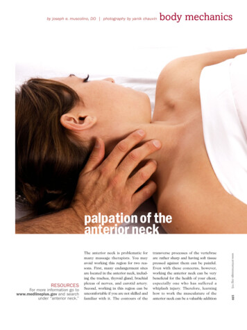
by joseph e. muscolino, DO photography by yanik chauvinbody mechanicspalpation of theanterior neck159transverse processes of the vertebraeare rather sharp and having soft tissuepressed against them can be painful.Even with these concerns, however,working the anterior neck can be verybeneficial for the health of your client,especially one who has suffered awhiplash injury. Therefore, learninghow to work the musculature of theanterior neck can be a valuable additionwww.amtamassage.org/mtjRESOURCESFor more information go towww.medlineplus.gov and searchunder “anterior neck.”The anterior neck is problematic formany massage therapists. You mayavoid working this region for two reasons. First, many endangerment sitesare located in the anterior neck, including the trachea, thyroid gland, brachialplexus of nerves, and carotid artery.Second, working in this region can beuncomfortable if you are not skilled andfamiliar with it. The contours of the
THE MUSCLES OF THE ANTERIOR NECKFIGURE 1A is asuperficial anteriorview. The platysmais shown on theright and removedon the left.SUPERIORHyoid musclesMandibleCommon carotid arteryHyoid boneInternal jugular tor Upper trapeziusOmohyoidINFERIORCommoncarotid arteryMastoidprocessLongus capitusInternaljugular veinFIGURE 1B is adeep anterior view.The right side illustrates the SCM, scalene group and theprevertebral group.The left side has theSCM removed for abetter view of theother musclesLongus colli160 mtj/massage therapy journal spring rtery1st ribBrachialplexusto a massage therapist’s practice. Andthe first step to learning how to safelyand effectively work the anterior neck islearning how to identify, locate and palpate the muscles of this region.The anterior neck is home to a number of important muscles, including thesternocleidomastoid (SCM), scalenegroup and the prevertebral group ofmuscles (Figures 1A and 1B).* Functionally, the muscles of the anteriorneck are flexors of the neck at the spinaljoints. Consequently, during a typicalwhiplash accident when a person’s headand neck are forcibly thrown back intoextension, the muscles of the anteriorneck are excessively stretched, triggering the muscle spindle stretch reflex.This results in tightness and spasmingof the muscles of the anterior neck.Beyond local pain from the tightness ofthese muscles, tightness of the SCM isassociated with proprioceptive disturbances of the neck, often resulting indizziness. Tightness of the scalenes canbe associated with compression uponnerves that provide innervation to theupper extremity. Finally, tightness ofthe prevertebral muscles can causereferral pain that is interpreted as a sorethroat. Given the prevalence ofwhiplash injuries, and the variety andextent of signs and symptoms that canresult, there can be tremendous valuein working the anterior neck musculature of our clients!The Scalene and Prevertebral MusclesWhile most of you are knowledgeableand comfortable working the SCM, thescalenes and prevertebral muscles areless often addressed. We will begin bylocating and palpating the SCM. TheSCM will then be used as a landmark forthe location and palpation of the scalene and prevertebral muscle groups.The SCM has two heads—a sternal* The hyoid group of muscles is also located in the anterior neck. This article will not address their palpation.Figure 1A courtesy The Muscular System Manual by Joseph Muscolino. Mosby, 2005. Figure 1B courtesy of Joseph Muscolino.
PALPATION OF THE SCMFIGURE 2B illustrates the therapistoffering resistanceto further lateralflexion of the headand neck toward thesame side; thisrequires the SCM tocontract, making itmore palpable.FIGURE 2C demonstrates supine palpation. The clientfirst rotates thehead and neck tothe opposite sideand then lifts thehead and neck offthe table, creating acontraction of theSCM.161* The clavicular head of the SCM is often not as visible as the sternal head and should be carefully palpated. Further, there is a great deal of variability regarding the relationship between the sternal and clavicular heads; often there is a gap between them, at times there is not.www.amtamassage.org/mtjand a clavicular head. Inferiorly, thesternal head attaches onto themanubrium of the sternum, and theclavicular head attaches onto the medialclavicle. Both heads conjoin and attachsuperiorly onto the mastoid process ofthe temporal bone and superior nuchalline of the occipital bone. The SCM canbe easily palpated with the client seatedor supine. With the client seated, standto the side that will be palpated. Ask theclient to first rotate the head and neck atthe spinal joints to the opposite side(contralateral rotation) and slightly laterally flex the head and neck to thesame side (ipsilateral lateral flexion)(Figure 2A). Now resist the client fromfurther lateral flexion (Figure 2B) andthe two heads of the SCM will be visibleand palpable. It is important to makesure that the client maintains the contralateral rotation; this is especially sofor the sternal head because this head ismore active in creating the rotationcomponent of the SCM’s actions. If theclavicular head is not readily palpable,ask the client to increase the force ofresistance of ipsilateral lateral flexionbecause the clavicular head is moreactive in creating the lateral flexioncomponent of the SCM’s actions.After palpating the entire length ofboth heads of the SCM* with the musclecontracted, palpate the SCM again whileit is relaxed so that its resting baselinetone can be assessed. When palpatingthe SCM, be careful not to place excessive pressure upon the carotid arterybecause this will stimulate a neurologicreflex that can lower blood pressure;you can tell if you are pressing upon thecarotid artery by feeling for a pulseunder your fingertips.Supine palpation of the SCM is alsostraightforward. The client is supinewhile you are seated at the head of thetable. First ask the client to contralater-FIGURE 2A showsthe beginningposition for seatedpalpation. Standbehind the clientand to the side thatwill be palpated; theclient rotates thehead and necktoward the oppositeside and slightlylaterally flexes thehead and necktoward the sameside.
162 mtj/massage therapy journal spring 2006SEATED PALPATION OF THE SCALENEGROUP OF MUSCLESally rotate the head and neck fully to oneside; then ask the client to lift the headand neck up off the table. The SCM willbe visible and palpable (Figure 2C).Now that the SCM has been located,its lateral border can be used as a landmark for palpation of the scalene group.The scalene group of muscles is composed of three muscles—the anterior,middle and posterior scalenes. (Theirnames reflect their positions relative toeach other.) As a group, the scalenesattach inferiorly to the first and secondribs; superiorly they attach to the transverse processes of the second throughseventh cervical vertebrae.To palpate the scalenes with theclient seated, stand behind the clientand locate the lateral border of the SCM(be sure that you have the lateral borderof the clavicular head) (Figure 3A).From there, move your palpating fingersslightly laterally off the SCM—you willbe over the scalenes. Now ask the clientto take in a short quick breath throughthe nose and feel for the contraction ofthe scalenes (Figure 3B). Taking in abreath requires elevation of the ribs,which is an action of the scalene group.Once located, palpate the scaleneswhile they are contracted, and thenwhile they are relaxed so that you canassess their baseline tone. Be sure toexplore the entire breadth of thescalenes within the anterior aspect ofthe posterior triangle of the neck (Figures4A, 4B). The posterior triangle of theneck is the region of the neck boundedanteriorly by the SCM, posteriorly bythe upper trapezius and inferiorly bythe clavicle. Superficial within the posterior triangle are the scalenes, levatorscapulae, splenius capitis and the inferior belly of the omohyoid. (These muscles are all deep to the platysma, whichis very thin and does not impede palpa-FIGURE 3A. Standbehind the clientand locate the lateral border of theclavicular head ofthe SCM.FIGURE 3B. Moveyour palpating fingers just lateral tothat and you will beover the scalenemuscles. By askingthe client to take ina short quick breaththrough the nose,the scalenes can befelt to contract.tion of the muscles that are deep to it.)When palpating the scalenes, be careful not to exert excessive pressure uponthe brachial plexus of nerves and/or thesubclavian artery. These structurestravel between the anterior and middlescalenes.* If you feel a pulse under yourfingertips or the client reports tinglingdown the upper extremity, move yourpalpating fingers.The prevertebral group of musclesconsists of the longus colli, longus capitis, rectus capitis anterior and rectuscapitis lateralis. Of these, the longuscolli and longus capitis can be easily palpated; these two muscles lie along theanterolateral vertebral column from thevertebral level of T3 to the skull. Tolocate the longus muscles, we will againuse the SCM as our landmark. This* When the anterior and middle scalenes are excessively tight, they can impinge upon the brachial plexus of nerves and/or the subclavian arterycausing symptoms anywhere within the upper extremity. This condition is called anterior scalene syndrome and is one of the three types of thoracicoutlet syndrome.
THE POSTERIOR TRIANGLE OF THE NECKSUPERIORFIGURE 4A shows alateral view of themuscles locatedsuperficially withinthe posterior triangle.Splenius CapitisPOSTERIORHyoid boneLevator scapulaeANTERIORSternocleidomastoidScalenesUpper trapeziusOmohyoidClavicleINFERIORFIGURE 4B showsan anterior viewof the posteriortriangle with theborders drawn. Eventhough it is namedthe posterior triangle, its location inthe neck is primarilyanterolateral.Outline ofposterior trianglewww.amtamassage.org/mtj163Figure 4A courtesy The Muscular System Manual by Joseph Muscolino. Mosby, 2005. Figure 4B courtesy of Joseph Muscolino.
SEATED PALPATION OF THE LONGUS COLLI AND LONGUSCAPITIS MUSCLES OF THE PREVERTEBRAL GROUP164 mtj/massage therapy journal spring 2006 To read tips on how to prevent neckpain, go to www.aapmr.org/condheat/pain/necktips.htm.time, locate the medial border of thesternal head of the SCM. Next, palpatejust medial to that. Because the longusmuscles are located deep against thespine, to access them you must gently,but firmly, sink into the tissue in theposterior direction aiming toward thespinal column. It is important that thisbe done slowly or it will be very uncomfortable for the client. To bring out acontraction of these muscles so thatthey are more palpable, resist the clientfrom flexing the head and neck againstyour hand. When palpating the longusmuscles, be careful not to exert toomuch pressure against the trachea.Otherwise, it will be irritated, and theclient may involuntarily cough. (Seeright, Figures 5A-5C for this series.)While many therapists are hesitant toapproach the muscles of the anteriorneck, it usually only takes a little practice to locate and palpate them, andonly a little more practice to becomesmooth and comfortable at it. Ofcourse, the more proficient youbecome, the more comfortable thesepalpations are for you and the client.Once you are proficient at palpatingthese muscles, working them in a therapeutic manner easily follows!IJoseph E. Muscolino,DC, is an instructor atthe Connecticut Centerfor Massage Therapyand the owner of LearnMuscles in Redding,Connecticut. He is alsothe author of The Muscular System Manual & Kinesiology textbook (Elsevier, 2006).FIGURE 5A. Beginby locating themedial border of thesternal head of theSCMFIGURE 5B. Thenmove just medial tothat. Because thesemuscles are deep,you must sink intothe tissue towardthe spine, slowly, ina gentle but firmmanner.FIGURE 5C. Thenask the client to tryto flex the head andneck against yourresistance. This willcause the musclesto contract, makingthem more easilypalpable.
many massage therapists. You may avoid working this region for two rea-sons. First, many endangerment sites are located in the anterior neck, includ-ing the trachea, thyroid gland, brachial plexus of nerves, and carotid artery. Second, working in this region can be uncomfortable if you are not skilled and familiar with it. The contours of the