Transcription
CHAPTER 13Normal Labor and DeliverySarah Kilpatrick and Etoi GarrisonLabor: Definition and PhysiologyMechanics of Labor 268267Uterine Activity (Powers) 270The Fetus (Passenger) 270The Maternal Pelvis (Passage) 272Cardinal Movements in LaborEngagement 275Descent 277275Flexion 277Internal Rotation 277Extension 277External Rotation 277Expulsion 277Normal Progress of Labor277Interventions Affecting Normal LaborOutcomes 279KEY ABBREVIATIONSAmerican College of Obstetriciansand GynecologistsCephalopelvic DisproportionLeft Occiput AnteriorOcciput AnteriorOcciput PosteriorOcciput TransverseProstaglandinsRandomized Controlled TrialRight Occiput AnteriorActive Management of Labor 279Second Stage of Labor 280ACOGCPDLOAOAOPOTPGsRCTROALABOR: DEFINITION AND PHYSIOLOGYLabor is defined as the process by which the fetus isexpelled from the uterus. More specifically, labor requiresregular, effective contractions that lead to dilation andeffacement of the cervix. This chapter describes thephysiology and normal characteristics of term labor anddelivery.The physiology of labor initiation has not been completely elucidated, but the putative mechanisms havebeen well reviewed by Liao and colleagues.1 Labor initiation is species-specific, and the mechanisms in humanlabor are unique. The four phases of labor from quiescenceto involution are outlined in Figure 13-1.2 The first phaseis quiescence and represents that time in utero beforelabor begins when uterine activity is suppressed by theaction of progesterone, prostacyclin, relaxin, nitric oxide,parathyroid hormone–related peptide, and possibly otherhormones. During the activation phase, estrogen begins tofacilitate expression of myometrial receptors for prostaglandins (PGs) and oxytocin, which results in ion channelactivation and increased gap junctions. This increase in thegap junctions between myometrial cells facilitates effective contractions.3 In essence, the activation phase readiesSpontaneous Vaginal Delivery 280Delivery of the Placenta and FetalMembranes 281Episiotomy, Perineal Injury,and Perineal Repair 281the uterus for the subsequent stimulation phase, whenuterotonics, particularly PGs and oxytocin, stimulateregular contractions. In the human, this process at termmay be protracted, occurring over days to weeks. The finalphase, uterine involution, occurs after delivery and ismediated primarily by oxytocin. The first three phases oflabor require endocrine, paracrine, and autocrine interaction between the fetus, membranes, placenta, and mother.The fetus has a central role in the initiation of term laborin nonhuman mammals; in humans, the fetal role is notcompletely understood (Figure 13-2).2-5 In sheep, termlabor is initiated through activation of the fetal hypothalamicpituitary-adrenal axis, with a resultant increase in fetaladrenocorticotrophic hormone and cortisol.4,5 Fetal cortisolincreases production of estradiol and decreases productionof progesterone by a shift in placental metabolism of cortisol dependent on placental 17α-hydroxylase. The changein the progesterone/estradiol ratio stimulates placental production of oxytocin and PG, particularly PGF2α.4 If thisincrease in fetal adrenocorticotrophic hormone and cortisolis blocked, parturition is delayed.5 In contrast, humans lackplacental 17α-hydroxylase and there is no increase in fetalcortisol near term. Rather, in humans, uterine activationmay be potentiated in part by increased fetal adrenal production of dehydroepiandrostenedione, which is convertedin the placenta to estradiol and estriol. Placental estriolstimulates an increase in maternal (likely decidual) PGF2α,PG receptors, oxytocin receptors, and gap junctions. Inhumans, there is no documented decrease in progesteronenear term and a fall in progesterone is not necessary forlabor initiation. However, some research suggests thepossibility of a “functional progesterone withdrawal” inhumans: Labor is accompanied by a decrease in the concentration of progesterone receptors, as well as a changein the ratio of progesterone receptor isoforms A and B inboth the myometrium6,7 and membranes.8 More researchis needed to elucidate the precise mechanism throughwhich the human parturition cascade is activated. Fetalmaturation may play an important role, as well as maternal267
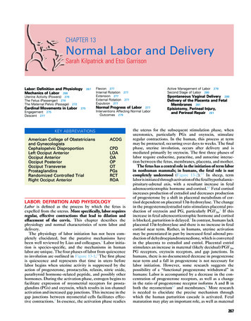
Uterine contractility268 Section III Intrapartum c oxideParathyroid hormonerelated peptide Corticotropinreleasing hormone Human placentallactogenUterotropinsEstrogen Progesterone Prostaglandins Corticotropinreleasing Oxytocin ThrombinTimePhase 0(Quiescence)Phase 1(Activation)Phase 2(Stimulation)Phase 3(Involution)ParturitionFIGURE 13-1. Regulation of uterine activity during pregnancy and labor. (Modified from Challis JRG, Gibb W: Control of parturition. Prenat NeonatMed 1:283, 1996.)cues that affect circadian cycling. There are distinct diurnalpatterns of contractions and delivery in most species, andin humans, the majority of contractions occur at night.2,9Oxytocin is used commonly for labor induction and augmentation; a full understanding of the mechanism of oxytocin action is helpful. Oxytocin is a peptide hormonesynthesized in the hypothalamus and released from theposterior pituitary in a pulsatile fashion. At term, oxytocinis a potent uterotonic agent that is capable of stimulatinguterine contractions at intravenous infusion rates of 1 to2 mIU/min.10 Oxytocin is inactivated largely in the liverand kidney, and during pregnancy, it is degraded primarilyby placental oxytocinase. Its biologic half-life is approximately 3 to 4 minutes, but appears to be shorter whenhigher doses are infused. Concentrations of oxytocin in thematernal circulation do not change significantly duringpregnancy or before the onset of labor, but they do riselate in the second stage of labor.10,11 Studies of fetal pituitary oxytocin production and the umbilical arteriovenousdifferences in plasma oxytocin strongly suggest that thefetus secretes oxytocin that reaches the maternal side ofthe placenta.10,12 The calculated rate of active oxytocinsecretion from the fetus increases from a baseline of1 mIU/min before labor to around 3 mIU/min after spontaneous labor.Significant differences in myometrial oxytocin receptordistribution have been reported, with large numbers offundal receptors and fewer receptors in the lower uterinesegment and cervix.13 Myometrial oxytocin receptorsincrease on average by 100- to 200-fold during pregnancy,reaching a maximum during early labor.10,11,14,15 This risein receptor concentration is paralleled by an increasein uterine sensitivity to circulating oxytocin. Specifichigh-affinity oxytocin receptors have also been isolatedfrom human amnion and decidua parietalis but not deciduavera.10,13 It has been suggested that oxytocin plays a dual rolein parturition. First, through its receptor, oxytocin directlystimulates uterine contractions. Second, oxytocin may actindirectly by stimulating the amnion and decidua toproduce PG.13,16,17 Indeed, even when uterine contractionsare adequate, induction of labor at term is successful onlywhen oxytocin infusion is associated with an increase inPGF production.13Oxytocin binding to its receptor activates phospolipaseC. In turn, phospholipase C increases intracellular calciumboth by stimulating the release of intracellular calcium andby promoting the influx of extracellular calcium. Oxytocinstimulation of phospholipase C can be blocked by increasedlevels of cyclic adenosine monophosphate. Increasedcalcium levels stimulate the calmodulin-mediated activation of myosin light-chain kinase. Oxytocin may alsostimulate uterine contractions via a calcium-independentpathway by inhibiting myosin phosphatase, which in turnincreases myosin phosphorylation. These pathways (PGF2αand intracellular calcium) have been the target of multipletocolytic agents: indomethacin, calcium channel blockers,beta mimetics (through stimulation of cyclic adenosinemonophosphate), and magnesium.MECHANICS OF LABORLabor and delivery are not passive processes in whichuterine contractions push a rigid object through a fixedaperture. The ability of the fetus to successfully negotiatethe pelvis during labor and delivery depends on thecomplex interactions of three variables: uterine activity,
Chapter 13 Normal Labor and Delivery 26916-hydroxy-dehydroepiandrostenedione sulfate from fetal adrenalFETUSPLACENTA/FETAL MEMBRANES? negativefeedback loop? FetaltriggerMOTHERCortisol11 oneHypothalamusProgesterone17 -hydroxypregnenoloneHypothalamusCortisone17 -hydroxylase/17,20-desmolasePlacentalsulfatase17 dione3 ropichormone17-oxidoreductase17 -estradiol16-hydroxylaseAdrenalglandFrom fetalzone pase A2CyclooxygenaseInhibited byprogesteroneacting E2(PGF2 )16-hydroxyprostaglandindehyrogenasePreparesfetal organsystems fordeliveryArachidonic acidCortisolProstaglandin receptorsOxytocin receptorsGap junctionsDecidualPGF2Membrane phospholipidsFromdefinitiveadrenalcortexPlacental PGFM)Placental corticotropinreleasing hormoneUterusRupture hydroepiandrostenedionesulfate to placenta/fetal membranesFIGURE 13-2. Proposed “parturition cascade” for labor induction at term. The spontaneous induction of labor at term in the human is regulatedby a series of paracrine/autocrine hormones acting in an integrated parturition cascade responsible for promoting uterine contractions. PGE2,Prostaglandin E2; PGEM, 13, 14-dihydro-15-keto-PGE2; PGF2α, prostaglandin F2α; PGFM, 13, 14-dihydro-15keto-PGF2α. (Modified from NorwitzER, Robinson JN, Repke JT: The initiation of parturition: a comparative analysis across the species. Curr Prob Obstet Gynecol Fertil 22:41, 1999.)
270 Section III Intrapartum Carethe fetus, and the maternal pelvis (Powers, Passenger,Passage).Uterine Activity (Powers)The powers refer to the forces generated by the uterinemusculature. Uterine activity is characterized by the frequency, amplitude (intensity), and duration of contractions.Assessment of uterine activity may include simple observation, manual palpation, external objective assessmenttechniques (such as external tocodynamometry), and directmeasurement via an internal uterine pressure catheter(IUPC). External tocodynamometry measures the changein shape of the abdominal wall as a function of uterinecontractions and, as such, is qualitative rather than quantitative. Although it permits graphic display of uterineactivity and allows for accurate correlation of fetal heartrate patterns with uterine activity, external tocodynamometry does not allow measurement of contraction intensityor basal intrauterine tone. The most precise method fordetermination of uterine activity is the direct measurement of intrauterine pressure with an IUPC. However, thisprocedure should not be performed unless indicated giventhe small but finite associated risks of uterine perforation,placental disruption, and intrauterine infection.Despite technologic improvements, the definition of“adequate” uterine activity during labor remains unclear.Classically, three to five contractions per 10 minutes hasbeen used to define adequate labor; this pattern has beenobserved in approximately 95% of women in spontaneouslabor. In labor, patients usually contract every 2 to 5minutes, with contractions becoming as frequent as every2 to 3 minutes in late active labor, as well as during thesecond stage. Abnormal uterine activity can also beobserved either spontaneously or resulting from iatrogenicinterventions. Tachysystole is defined as more than five contractions in 10 minutes, averaged over 30 minutes. If tachysytole occurs, documentation should note the presence orabsence of fetal heart rate (FHR) decelerations. The termhyperstimulation should no longer be used.18Various units have been devised to objectively measureuterine activity, the most common of which is the Montevideo unit (MVU), a measure of average frequency andamplitude above basal tone (the average strength of contractions in millimeters of mercury multiplied by thenumber of contractions per 10 minutes). Although 150 to350 MVU has been described for adequate labor, 200 to250 MVU is commonly accepted to define adequate laborin the active phase of labor.19,20 There are no data thatidentify adequate forces during latent labor. Although it isgenerally believed that optimal uterine contractions areassociated with an increased likelihood of vaginal delivery,there are limited data to support this assumption. If uterinecontractions are “adequate” to effect vaginal delivery, oneof two things will happen: either the cervix will efface anddilate, and the fetal head will descend, or there will beworsening caput succedaneum (scalp edema) and moldingof the fetal head (overlapping of the skull bones) withoutcervical effacement and dilation. The latter situation suggests the presence of cephalopelvic disproportion (CPD),which can be either absolute (in which a given fetus issimply too large to negotiate a given pelvis) or relative (inwhich delivery of a given fetus through a given pelvisLongitudinal lieOblique lieTransverse lieFIGURE 13-3. Examples of different fetal lie.would be possible under optimal conditions, but is precluded by malposition or abnormal attitude of the fetalhead), or pelvic outlet obstruction such as with uterinefibroids.The Fetus (Passenger)The passenger, of course, is the fetus. Several fetal variables influence the course of labor and delivery.1. Fetal size can be estimated clinically by abdominalpalpation or with ultrasound, but both are subject to alarge degree of error. Fetal macrosomia (defined by theAmerican College of Obstetricians and Gynecologists[ACOG] as actual birth weight greater than 4500 g21)is associated with an increased likelihood of failedtrial of labor and may be associated with laborabnormalities.222. Lie refers to the longitudinal axis of the fetus relative tothe longitudinal axis of the uterus. Fetal lie can belongitudinal, transverse, or oblique (Figure 13-3). In asingleton pregnancy, only fetuses in a longitudinal liecan be safely delivered vaginally.3. Presentation refers to the fetal part that directlyoverlies the pelvic inlet. In a fetus presenting in thelongitudinal lie, the presentation can be cephalic(vertex) or breech. Compound presentation refers tothe presence of more than one fetal part overlyingthe pelvic inlet, such as a fetal hand and the vertex.Funic presentation refers to presentation of theumbilical cord and is rare at term. In a cephalicfetus, the presentation is classified according tothe leading bony landmark of the skull, which canbe either the occiput (vertex), the chin (mentum),or the brow (Figure 13-4). Malpresentation, referringto any presentation other than vertex, is seen inapproximately 5% of all term labors.4. Attitude refers to the position of the head withregard to the fetal spine (the degree of flexion and/orextension of the fetal head). Flexion of the head isimportant to facilitate engagement of the head in thematernal pelvis. When the fetal chin is optimally
Chapter 13 Normal Labor and Delivery 271BROWOCCIPUTMENTUM (CHIN)FIGURE 13-4. Landmarks of fetal skull for determination of fetal position.flexed onto the chest, the suboccipitobregmaticdiameter (9.5 cm) presents at the pelvic inlet (Figure13-5). This is the smallest possible presentingdiameter in the cephalic presentation. As the headdeflexes (extends), the diameter presenting to thepelvic inlet progressively increases even before themalpresentations of brow and face are encountered(see Figure 13-5), and may contribute to failure toprogress in labor. The architecture of the pelvic flooralong with increased uterine activity may correctdeflexion in the early stages of labor.5. Position of the fetus refers to the relationship of thefetal presenting part to the maternal pelvis, and it canbe assessed most accurately on vaginal examination.For cephalic presentations, the fetal occiput is thereference. If the occiput is directly anterior, theposition is occiput anterior (OA). If the occiput isturned toward the mother’s right side, the positionis right occiput anterior (ROA). In the breechpresentation, the sacrum is the reference (rightsacrum anterior). The various positions of a cephalicpresentation are illustrated in Figure 13-6. In a vertexpresentation, position can be determined by palpationof the fetal sutures. The sagittal suture is the easiestto palpate. Palpation of the distinctive lamdoidsutures should identify the position of the fetalFIGURE 13-5. Presenting diameters of the average term fetal skull.occiput. The frontal suture can also be used todetermine the position of the front of the vertex.Most commonly, the fetal head enters the pelvis in atransverse position and, then as a normal part of labor,rotates to an OA position. Most fetuses deliver in theOA, LOA, or ROA position. In the past, less than10% of presentations were occiput posterior (OP) atdelivery.23 However, epidural analgesia is associatedwith an increased risk of OP presentation (observedin 12.9% of women with epidural analgesia).24
272 Section III Intrapartum CareFIGURE 13-6. Fetal presentations and positions inlabor. LOA, Left occiput anterior; LOP, left occiputposterior; LOT, left occiput transverse; ROA, rightocciput anterior; ROT, right occiput transverse;ROP, right occiput posterior. (Modified fromNorwitz ER, Robinson J, Repke JT: The initiationand management of labor. In Seifer DB, SamuelsP, Kniss DA [eds]: The Physiologic Basis ofGynecology and Obstetrics. Philadelphia, LippincottWilliams & Wilkins, 2001.)Asynclitism occurs when the sagittal suture is notdirectly central relative to the maternal pelvis. If thefetal head is turned such that more parietal bone ispresent posteriorly, the sagittal suture is more anteriorand this is referred to as posterior asynclitism.Anterior asynclitism occurs when there is moreparietal bone presenting anteriorly. The occiputtransverse (OT) and OP positions are less common atdelivery and more difficult to deliver. Malpositionrefers to any position in labor that is not ROA, OA, orLOA.6. Station is a measure of descent of the bony presentingpart of the fetus through the birth canal (Figure 13-7).The current standard classification ( 5 to 5) is basedon a quantitative measure in centimeters of thedistance of the leading bony edge from the ischialspines. The midpoint (0 station) is defined as theplane of the maternal ischial spines. The ischialspines can be palpated on vaginal examination atapproximately 8 o’clock and 4 o’clock. For theright-handed person, they are most easily felt on thematernal right.An abnormality in any of these fetal variables may affectboth the course of labor and the likelihood of vaginal delivery. For example, OP presentation is well known to beassociated with longer labor.25The Maternal Pelvis (Passage)The passage consists of the bony pelvis (composed ofthe sacrum, ilium, ischium, and pubis) and the resistance–2–10 1 2 3OLD CLASSIFICATION(Subjective)–5–4–3–2–10 1 2 3 4 5NEW CLASSIFICATION(Estimated distance incentimeters from theischial spines)FIGURE 13-7. The relationship of the leading edge of the presenting partof the fetus to the plane of the maternal ischial spines determinesthe station. Station 1/ 3 (old classification) or 2/ 5 (new classification) is illustrated.
Chapter 13 Normal Labor and Delivery 273FIGURE 13-8. Superior (A) and anterior (B) view of thefemale pelvis. (From Repke JT: Intrapartum Obstetrics.New York, Churchill Livingstone, 1996, p 68.)provided by the soft tissues. The bony pelvis is dividedinto the false (greater) and true (lesser) pelvis by the pelvicbrim, which is demarcated by the sacral promontory, theanterior ala of the sacrum, the arcuate line of the ilium, thepectineal line of the pubis, and the pubic crest culminatingin the symphysis (Figure 13-8). Measurements of thevarious parameters of the bony female pelvis have beenmade with great precision, directly in cadavers and usingradiographic imaging in living women. Such measurements have divided the true pelvis into a series of planesthat must be negotiated by the fetus during passagethrough the birth canal, which can be broadly classifiedinto the pelvic inlet, midpelvis, and pelvic outlet. X-raypelvimetry and computed tomography (CT) have beenused to define average and critical limit values for thevarious parameters of the bony pelvis (Table 13-1).26,27Critical limit values are measurements that are associatedwith a significant probability of CPD.26 However, CT andx-ray pelvimetry are rarely used, having been replaced by aclinical trial of the pelvis (labor). The remaining indications for x-ray or CT pelvimetry are evaluation for vaginalbreech delivery or evaluation of a woman who has suffereda significant pelvic fracture.28Clinical pelvimetry is currently the only method ofassessing the shape and dimensions of the bony pelvis inlabor. A useful protocol for clinical pelvimetry is detailedin Figure 13-9 and involves the assessment of the pelvicinlet, midpelvis, and pelvic outlet. The inlet of the truepelvis is largest in its transverse diameter (usually greaterthan 12.0 cm). The diagonal conjugate (the distance fromthe sacral promontory to the inferior margin of the symphysis pubis as assessed on vaginal examination) is a clinical representation of the anteroposterior diameter of thepelvic inlet. The true conjugate (or obstetric conjugate) of thepelvic inlet is the distance from the sacral promontory toAVERAGE AND CRITICAL LIMIT VALUES FORTABLE 13-1 PELVIC MEASUREMENTS BY X-RAY PELVIMETRYDIAMETERPelvic InletAnteroposterior (cm)Transverse (cm)Sum (cm)Area (cm2)Pelvic MidcavityAnteroposterior (cm)Transverse (cm)Sum (cm)Area (cm2)AVERAGE VALUE CRITICAL .0125.010.09.519.5106.0Modified from O’Brien WF, Cefalo RC: Labor and delivery. In Gabbe SG, NiebylJR, Simpson JL (eds): Obstetrics: Normal and Problem Pregnancies,ed 3. New York, Churchill Livingstone, 1996, p 377.*The critical limit values cited imply a high likelihood of cephalopelvicdisproportion.the superior aspect of the symphysis pubis. This measurement cannot be made clinically but can be estimated bysubtracting 1.5 to 2.0 cm from the diagonal conjugate. Thisis the smallest diameter of the inlet, and it usually measures approximately 10 to 11 cm. The limiting factor in themidpelvis is the interspinous diameter (the measurementbetween the ischial spines), which is usually the smallestdiameter of the pelvis but should be greater than 10 cm.The pelvic outlet is rarely of clinical significance. Theanteroposterior diameter from the coccyx to the symphysispubis is approximately 13 cm in most cases, and the transverse diameter between the ischial tuberosities is approximately 8 cm.The shape of the female bony pelvis can be classifiedinto four broad categories: gynecoid, anthropoid, android,and platypelloid (Figure 13-10). This classification, based
274 Section III Intrapartum Care1 Estimation of prominence ofsacral promontory2 Estimation of obstetric conjugate3 Assessment of transversediameter of pelvic inletTrue conjugatePELVIC ransverse diameter2 Assess curvature of the sacrum3 Assessment of interspinousdiameterPELVIC MIDCAVITY1 Estimation of prominence ofischial spinesSacral curvature2 Estimation of subpelvic angle3 Estimation of intertuberousdiameterPELVIC OUTLET1 Estimation of prominence ofcoccyxInterspinous diameterSubpelvicangleJWKOICoccyxFIGURE 13-9. A protocol for clinical pelvimetry.
Chapter 13 Normal Labor and Delivery 275FIGURE 13-10. Characteristics of the four types of female bony pelvis. (Modified from Callahan TL, Caughey AB, Heffner LJ [eds]: Blueprints inObstetrics and Gynecology. Malden, MA, Blackwell Science, 1998, p 45.)on the radiographic studies of Caldwell and Moloy, separates those with more favorable characteristics (gynecoid,anthropoid) from those that are less favorable for vaginaldelivery (android, platypelloid).29 In reality, however, manywomen fall into intermediate classes, and the distinctionsbecome arbitrary. The gynecoid pelvis is the classic femaleshape. The anthropoid pelvis with its exaggerated ovalshape of the inlet, largest anterioposterior diameter, andlimited anterior capacity is more often associated withdelivery in the OP position. The android pelvis is male inpattern and theoretically has an increased risk of CPD, andthe platypelloid pelvis with its broad, flat pelvis theoretically predisposes to a transverse arrest. Although theassessment of fetal size along with pelvic shape and capacity is still of clinical utility, it is a very inexact science. Anadequate trial of labor is the only definitive method todetermine whether a given fetus will be able to safelynegotiate a given pelvis.Pelvic soft tissues may provide resistance in boththe first and second stages of labor. In the first stage, resistance is offered primarily by the cervix, whereas in thesecond stage, it is offered by the muscles of the pelvic floor.In the second stage of labor, the resistance of the pelvicmusculature is believed to play an important role in therotation and movement of the presenting part through thepelvis.CARDINAL MOVEMENTS IN LABORThe mechanisms of labor, also known as the cardinalmovements, refer to the changes in position of fetal headduring its passage through the birth canal. Because ofthe asymmetry of the shape of both the fetal head and thematernal bony pelvis, such rotations are required for thefetus to successfully negotiate the birth canal. Althoughlabor and birth comprise a continuous process, seven discrete cardinal movements of the fetus are described: engagement, descent, flexion, internal rotation, extension, externalrotation or restitution, and expulsion (Figure 13-11).EngagementEngagement refers to passage of the widest diameter of thepresenting part to a level below the plane of the pelvic inlet(Figure 13-12). In the cephalic presentation with a wellflexed head, the largest transverse diameter of the fetalhead is the biparietal diameter (9.5 cm). In the breech, thewidest diameter is the bitrochanteric diameter. Clinically,engagement can be confirmed by palpation of the presenting part both abdominally and vaginally. With a cephalicpresentation, engagement is achieved when the presenting part is at 0 station on vaginal examination. Engagementis considered an important clinical prognostic sign becauseit demonstrates that, at least at the level of the pelvic inlet,
276 Section III Intrapartum CareFIGURE 13-11. Cardinal movements of labor.
Chapter 13 Normal Labor and Delivery 277pelvic inlet, the fetal head engages in an asynclitic fashion(i.e., with one parietal eminence lower than the other).With uterine contractions, the leading parietal eminencedescends and is first to engage the pelvic floor. As theuterus relaxes, the pelvic floor musculature causes the fetalhead to rotate until it is no longer asynclitic.ExtensionExtension occurs once the fetus has descended to the levelof the introitus. This descent brings the base of the occiputinto contact with the inferior margin at the symphysispubis. At this point, the birth canal curves upward. Thefetal head is delivered by extension and rotates around thesymphysis pubis. The forces responsible for this motionare the downward force exerted on the fetus by the uterinecontractions along with the upward forces exerted by themuscles of the pelvic floor.External RotationFIGURE 13-12. Engagement of the fetal head.the maternal bony pelvis is sufficiently large to allowdescent of the fetal head. In nulliparas, engagement of thefetal head usually occurs by 36 weeks’ gestation. In multiparas, however, engagement can occur later in gestationor even during the course of labor.DescentDescent refers to the downward passage of the presentingpart through the pelvis. Descent of the fetus is not continuous; the greatest rates of descent occur during thedeceleration phase of the first stage of labor and during thesecond stage of labor.FlexionFlexion of the fetal head occurs passively as the headdescends owing to the shape of the bony pelvis and theresistance offered by the soft tissues of the pelvic floor.Although flexion of the fetal head onto the chest is presentto some degree in most fetuses before labor, completeflexion usually occurs only during the course of labor. Theresult of complete flexion is to present the smallest diameter of the fetal head (the suboccipitobregmatic diameter)for optimal passage through the pelvis.Internal RotationInternal rotation refers to rotation of the presenting partfrom its original position as it enters the pelvic inlet (usuallyOT) to the anteroposterior position as it passes through thepelvis. As with flexion, internal rotation is a passive movement resulting from the shape of the pelvis and the pelvicfloor musculature. The pelvic floor musculature, includingthe coccygeus and ileococcygeus muscles, forms a V-shapedhammock that diverges anteriorly. As the head descends,the occiput of the fetus rotates toward the symphysis pubis(or, less commonly, toward the hollow of the sacrum),thereby allowing the widest portion of the fetus to negotiate the pelvis at its widest dimension. Owing to the angleof inclination between the maternal lumbar spine andExternal rotation, also known as restitution, refers to thereturn of the fetal head to the correct anatomic position inrelation to the fetal torso. This can occur
physiology and normal characteristics of term labor and delivery. The physiology of labor initiation has not been com-pletely elucidated, but the putative mechanisms have been well reviewed by Liao and colleagues. 1. Labor initia-tion is species-specific, and the mechanisms in human labor are unique. The four phases of labor from quiescence